Circulatory system
1/39
There's no tags or description
Looks like no tags are added yet.
Name | Mastery | Learn | Test | Matching | Spaced |
|---|
No study sessions yet.
40 Terms
Respiratory and Circulatory systems
Bring oxygen and nutrients to the cells
Work together to maintain homeostasis
Circulatory system
A transport system to supply all tissues with nutrients and to transport waste materials away from the tissues.
Including the cardiovascular and lymphatic systems
Transports nutrients, hormones, and metabolic wastes to different parts of the body
Closed system
Contains 2 fluids, blood and lymph
Components of Blood
Plasma (55%)
Platelets
Blood cells:
Red blood cells (erythrocytes): 45%
White blood cells (leucocytes)
Plasma
55% of blood volume
Pale yellow, clear liquid that is about 90% water
Includes transporting nutrients, gases and vitamins
=> Functions:
Transport medium for blood
Regulates fluid and electrolyte (Na+, K+, Ca2+) balance and maintains pH
red blood cells (erythrocytes)
Circular, flattened, biconcave discs with the centre of each cell thinner than its edge
No nucleus and mitochondria
Contain hemoglobin (T and R states)
Transport oxygen from the lung to the other tissue
From the stem cells in the red bone marrow of long bones
red blood cell production and its control
Low blood oxygen causes the kidneys and the liver to release erythropoietin (EPO) which stimulates RBC production
This is a negative feedback mechanism
White blood cells (leucocytes)
-White blood cells have nucleus, lack hemoglobin and colorless
-Protection against disease
Phagocytes: protect the body by ingesting harmful foreign particles, bacteria and dead or dying cells
Lymphocytes: eliminate the antigen by releasing antibodies (B cells) and cytotoxic granules (cytotoxic T cells)
Signaling to other cells of the immune system
Platelets (thrombocytes)
not true cells
fragments of cytoplasm from bone marrow cells
function in blood clotting.
Functions of blood
2 main functions:
Transport: substances such as oxygen, carbon dioxide, nutrients, waste products, hormones
Protection:
Ingest foreign particles through phagocytosis
Produce antibodies to combine with antigens from pathogen to destroy them
Clotting to protect humans from bleeding continuously
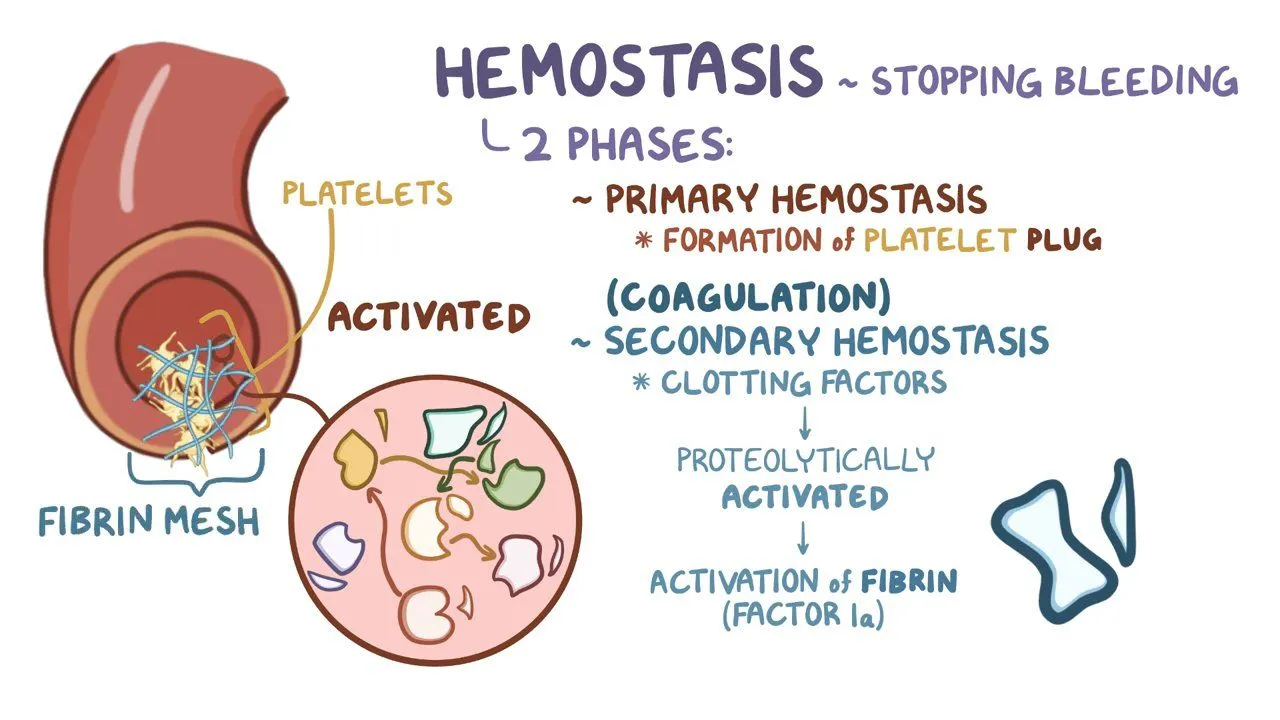
Hemostasis and Blood Coagulation
Following an injury, 4 events are available to stop the bleeding:
1) Blood Vessel Spasm: Vessels decrease in size to stop bleeding in small vessels.
2) Platelet Plug Formation: Inner torn layer of the vessels releases chemical signals that call platelets to the site of injury
3) Blood clotting: Requires the presence of certain clotting factors to form fibrin (Prothrombin + Ca2+→ Thrombin + Fibrinogen + Ca2+→ Fibrin)
4) Fibrinolysis: Begins the repair process
Agglutination
Clumping of red blood cells in response to a reaction between an antibody and an antigen
Antigens
A chemical that stimulates cells to produce antibody
Antibodies
A protein that reacts against a specific antigen
Cardiovascular system
consists of the heart and all blood vessels
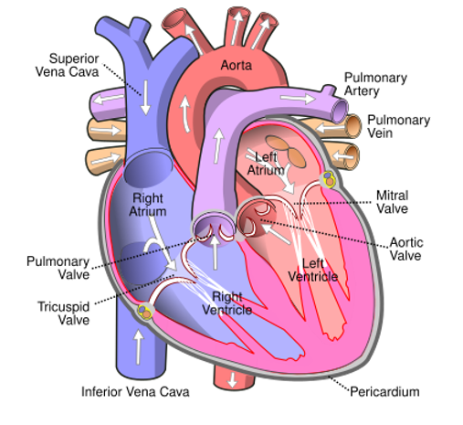
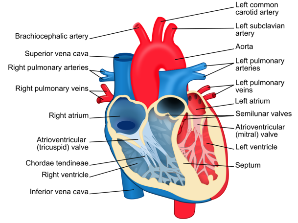
Heart
The heart lies in the thoracic cavity
coverings: pericardium
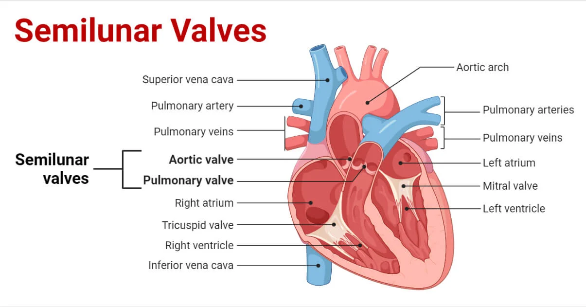
4 chambers (cavities):
Right and left atrium
Right and left ventricle
An atrioventricular valve connects each atrium to the ventricle below it
For the right: Tricuspid valve
For the left: Bicuspid valve (Mitral valve)
The septum: prevents mixing of blood from the 2 sides of the heart
The valves ensure that blood flows in only one direction
Atrium
Thin-walled, elastic
Expands as it collects blood, only pump blood the short distance to the ventricle
Ventricle
much thicker muscular wall, as it pumps blood either to the lungs or the rest of the body
The left-hand side of the heart
deals with the oxygenated blood from the lungs
The right-hand side of the heart
deals with the deoxygenated blood from the body
Blood vessels
make up 2 circuits:
+) Pulmonary circuit: eliminates carbon dioxide via lungs and oxygenates the blood
+) Systematic circuit: delivers oxygen to all body cells and carries away wastes
3 types of blood vessels:
Artery ( 3 layers of tissue)
Vein (3 layers of tissue)
Capillary ( 1 layer of tissue)
Arteries carry blood away from the heart
Veins carry blood toward the heart
The capillaries are exchanging vessels located between the arterial and venous systems
=> allow the exchange of gases, nutrients, hormones, and other molecules in the blood.
Aorta → Arteries → Arterioles → Capillaries → Venules → Veins → Vena Cava
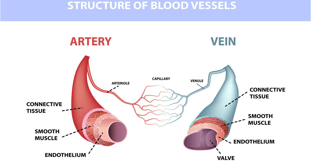
Arteries
Carry blood away from the heart
Bright red due to high oxygen levels
Thick muscle and elastic fibres:
the thick muscle can contract to push the blood along
the elastic fibres allow the artery to stretch under pressure
Veins
Carry blood towards the heart
Blood appears darker in color
Have valves which prevent the blood from going in the wrong direction
Thin muscle and elastic fibres
Capillaries
Connect the smallest arteriole and the smallest venule
Walls are very thin to exchange materials between the blood and other body cells.
One thick and permeable wall
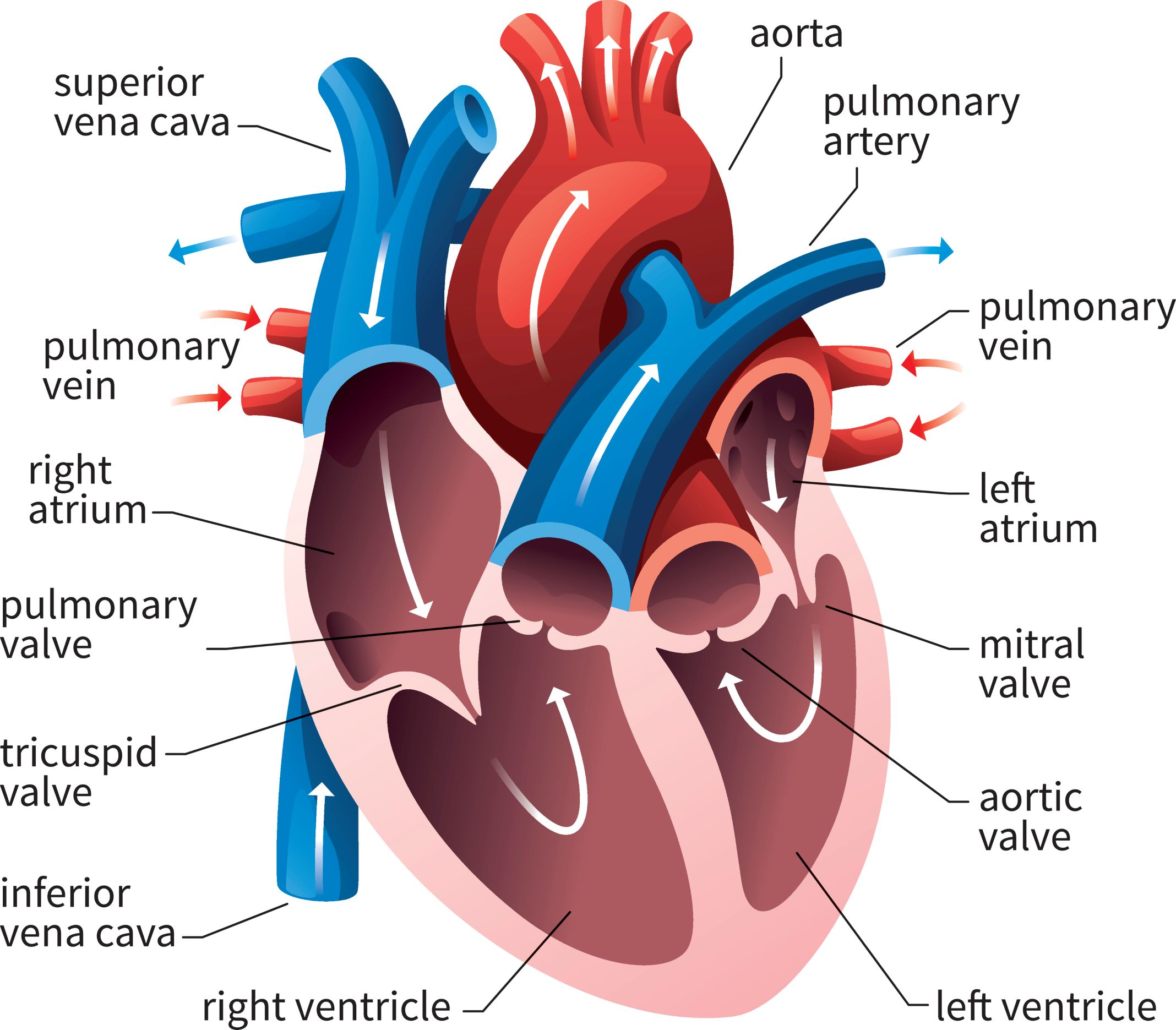
The flow of blood through the heart
Blood travels to the heart through the superior vena cava (head, neck, and forelimbs) and inferior vena cava (trunk and hind limbs)
The superior and inferior vena cava flow into the right atrium, moves into right ventricle, finally pushed into lungs in the pulmonary arteries.
The blood then picks up oxygen and travels back to the heart into left atrium through the pulmonary veins
The oxygen-rich blood travels through the left atrium to the left ventricle and exits to the body through the aorta
The aorta provides blood to the heart through the coronary arteries
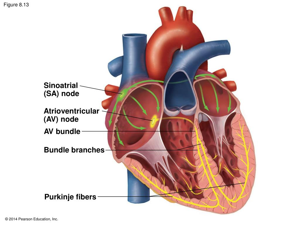
Initiating Contraction
Contraction of the heart is initiated by a small cluster of cardiac muscle cells called the sinoatrial (SA) node
In the upper wall of the right atrium
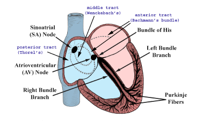
Contraction of the ventricle
The electrical impulse initiated by the SA node subsequently reaches another special area of the heart, known as the atrioventricular (AV) node.
The AV node is located in the septum between the atria
The AV node relays the electrical impulse to the muscle cells that make up the ventricles
=> The ventricles contract a fraction of a second after the atria
=> Completing one full heartbeat
Cardiac cycle
The heart contracts and relaxes in a rhythmic cycle called the cardiac cycle
Contains: systole and diastole
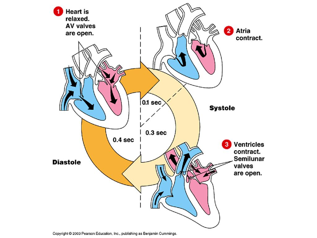
Systole
The contraction (pumping) phase
Occurs when the ventricles contract, closing the AV valves and opening the semilunar (SL) valves to pump blood into the 2 major vessels that exit the heart
Diastole
The relaxation, or filling, phase
Occurs when the ventricles relax, allowing the back pressure of the blood to close the SL valves and open the AV valves
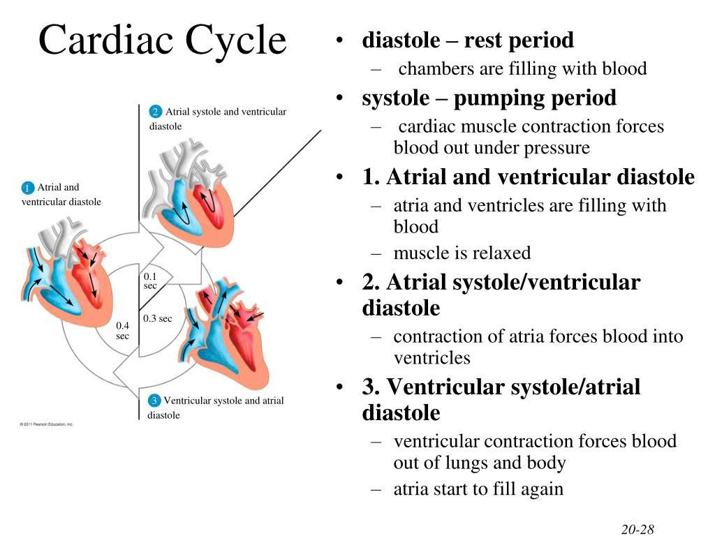
In atrial systole and ventricular diastole
Blood flows passively into the ventricles
The remaining 30% of blood is pushed into the ventricles.
The A-V valves open and the semilunar valves close
The ventricles relax
This causes an increase in ventricular pressure
In ventricular systole and atrial diastole:
The A-V valves close and semilunar valves open
The chordae tendineae prevent the cusps of the valves from bulging too far into the atria
The atria relax
The blood flows into atria
The ventricular pressure increases
The blood flows into pulmonary trunk and aorta
Electrocardiogram
In an average adult at rest, the heart beats about 70 times each minute.
The heart rate
Also called pulse, is the number of beats per minute
The stroke volume
the amount of blood pumped in a single contraction
The cardio output
the volume of blood pumped into the systemic circulation per minute
depends on both the heart rate and stroke volume
Regulation of the cardiac cycle
The SA node controls the heart rate
Factors that influence heart rate
Physical exercise
Body temperature
Concentration of ions: K+, Ca2+
Parasympathetic impulses decrease heart action
Sympathetic impulses increase heart action
Blood pressure
force exerted by blood as it moves through blood vessels
Heart attack
Occurs when an area of the heart muscle stops working and dies.
When an area of the brain dies the result is a stroke
Lymphatic system
Collects and recycles fluids leaked from the cardiovascular system, involved in fighting infections
Made up of a network of vessels called lymphatic vessels and tiny bean-shaped structures called lymph nodes
Lymph nodes may become swollen when they are fighting infection