CSB063 - MRI Spine
1/26
There's no tags or description
Looks like no tags are added yet.
Name | Mastery | Learn | Test | Matching | Spaced |
|---|
No study sessions yet.
27 Terms
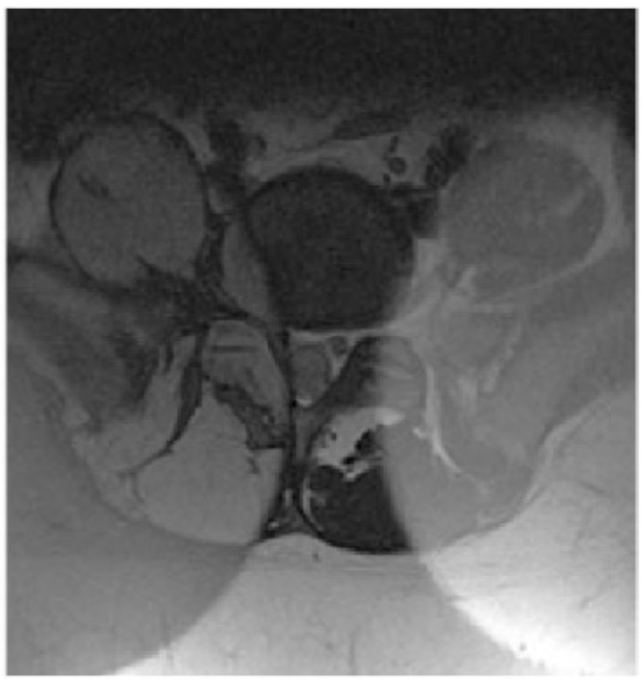
What weighting is this image? What structures appear hypointense in these sequences?
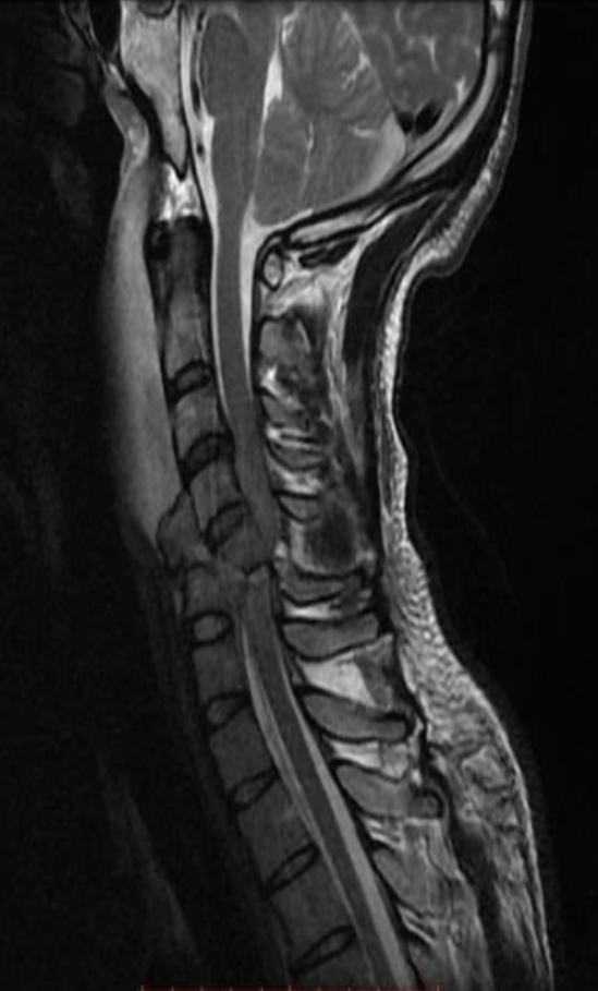
T2
Tendons
Ligaments
Cortical bone

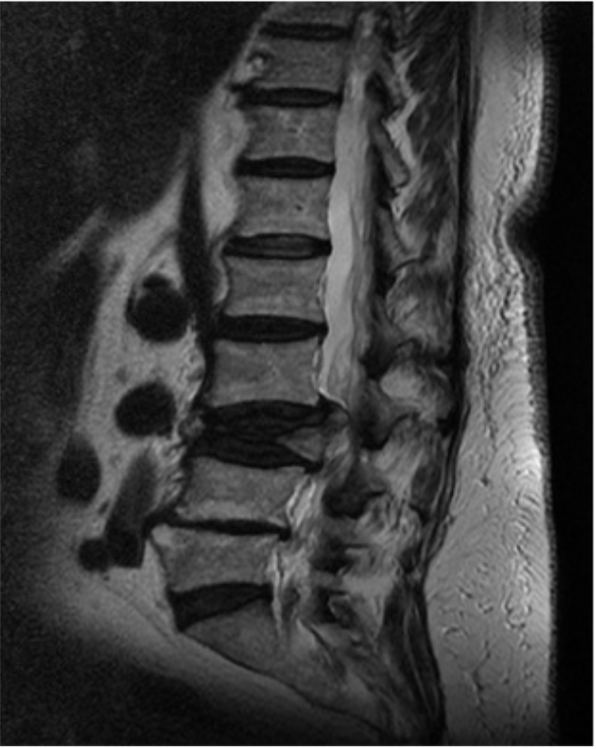
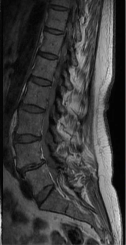
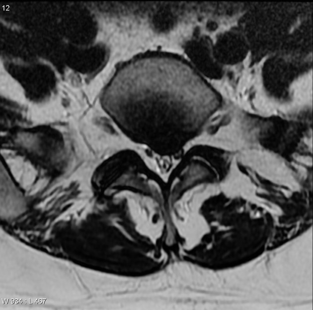
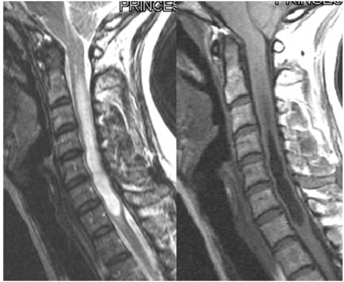
What weighting is this image? How can you tell?
T1 - CSF is dark/hypointense
Why is there a saturation band on this thoracic spine image?
To decrease respiratory motion and cardiac/aortic pulsation
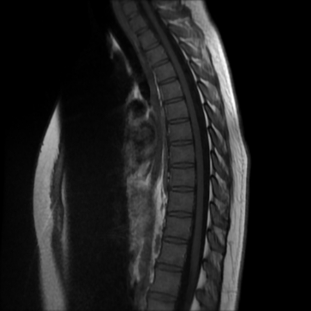
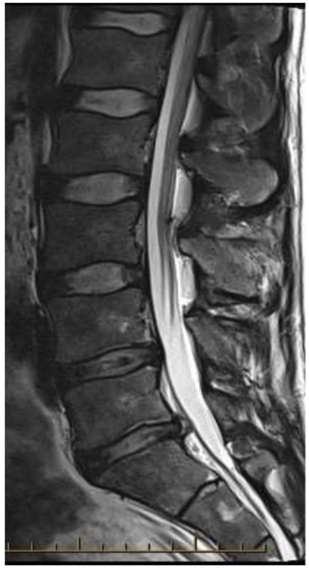
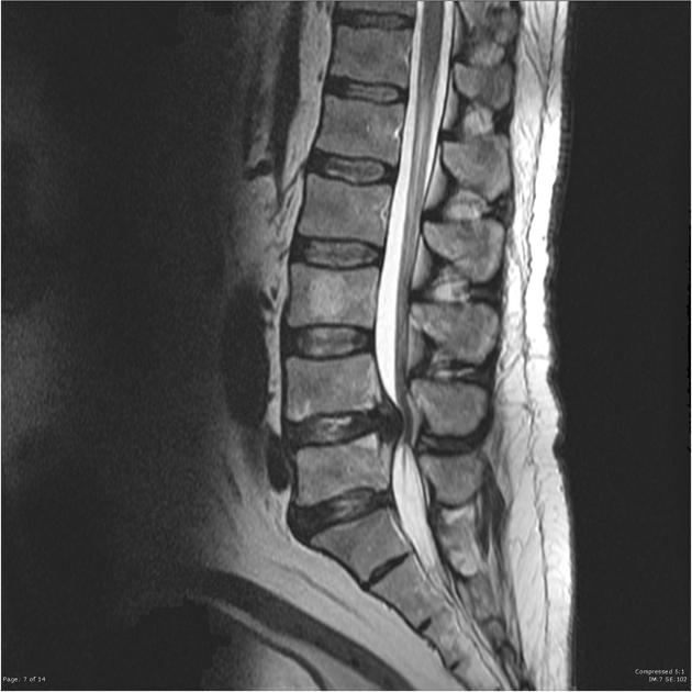
What weighting is this image? How can you tell?
T1 FS - CSF is dark/hypointense and fat is nulled
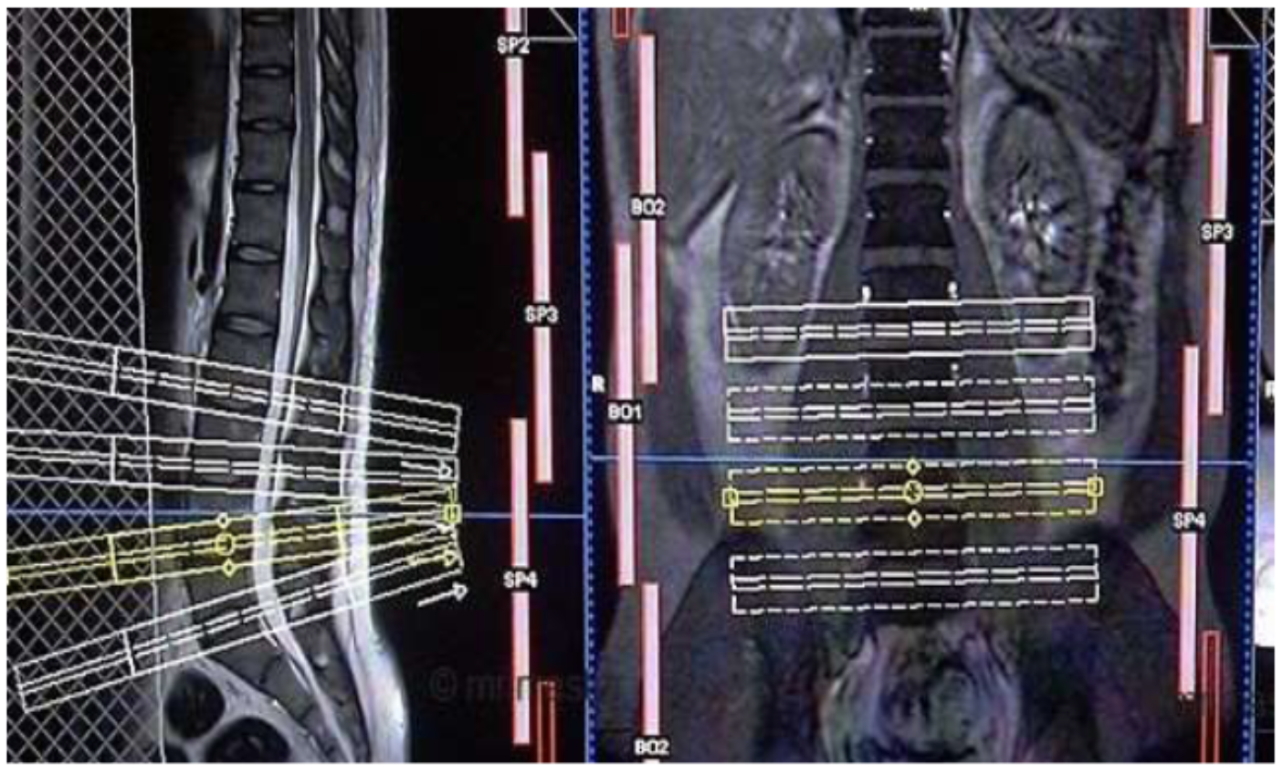
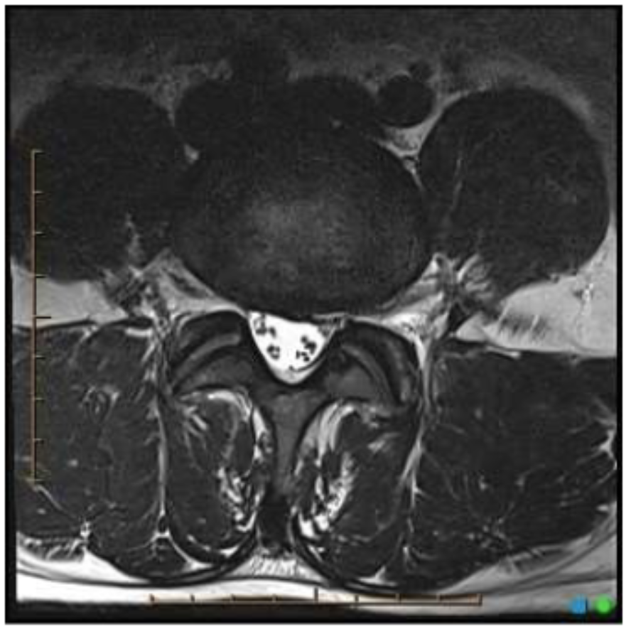
Why are the axial slices on these lumbar spine planning scans angled?
Due to the curvature of the lower lumbar vertebrae
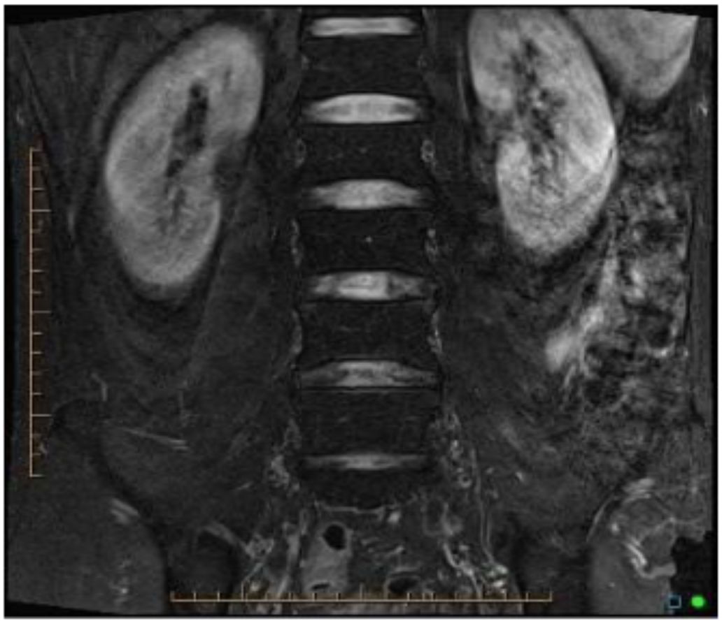
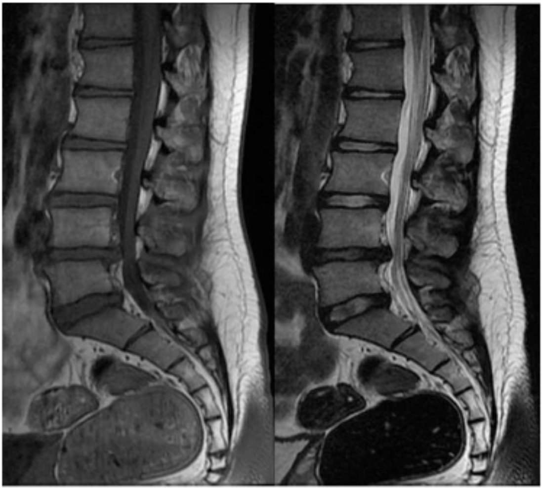
How was this image created? For what clinical indication would you perform this technique?
Each stack/region of spine was acquired separately (using the same FOV) and then stitched together
For scoliosis
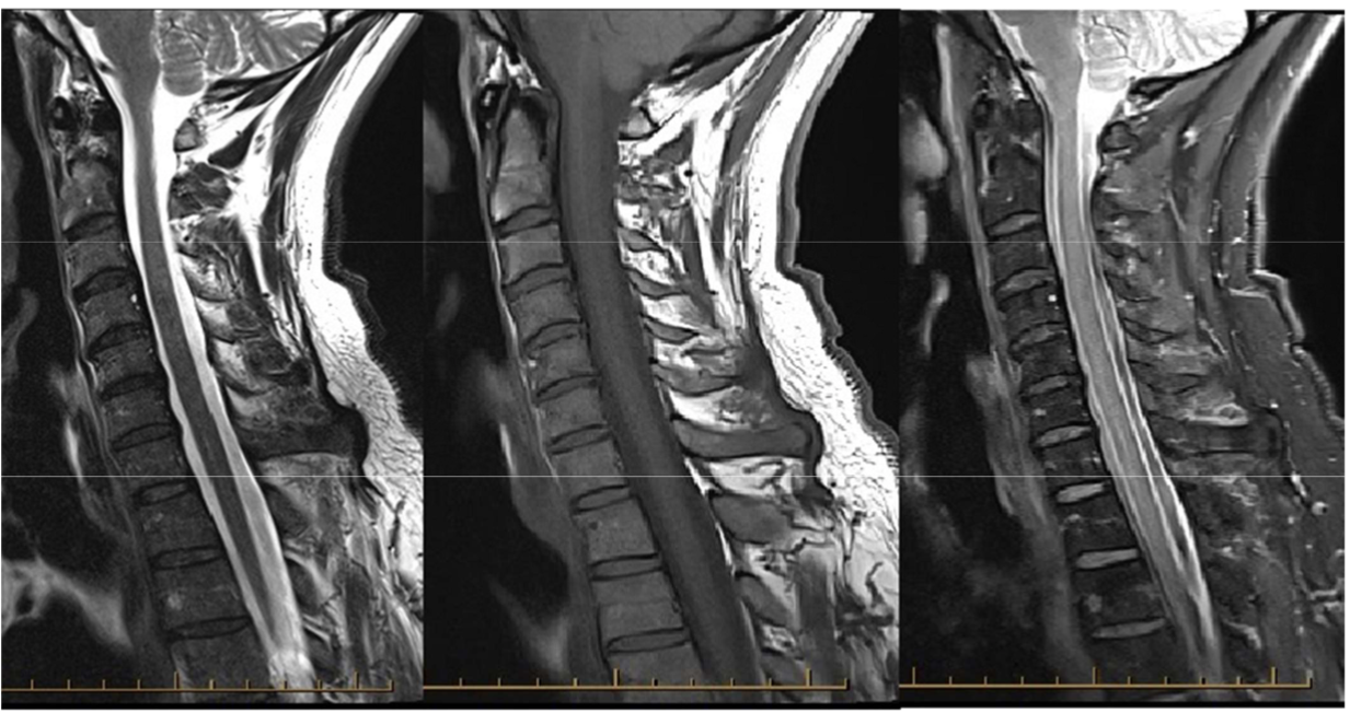
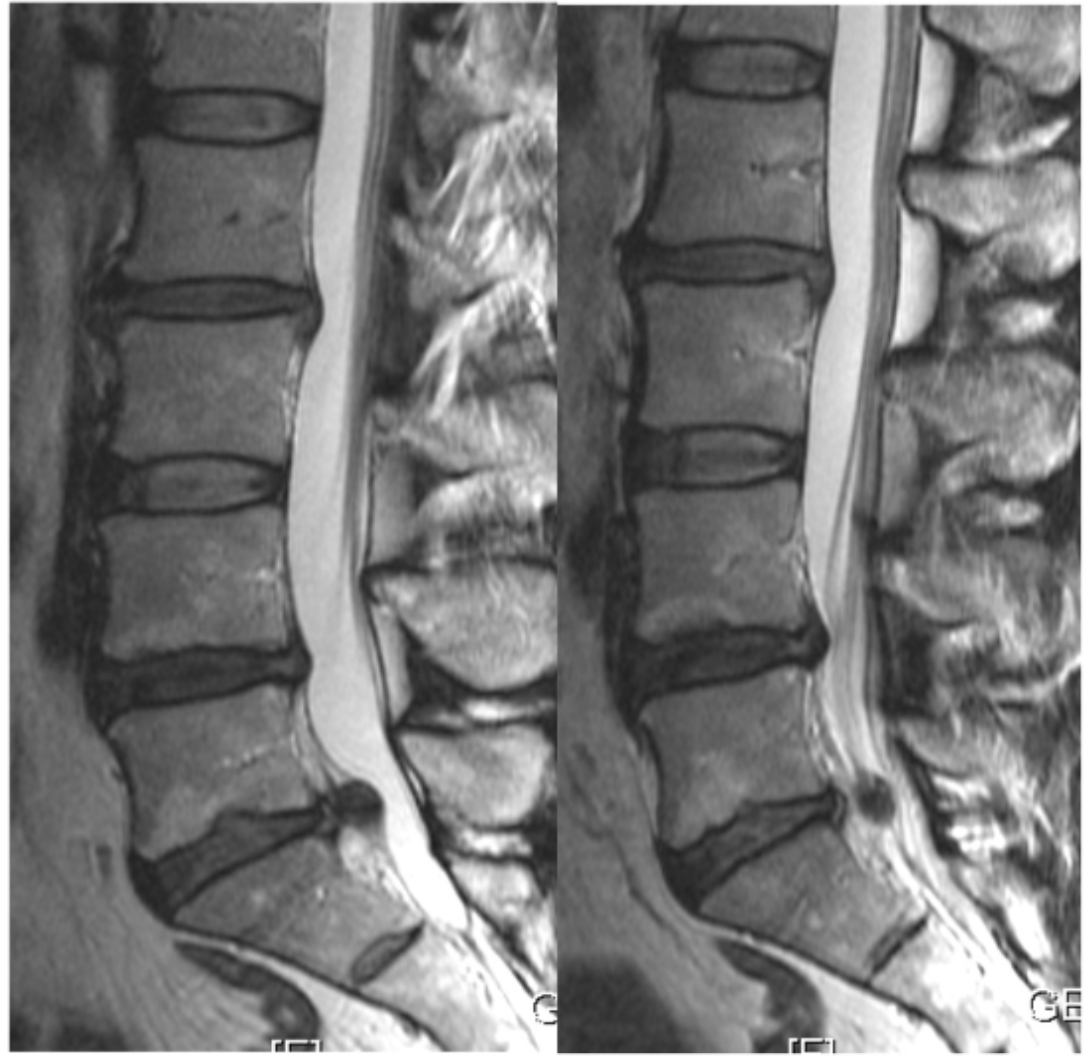
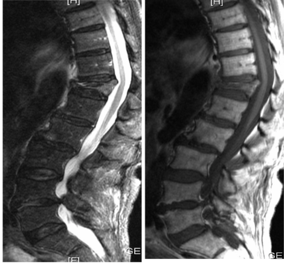
List the image weightings shown.
T2, T1, T2 STIR
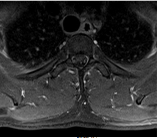
What is the image weighting of this slice?
T2
What is the image weighting of this slice?
T1
What is the image weighting of this slice?
T2
What is the image weighting of this slice?
T2 STIR
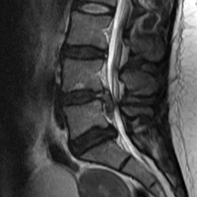
Name the main pathology demonstrated.
Spondylolisthesis of L5 (posteriorly)
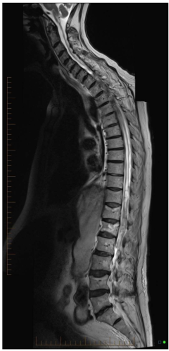
Name the main pathology demonstrated.
T5 vertebral body fracture causing cord impingement
Name this pathology.
Disc protrusion, impinging on left nerve root (S1)
Name this pathology.
Disc extrusion at L4/5
Name this pathology.
Disc protrusion at L4/5 and L5/S1 with canal involvement
Name the main pathology.
Disc sequestration posterior to L3 vertebral body
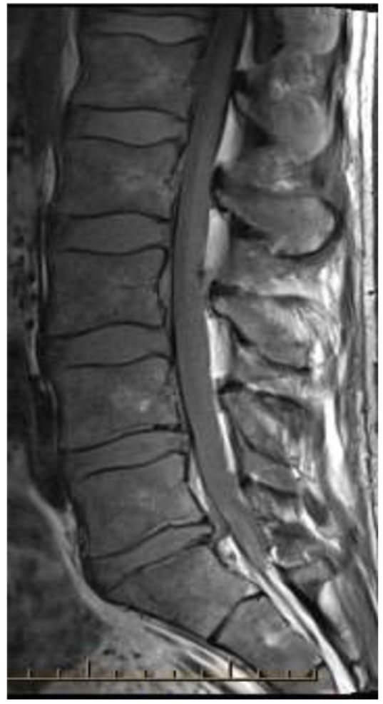
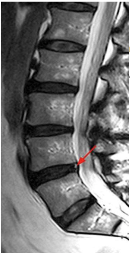
Name the pathology.
L5/S1 disc sequestration
Name the pathology (indicated by arrows).
Intravertebral herniation
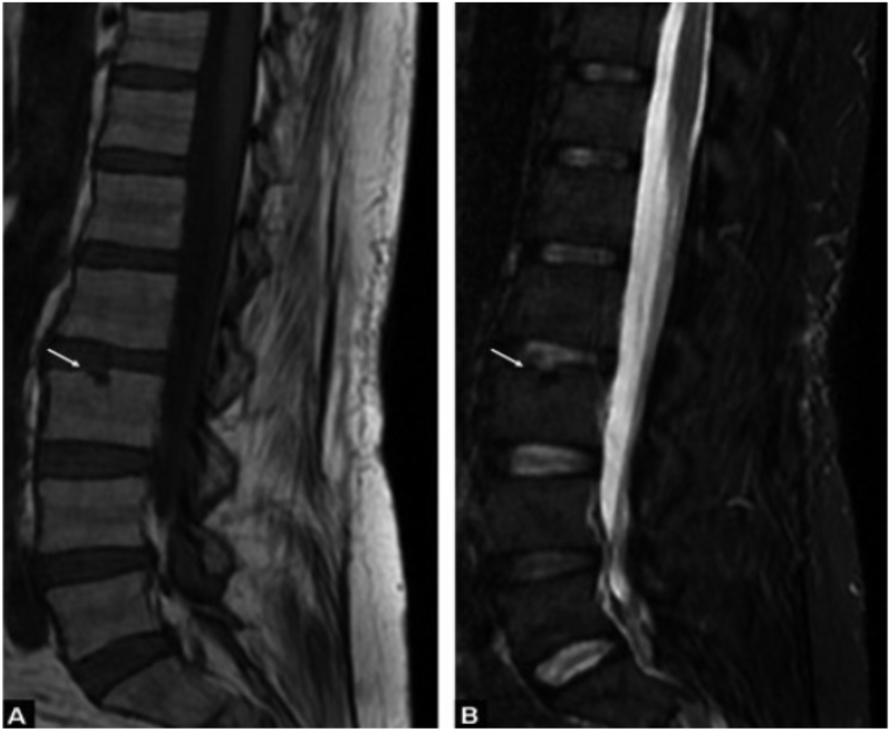
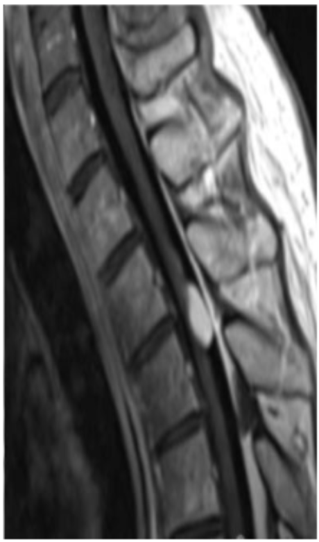
What might the high signal at the posterior aspect of the disc (indicated by red arror) be?
Annular tear
What might the high signal at the posterior aspect of the disc be?
Annular tear
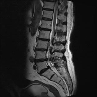
Name the main (bony) pathology.
Compression fracture, with canal involvement
Name the pathology and explain what is it.
Syrinx - fluid-filled cyst within the spinal cord
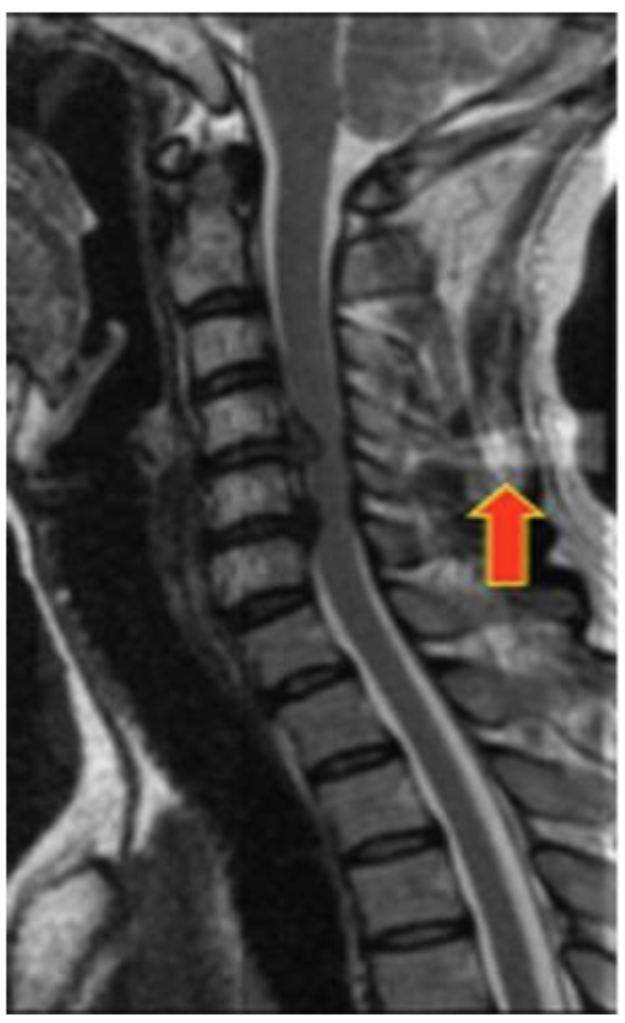
Name the weighting of this sequence and the pathology shown.
T1, hyperintense lesion around the spinal cord (tumour)
What artefact is present (red arrow)?
Aliasing artefact
What techniques can be utilised to overcome this artefacts?
Arms above head
Phase oversampling
Increase FOV
Saturation bands
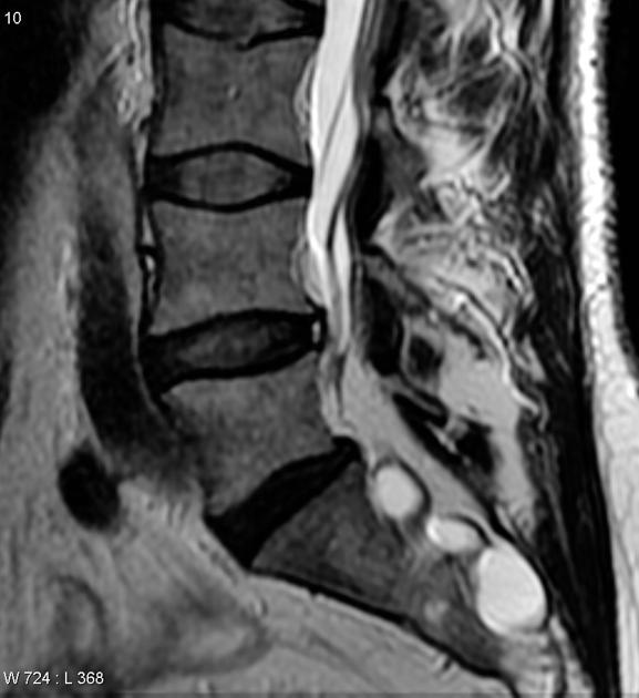
Name this pathology. What structure is it impinging on?
L4/5 disc bulge, impinging on the cauda equina