Testes: Stroma & Parenchyma

0.0(0)
studied byStudied by 0 people
0.0(0)
full-widthCall Kai
learnLearn
examPractice Test
spaced repetitionSpaced Repetition
heart puzzleMatch
flashcardsFlashcards
GameKnowt Play
Card Sorting

1/11

encourage image

There's no tags or description

Looks like no tags are added yet.

Study Analytics
Name
Mastery
Learn
Test
Matching
Spaced
Call with Kai

No study sessions yet.

12 Terms

1
New cards

What are the 2 structures that form testes?

  • Stroma (supporting tissue) #00a5ff

  • Parenchyma (functional tissue) #ff8300

2
New cards

What are the structures of the stroma? #00a5ff

  • Tunica vaginalis

  • Tunica albungiea

  • Septila testis

  • Mediastinum testis

  • Interstitial tissue

3
New cards

Stroma: What is

  • Tunica vaginalis

  • Tunica albungiea

  • Septila testis

  • Mediastinum testis

#00a5ff

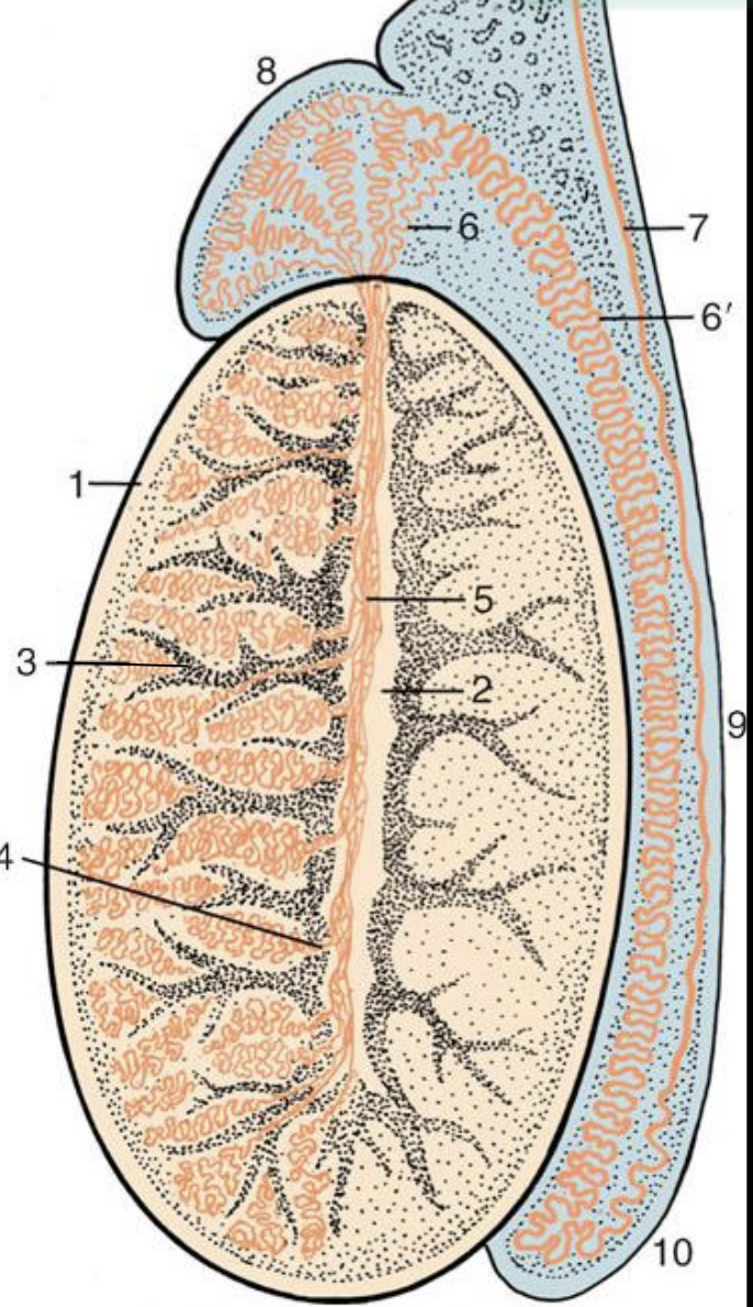
Tunica vaginalis: Serosal peritoneal layer of scrotum

Tunica albungiea: Dense, irregular collagenous CT (1)

Septila testis: Internally radiated tunica albunginea to form lobuli testis (3)

Mediastinum testis: Central emerging of tunica albuginea to form tiny cavity (2)

<p><span style="color: rgb(0, 0, 0);"><strong><u><span>Tunica vaginalis:</span></u></strong><span> </span></span><span style="color: rgb(0, 165, 255);"><strong><span>Serosal peritoneal</span></strong></span><span style="color: rgb(0, 0, 0);"><strong><span> </span></strong><span>layer of </span></span><span style="color: rgb(0, 165, 255);"><strong><span>scrotum</span></strong></span></p><p><span style="color: rgb(0, 0, 0);"><strong><u><span>Tunica albungiea:</span></u><span> </span></strong></span><span style="color: rgb(0, 165, 255);"><strong><span>Dense, irregular collagenous</span></strong></span><span style="color: rgb(0, 0, 0);"><strong><span> </span></strong><span>CT (1)</span></span></p><p><span style="color: rgb(0, 0, 0);"><strong><u><span>Septila testis:</span></u></strong></span><span style="color: rgb(0, 165, 255);"><strong><span> Internally radiated tunica albunginea</span></strong></span><span style="color: rgb(0, 0, 0);"><strong><span> </span></strong><span>to form </span></span><span style="color: rgb(0, 165, 255);"><strong><span>lobuli testis</span></strong></span><span style="color: rgb(0, 0, 0);"><strong><span> </span></strong></span><span style="color: rgb(0, 0, 0);"><span>(3)</span></span></p><p><span style="color: rgb(0, 0, 0);"><strong><u><span>Mediastinum testis:</span></u><span> </span></strong></span><span style="color: rgb(0, 165, 255);"><strong><span>Central</span></strong></span><span style="color: rgb(0, 0, 0);"><strong><span> </span></strong><span>emerging of </span></span><span style="color: rgb(0, 165, 255);"><strong><span>tunica albuginea</span></strong></span><span style="color: rgb(0, 0, 0);"><strong><span> </span></strong><span>to form</span></span><span style="color: rgb(0, 165, 255);"><span> </span><strong><span>tiny cavity</span></strong></span><span style="color: rgb(0, 0, 0);"><strong><span> </span></strong><span>(2)</span></span></p>
4
New cards

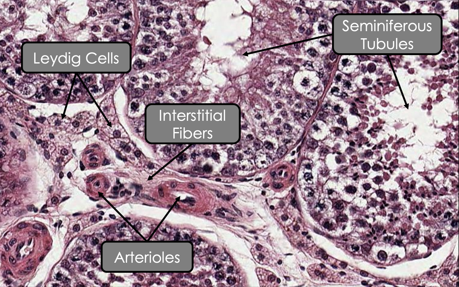
Interstitial tissue of stroma #00a5ff

  • Interstitial fibers

    • What

    • From 

    • Surrounds 

  • Interstitial cell 

    • Name 

    • Arrangement 

    • Adjacent to 

      • How they connect 

    • Cytoplasm 

    • Shape

    • Nucleus 

    • Abundant of which organelles 

    • Function 

Interstitial fibers

  • What: Infiltration of loose connective tissue

  • From: Septula testis

  • Surrounds: Seminiferous tubules

Interstitial cell

  • Name: Leydig cells

  • Arrangement: Clusters or cords

  • Adjacent to: Capillaries

    • How they connect: Gap junctions

  • Cytoplasm:

    • Pale due to acidophilical cytoplasm

  • Shape: Polyhedral 

  • Nucleus: 

    • Spherical 

    • Euchromatic

  • Abundant of which organelles:

    • Lopid vacuoles

    • sER

    • Mitochondria

  • Function: Synthesise testosterone under influenced of LH

<p><span style="color: rgb(0, 165, 255);"><strong><u><mark data-color="blue" style="background-color: blue; color: inherit;">Interstitial fibers</mark></u></strong></span></p><ul><li><p><strong><u>What:</u>&nbsp;</strong><span style="color: rgb(0, 165, 255);"><strong>Infiltration</strong></span><strong> </strong>of <span style="color: rgb(0, 165, 255);"><strong>loose connective tissue </strong></span></p></li><li><p><strong><u>From:</u> </strong><span style="color: rgb(0, 165, 255);"><strong>Septula testis</strong></span></p></li><li><p><strong><u>Surrounds:</u> </strong><span style="color: rgb(0, 165, 255);"><strong>Seminiferous tubules</strong></span></p></li></ul><p><span style="color: rgb(0, 165, 255);"><strong><u><mark data-color="blue" style="background-color: blue; color: inherit;">Interstitial cell</mark></u></strong></span></p><ul><li><p><strong><u>Name:</u> </strong><span style="color: rgb(0, 165, 255);"><strong>Leydig cells</strong></span></p></li><li><p><strong><u>Arrangement:</u> </strong><span style="color: rgb(0, 165, 255);"><strong>Clusters</strong></span><strong> </strong>or <span style="color: rgb(0, 165, 255);"><strong>cords</strong></span></p></li><li><p><strong><u>Adjacent to:</u> </strong><span style="color: rgb(0, 165, 255);"><strong>Capillaries</strong></span></p><ul><li><p><strong><u>How they connect:</u> </strong><span style="color: rgb(0, 165, 255);"><strong>Gap junctions</strong></span></p></li></ul></li><li><p><strong><u>Cytoplasm:</u></strong></p><ul><li><p><span style="color: rgb(0, 165, 255);"><strong>Pale</strong></span> due to<strong> </strong><span style="color: rgb(0, 165, 255);"><strong>acidophilical cytoplasm</strong></span></p></li></ul></li><li><p><strong><u>Shape:</u></strong>&nbsp;<span style="color: rgb(0, 165, 255);"><strong>Polyhedral</strong></span><strong>&nbsp;</strong></p></li><li><p><strong><u>Nucleus:</u>&nbsp;</strong></p><ul><li><p><span style="color: rgb(0, 165, 255);"><strong>Spherical&nbsp;</strong></span></p></li><li><p><span style="color: rgb(0, 165, 255);"><strong>Euchromatic </strong></span></p></li></ul></li><li><p><strong><u>Abundant of which organelles:</u></strong></p><ul><li><p><span style="color: rgb(0, 165, 255);"><strong>Lopid vacuoles </strong></span></p></li><li><p><span style="color: rgb(0, 165, 255);"><strong>sER </strong></span></p></li><li><p><span style="color: rgb(0, 165, 255);"><strong>Mitochondria</strong></span></p></li></ul></li><li><p><strong><u>Function:</u> </strong><span style="color: rgb(0, 165, 255);"><strong>Synthesise testosterone</strong></span><strong> </strong>under influenced of<span style="color: rgb(0, 165, 255);"> <strong>LH</strong></span></p></li></ul><p></p>
5
New cards

What are the structures of parenchyma? #ff8300

  • Convoluted seimiferous tubules (forms sperm

  • Straight seminiferous tubules (forms sperm

  • Rete testis (sperm transport)

  • Efferent ductules (sperm transport

6
New cards

Parenchyma: Seminiferous tubules #ff8300

  • Shape

  • Location

  • Straight seminiferous tubule is formed

  • What is ductuli efferentes

  • What is lamina propria

  • Myofibroblaat

    • What

    • Why needed

  • Function

  • Lined by

Shape: Tightly convoluted

Location: Within pyramidal lobuli testis

Straight seminiferous tubule is formed: Prior merging with rete testis within mediastinum testis 

What is ductuli efferentes: Duct that empties the spermatozoa into epididymis

What is lamina propria: Fine collagen and elastic fibers

Myofibroblast:

  • What: Peritubular contractile cell with actin bundles

  • Why needed: For contraction

Function: Important for spermatogenesis

Lined by:

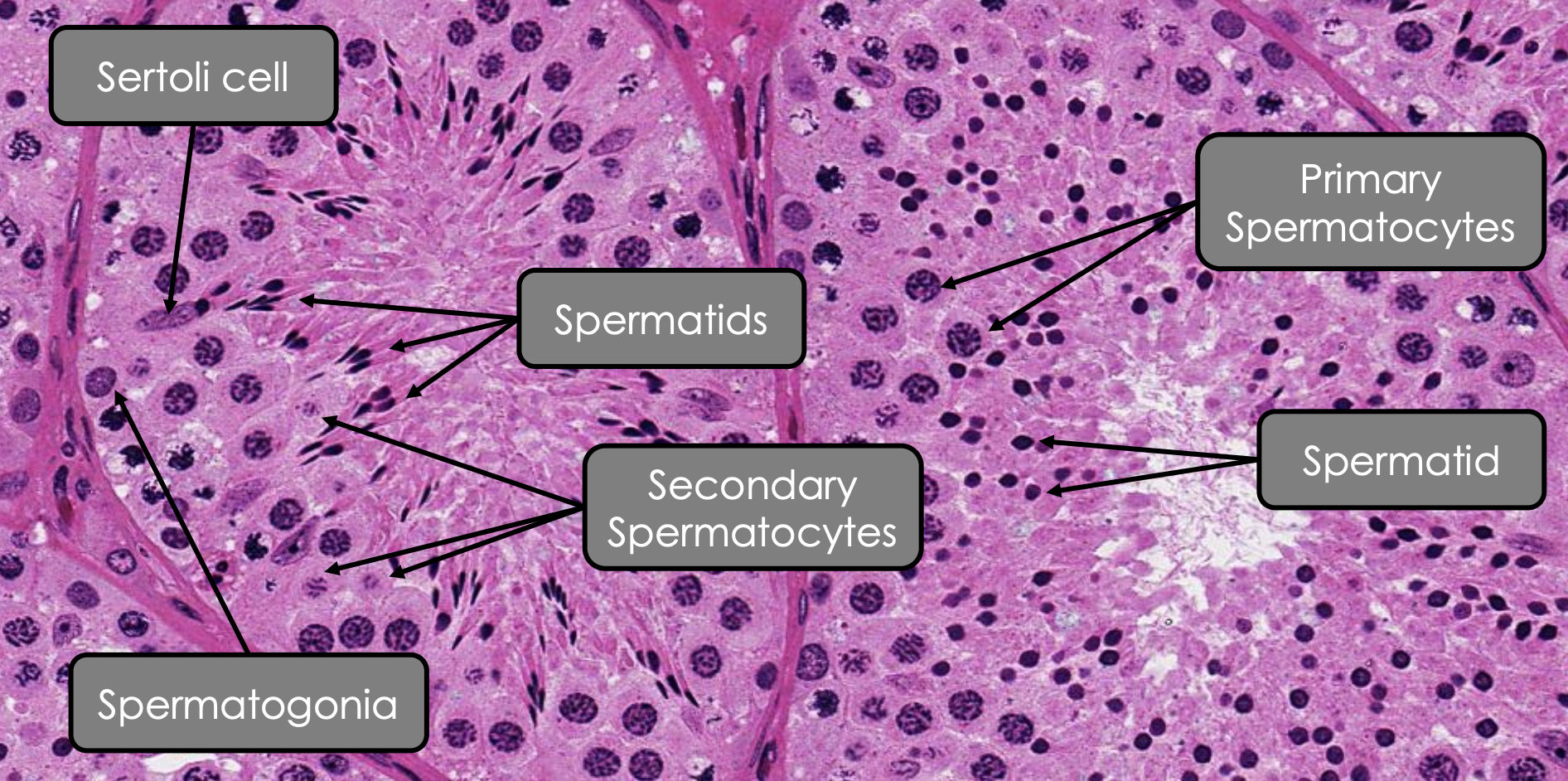
  • Stratified spermatogenic epithelium

  • With Sertoli cells

7
New cards

Parenchyma: How is secondary spermatocytes produced? #ff8300

Primary spermatocytes undergo meiosis I 

8
New cards

Parenchyma: How is spermatid produced?

Secondary spermatocytes undergo meiosis II

9
New cards

Parenchyma: Spermatogonia #ff8300

  • Closely associated with

  • Shape

  • Nucleus

Closely associated with: Basement membrane

Shape: Round, pear-shaped

Nucleus: Heterochormatic 

10
New cards

Parenchyma: Primary spermatocytes #ff8300

  • Nucleus

  • Abundant of what organelles

Nucleus: Varying chromatin density of nucleus

Abundant of what organelles:

  • Mitochondria

  • rER

  • Large GA

11
New cards

Parenchyma: Secondary spermatocytes #ff8300

  • Location 

  • Shape 

  • Nucleus 

Location: Near luminal surface of seminiferous tubule

Shape: Spherical

Nucleus: Weakly stained

12
New cards

Parenchyma: Sertoli Cell #ff8300

  • Shape

  • Location 

  • Nucleus 

  • Abundant organelles 

  • How do they communicate with adjacent cells

  • Function 

Shape: Pyramidal cell 

Location: Extend the entire height of spermatogenic epithelium

Nucleus: Ovoid and euchromatic 

Abundant organelles:

  • Cytoplasmic projections to all spermatogenic epithelium

  • GA

  • Mitochondria

  • sER (surrounds periacrosomal region of spermatid)

  • Fat vacuoles (at cellular base

How do they communicate with adjacent cells: Tight junction and desmosomes

Function: 

  • Structural and nutiritonal support 

  • Intra-tubular fluid secretion 

  • Formation of blood testis barrier 

<p><strong><u>Shape:</u>&nbsp;</strong><span style="color: rgb(255, 131, 0);"><strong>Pyramidal cell&nbsp;</strong></span></p><p><strong><u>Location:</u>&nbsp;</strong><span style="color: rgb(255, 131, 0);"><strong>Extend</strong></span> the<strong> </strong><span style="color: rgb(255, 131, 0);"><strong>entire height</strong> </span>of<strong> </strong><span style="color: rgb(255, 131, 0);"><strong>spermatogenic epithelium</strong></span></p><p><strong><u>Nucleus:</u>&nbsp;</strong><span style="color: rgb(255, 131, 0);"><strong>Ovoid</strong></span>&nbsp;and<strong>&nbsp;</strong><span style="color: rgb(255, 131, 0);"><strong>euchromatic</strong></span><strong>&nbsp;</strong></p><p><strong><u>Abundant organelles:</u></strong></p><ul><li><p><span style="color: rgb(255, 131, 0);"><strong>Cytoplasmic projections</strong></span>&nbsp;to all spermatogenic epithelium</p></li><li><p><span style="color: rgb(255, 131, 0);"><strong>GA</strong></span></p></li><li><p><span style="color: rgb(255, 131, 0);"><strong>Mitochondria </strong></span></p></li><li><p><span style="color: rgb(255, 131, 0);"><strong>sER</strong></span><strong> </strong>(<em>surrounds periacrosomal region of spermatid</em>) </p></li><li><p><span style="color: rgb(255, 131, 0);"><strong>Fat vacuoles</strong></span>&nbsp;(<em>at cellular base</em>)&nbsp;</p></li></ul><p><strong><u>How do they communicate with adjacent cells:</u> </strong><span style="color: rgb(255, 131, 0);"><strong>Tight junction</strong></span><strong> </strong>and <span style="color: rgb(255, 131, 0);"><strong>desmosomes</strong></span></p><p><strong><u>Function:</u>&nbsp;</strong></p><ul><li><p><span style="color: rgb(255, 131, 0);"><strong>Structural</strong></span> and<strong> </strong><span style="color: rgb(255, 131, 0);"><strong>nutiritonal support&nbsp;</strong></span></p></li><li><p><span style="color: rgb(255, 131, 0);"><strong>Intra-tubular fluid secretion&nbsp;</strong></span></p></li><li><p><span style="color: rgb(255, 131, 0);"><strong>Formation</strong></span> of<span style="color: rgb(255, 131, 0);"><strong> blood testis barrier</strong></span><strong>&nbsp;</strong></p></li></ul><p></p>