RCA Midterm
1/267
There's no tags or description
Looks like no tags are added yet.
Name | Mastery | Learn | Test | Matching | Spaced | Call with Kai |
|---|
No analytics yet
Send a link to your students to track their progress
268 Terms
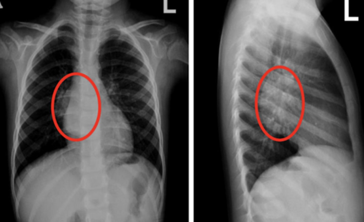
Azygous fissure
Right sided aortic arch
Dextrocardia with situs inversus
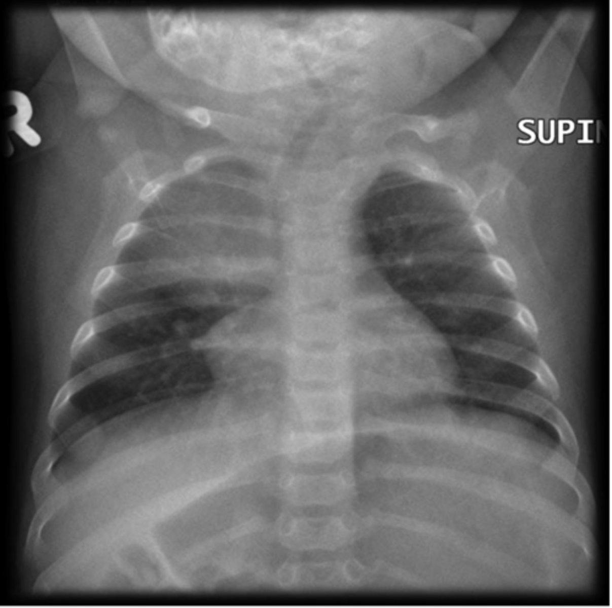
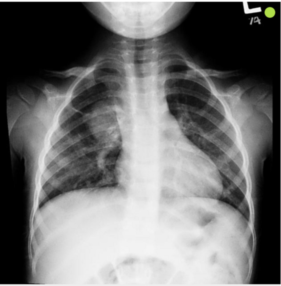
Thymus gland (in child), creates the "sail sign"
Pulmonary sequestration
Congenital pulmonary airway malformation (CPAM)
very lucent in front of heart shadow
CPAM
Bronchogenic cyst
Bronchogenic cyst
arteriovenous malformation
arteriovenous malformation
arteriovenous malformation
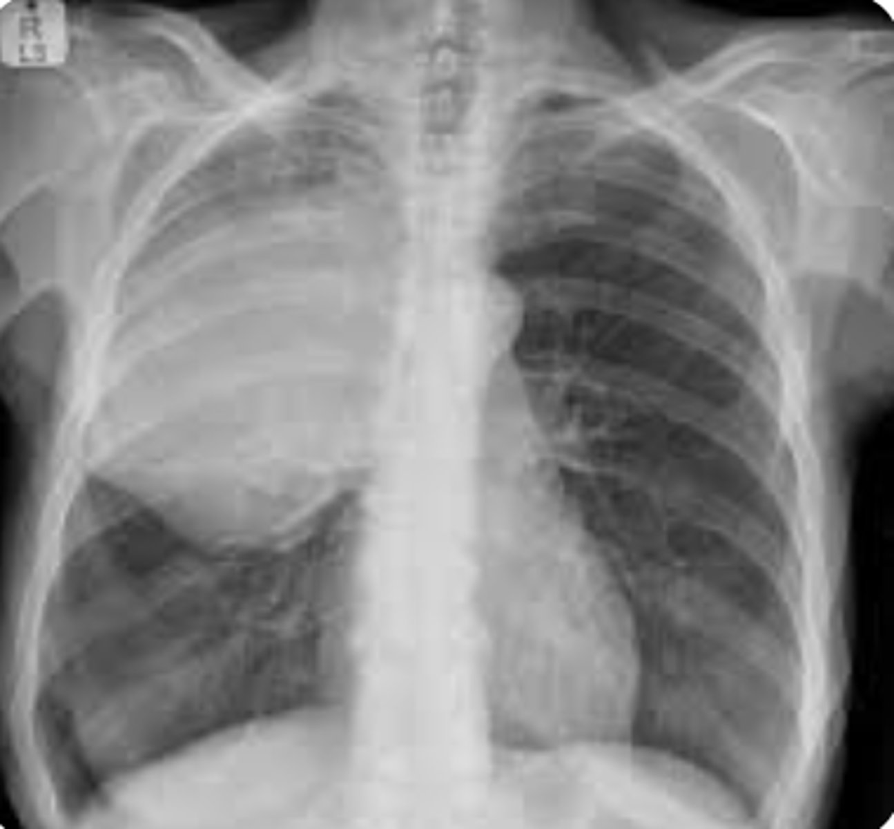
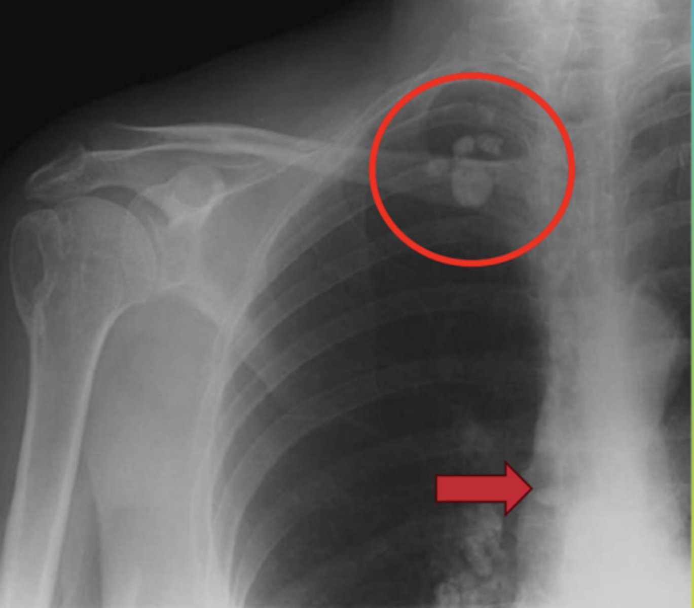
Scimitar Syndrome
Congenital diaphragmatic hernia
diaphragmatic eventration
pleural tenting
(juxtaphrenic peak sign)
Name the sign present in this case
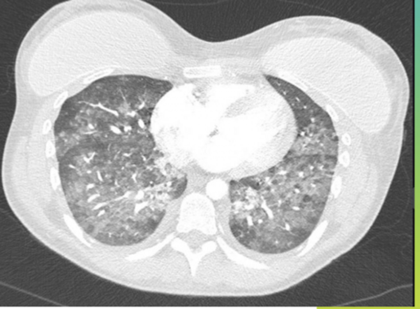
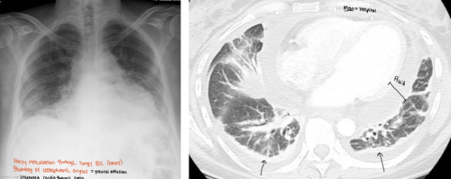
interstitial
What is this pattern of lung disease?
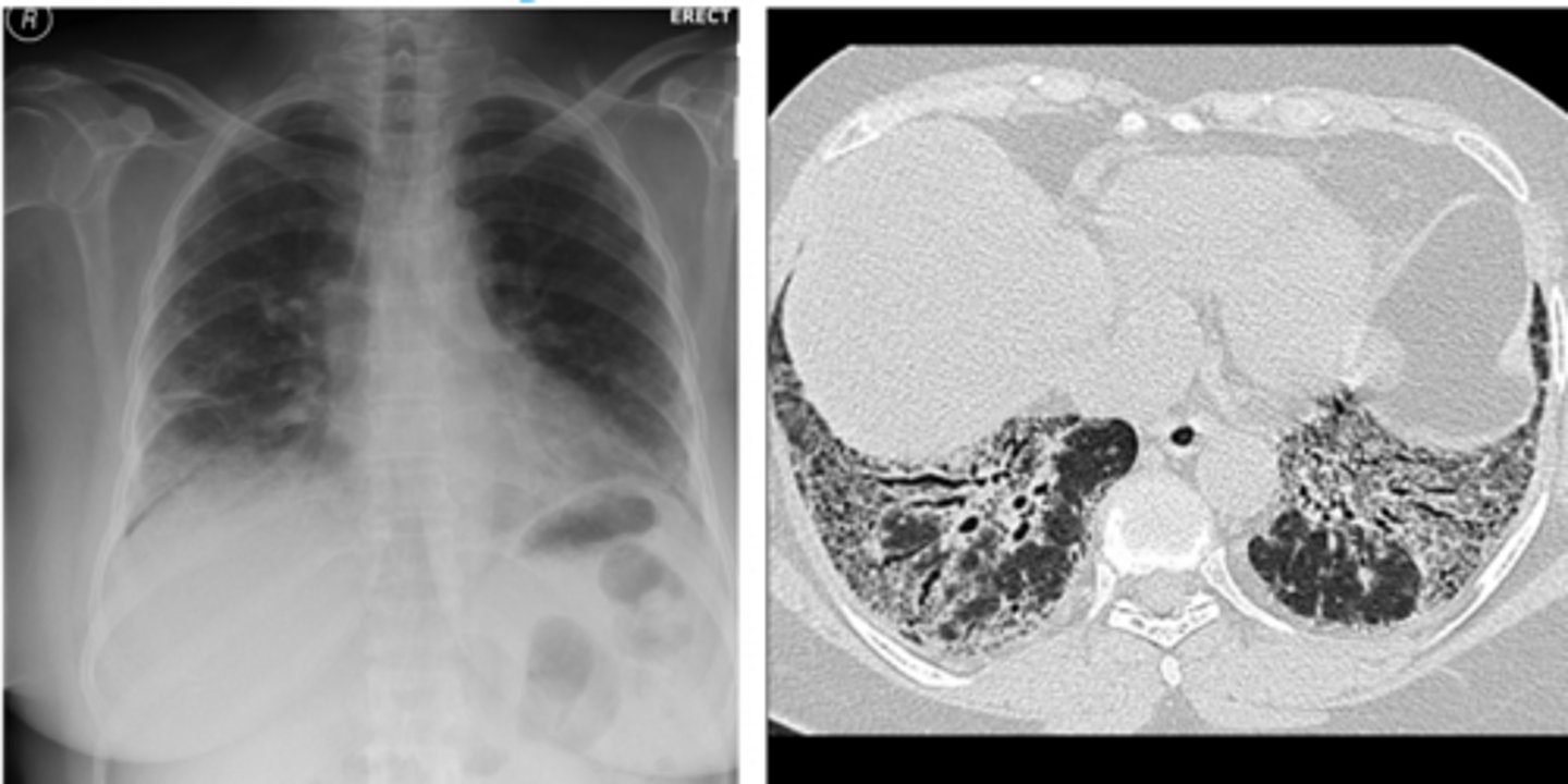
air space/consolidation
What is this pattern of lung disease?
overinflation
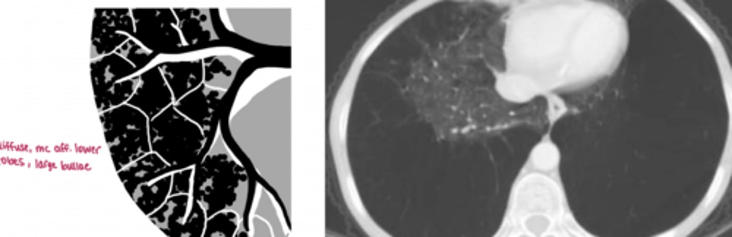
-Bullous emphysema, COPD, asthma
What is this pattern of lung disease? What are the 3 most common diseases this is associated with?
underinflation
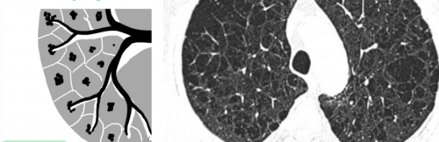
What is this pattern of lung disease?
underinflation (triangular opacity near heart shadow)
What is this pattern of lung disease?
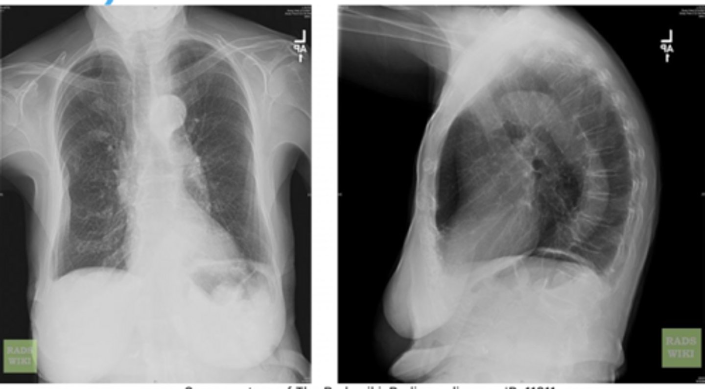
extra-parenchymal
What is this pattern of lung disease?
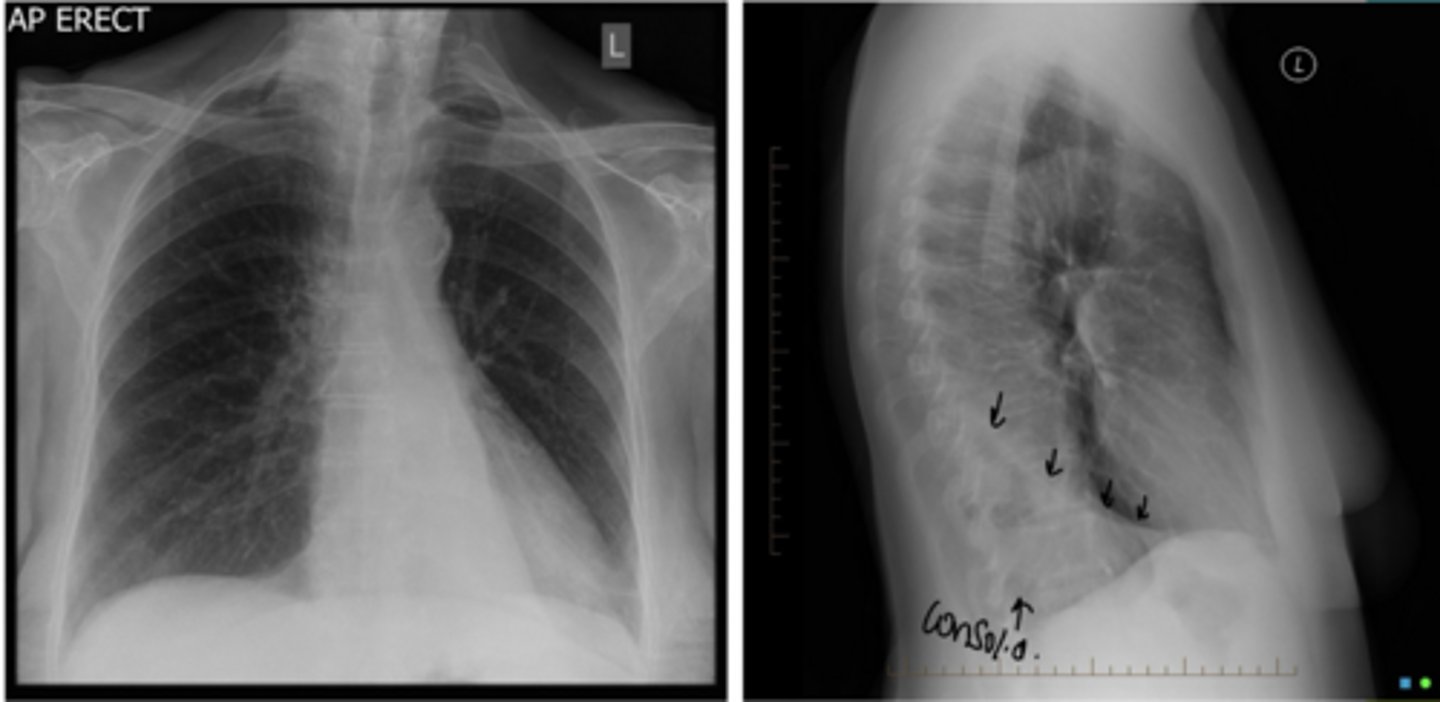
silhouette sign, affecting R lower lobe, obliterating the R hemidiaphragm
What sign is present? What lobe is affected?
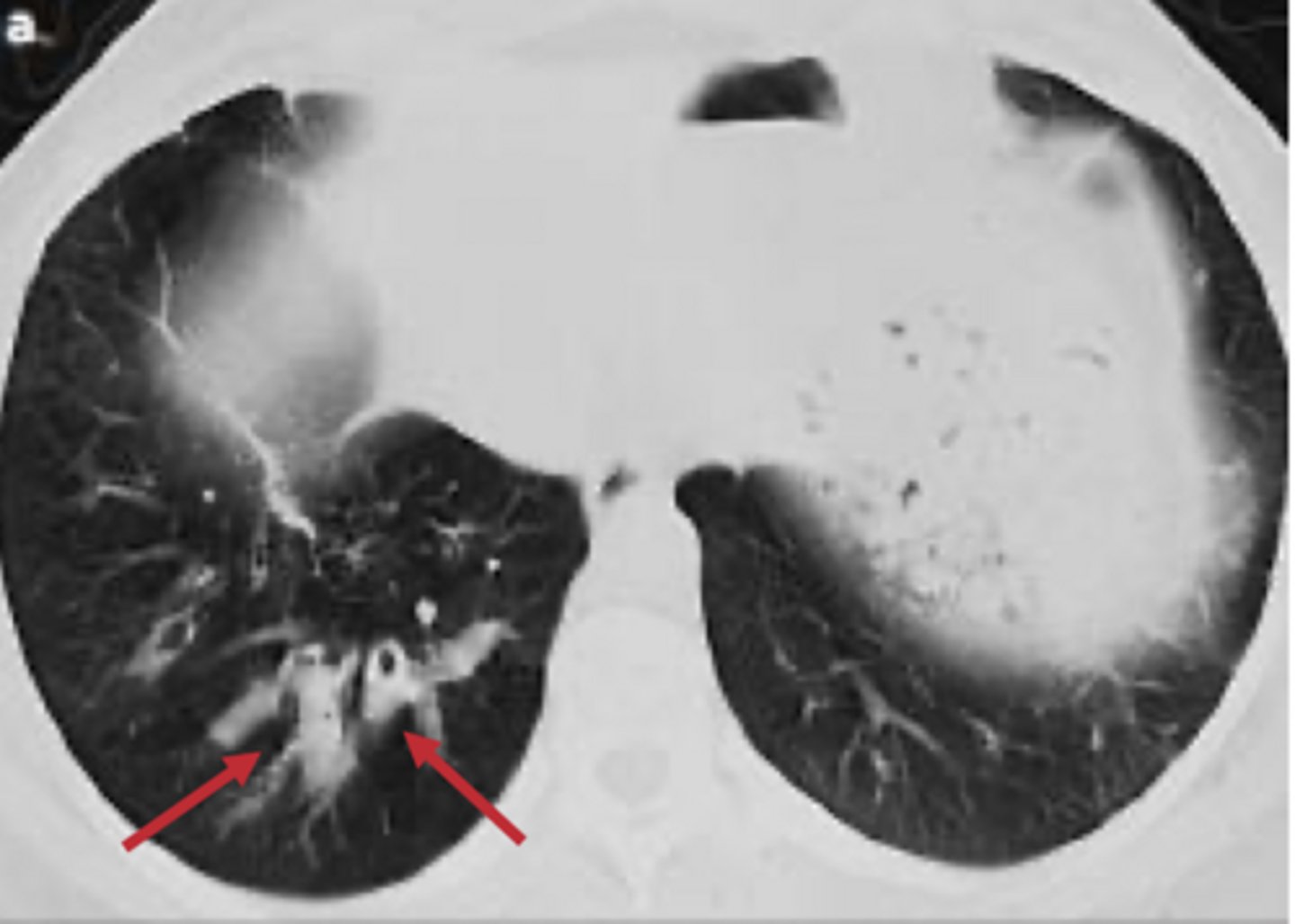
air bronchogram sign
What sign is present?
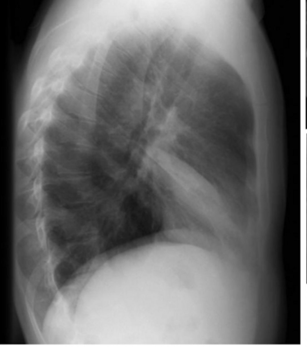
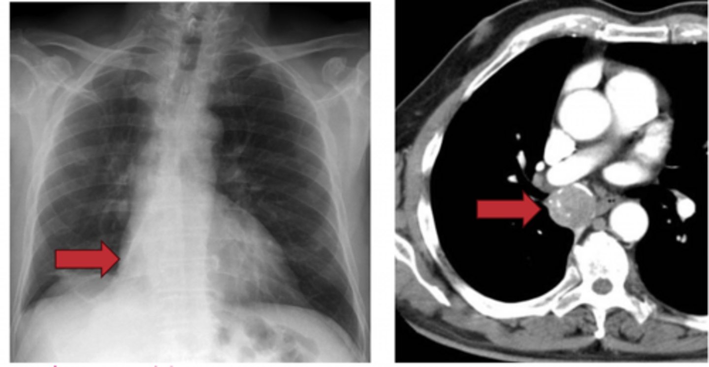
Hampton hump sign
represents a lung infarction
What sign is present? What does this represent?
Hampton hump sign
What sign is present?
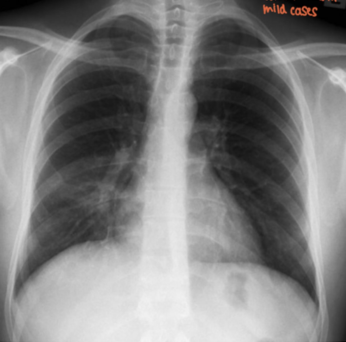
Butterfly (bat wing) opacification
mc cause is pulmonary edema
What "sign" is this? What is the most common cause?
bulging fissure sign
-not commonly seen d/t rapid administration of antibiotics
What sign is present? Is this commonly seen?
Reverse Halo (atoll) sign
What sign is present?
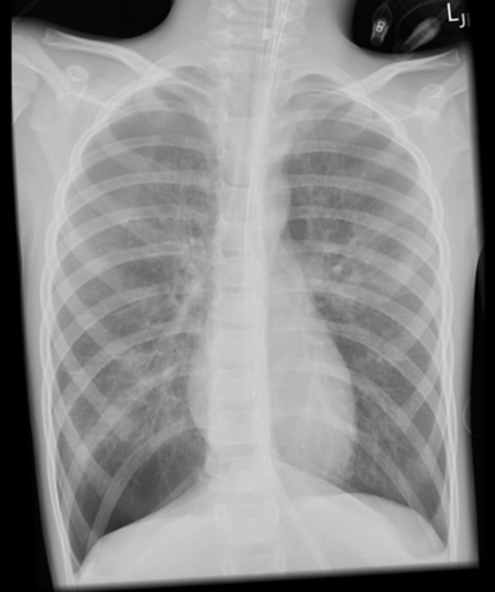
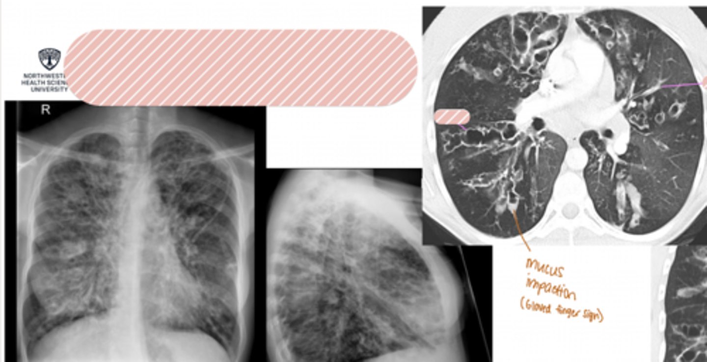
gloved finger sign
What sign is present?
Reticular pattern
What is this pattern of interstitial lung disease?
Honeycombing
What is this pattern of interstitial lung disease?
Tree-in-bud sign
What is this sign?
Crazy paving pattern
What is this sign?
Signet ring sign (traction bronchiectasis)
What sign is present?
Adult Respiratory Distress Syndrome
Name the condition
Adult Respiratory Distress Syndrome
Name the condition
Pneumonia
Name the condition
Pneumonia
Name the condition
Tuberculosis
Name the condition
Tuberculosis
Name the condition
Tuberculosis
Name the condition
Ghon lesion
Name this calcification that is associated with Tuberculosis
Vanishing tumor
treated w/ diuretics + it goes away
Name this appearance that is associated with Pulmonary Edema. How is this treated?
Pulmonary hemorrhage
Name the condition
Pulmonary Hemorrhage
Name the condition
Pulmonary contusion
Name the condition (pt has hx of trauma to chest)
Pulmonary contusio
Name the condition (pt has hx of trauma to chest)
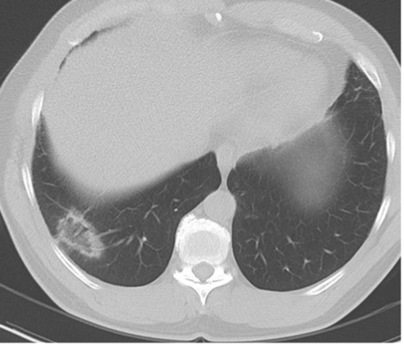
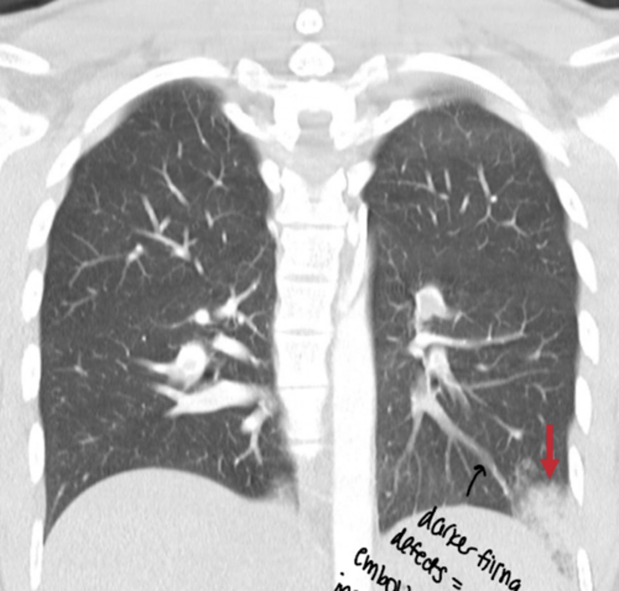
Lung infarction
Name the condition, patient has recent diagnosis of pulmonary embolism
Lung infarction
Name the condition of this patient who had a recent pulmonary embolism
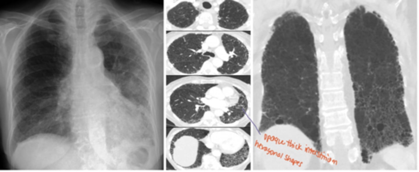
Alveolar proteinosis
Name the condition
Alveolar proteinosis
Name the condition
abscess
Name the condition
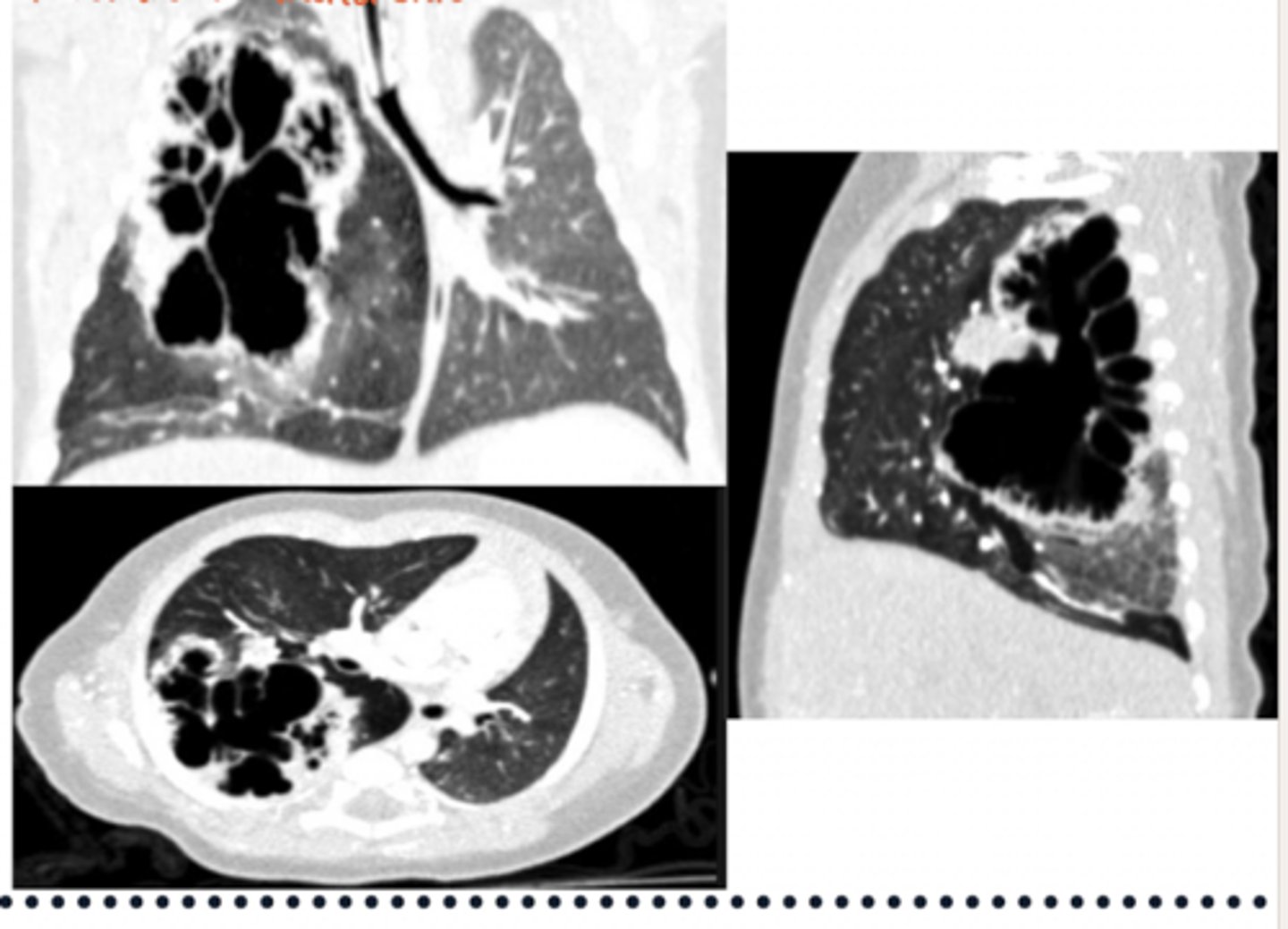
aspergilloma
A 58-year-old patient with a history of a healed cavitary lung lesion from prior tuberculosis undergoes routine chest imaging. The patient feels well overall but reports occasional episodes of hemoptysis.
Abscess
An 80-year-old immunocompromised patient presents with a 2-month history of fever, cough, shortness of breath, fatigue, and unintentional weight loss. Chest imaging shows:
Granulomatous disease
round pneumonia
A 7-year-old child presents with fever, sweats, and cough. Chest imaging shows a round, well-circumscribed pulmonary opacity. The infection remains localized due to underdeveloped pores of Kohn and canals of Lambert, limiting spread between lung subsegments.
Hydatid cyst aka echinococcosis
A patient with close contact with dogs and livestock presents with a pulmonary mass lesion on imaging. Further evaluation shows possible spread from a primary hepatic lesion through the diaphragm. Chest imaging reveals a solid mass with possible cavitation, described as a “water lily” sign.
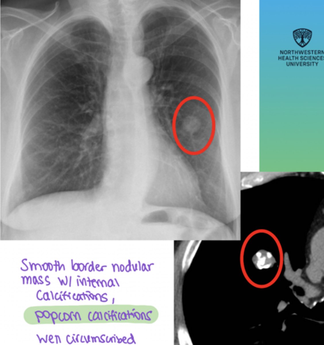
hamartoma
A 45-year-old adult has an incidental finding on chest imaging of a peripherally located solitary pulmonary nodule. The lesion is asymptomatic and shows fat density with “popcorn” calcifications on CT.
bronchogenic carcinomas
get chest x-ray
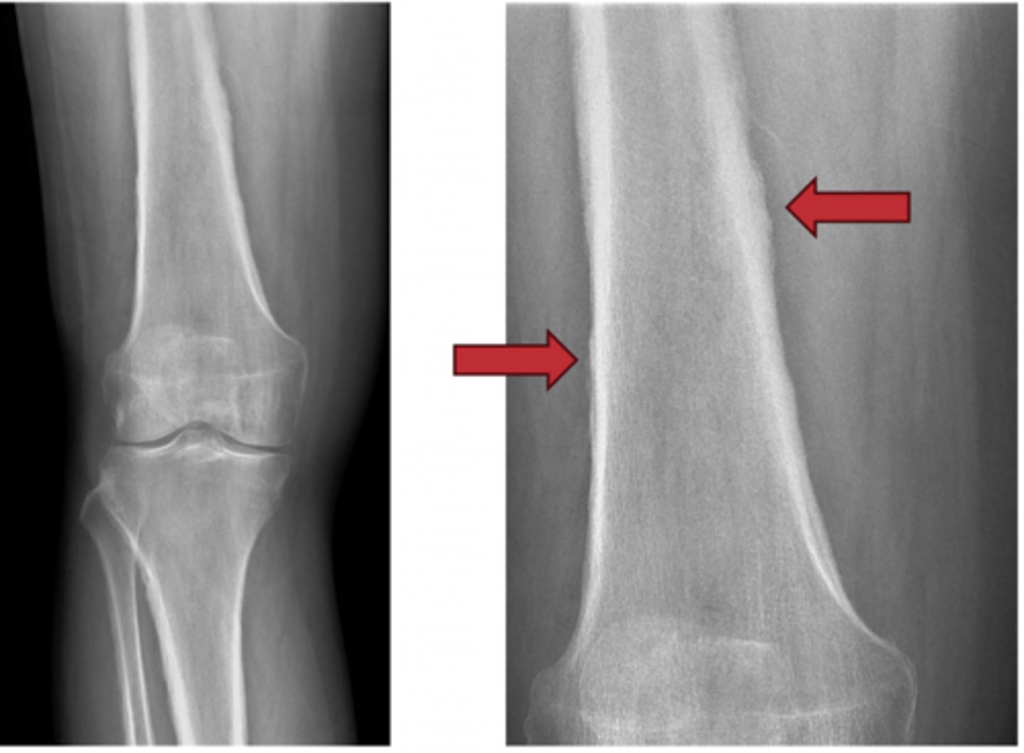
This condition, Hypertrophic Pulmonary Osteoarthropathy (HPOA) is very common with ____ _____. What should the next step for management be?
bronchogenic carcinoma
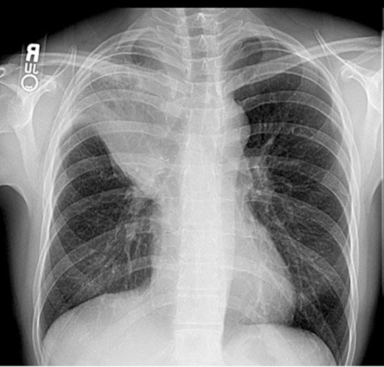
A 67-year-old man presents with a 4-month history of persistent cough and increasing fatigue. He reports coughing up small amounts of blood on several occasions over the past month. He has also noticed an unintentional 15-pound weight loss and decreased appetite. He smoked 1.5 packs of cigarettes daily for 45 years.
Lymphoma
A 24-year-old man presents with a 2-month history of painless swelling in his neck/lymphadenopathy. He reports increasing fatigue, intermittent fevers, and drenching night sweats. Over the past 3 months, he has unintentionally lost 12 pounds.
Physical examination reveals:
Enlarged, firm, non-tender cervical lymph nodes
No signs of infection
Mild pallor
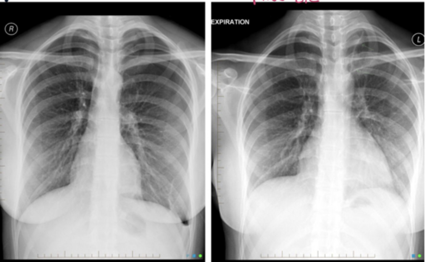
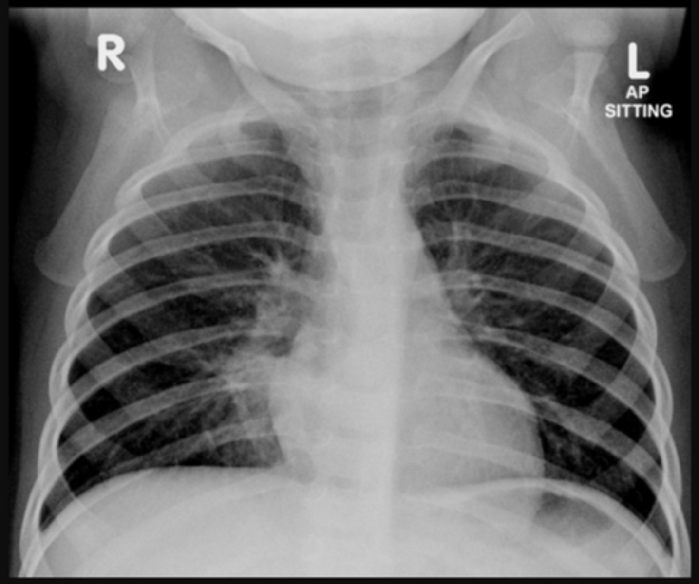
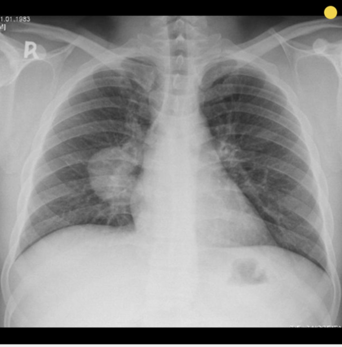
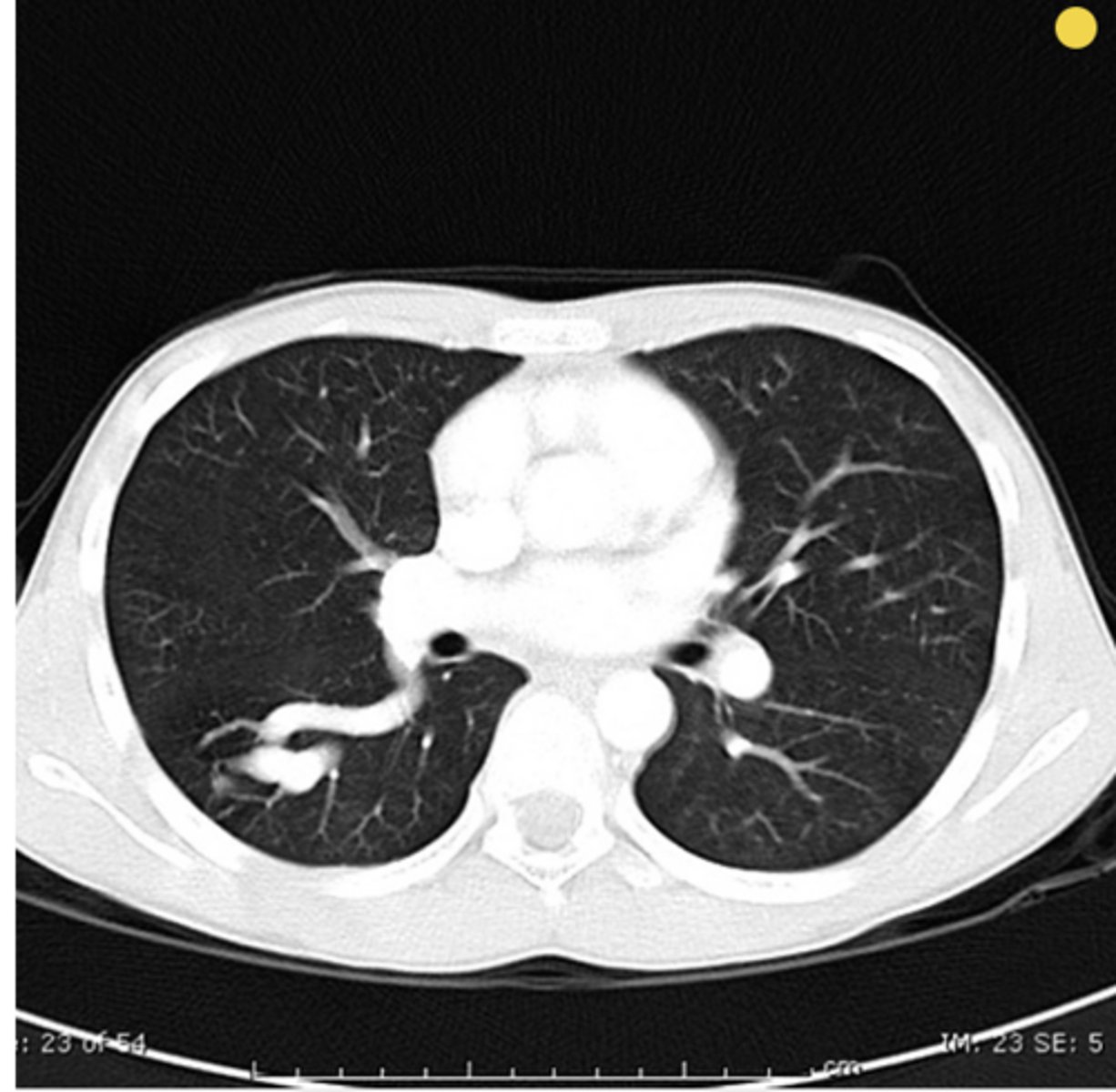
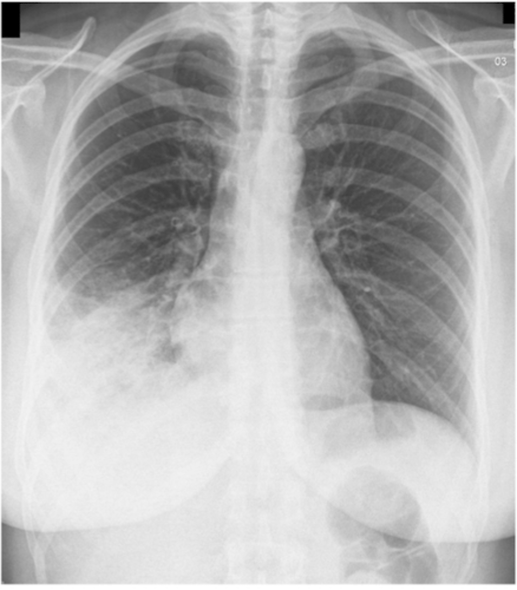
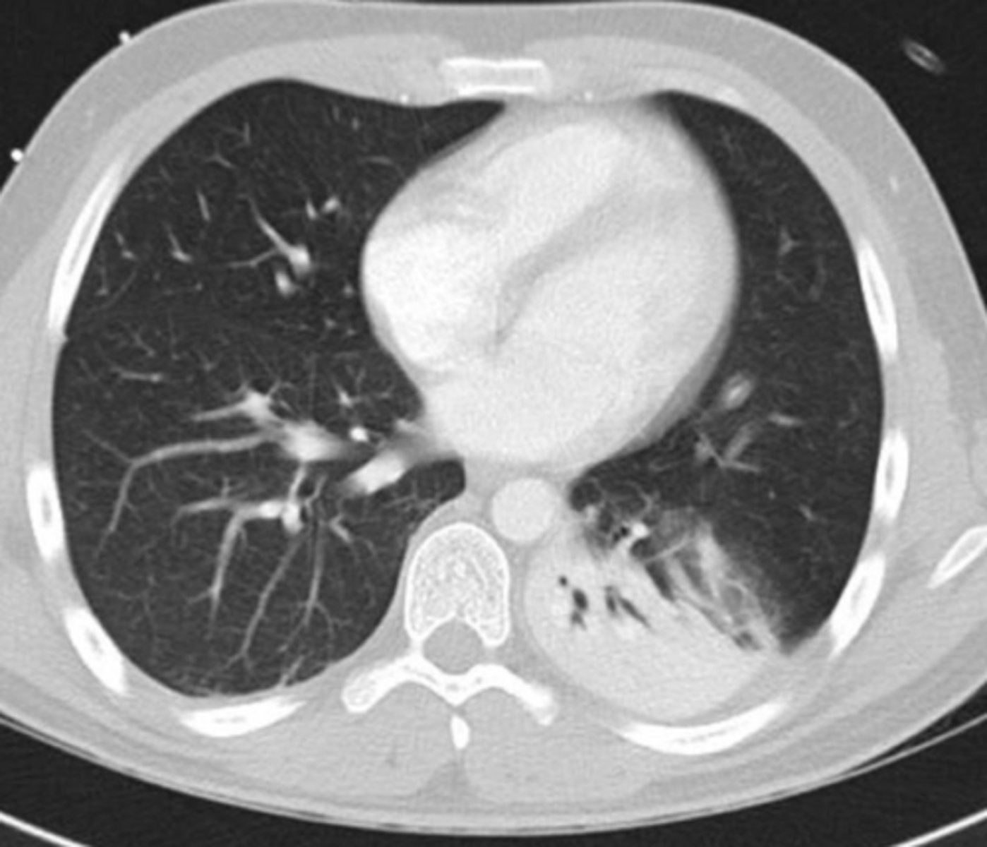
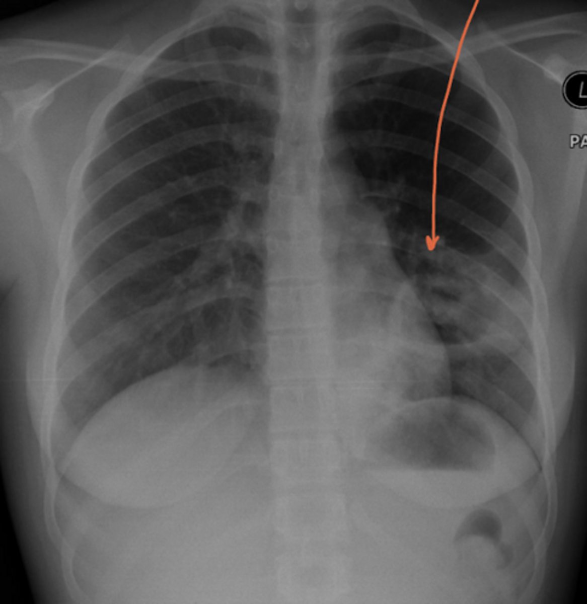
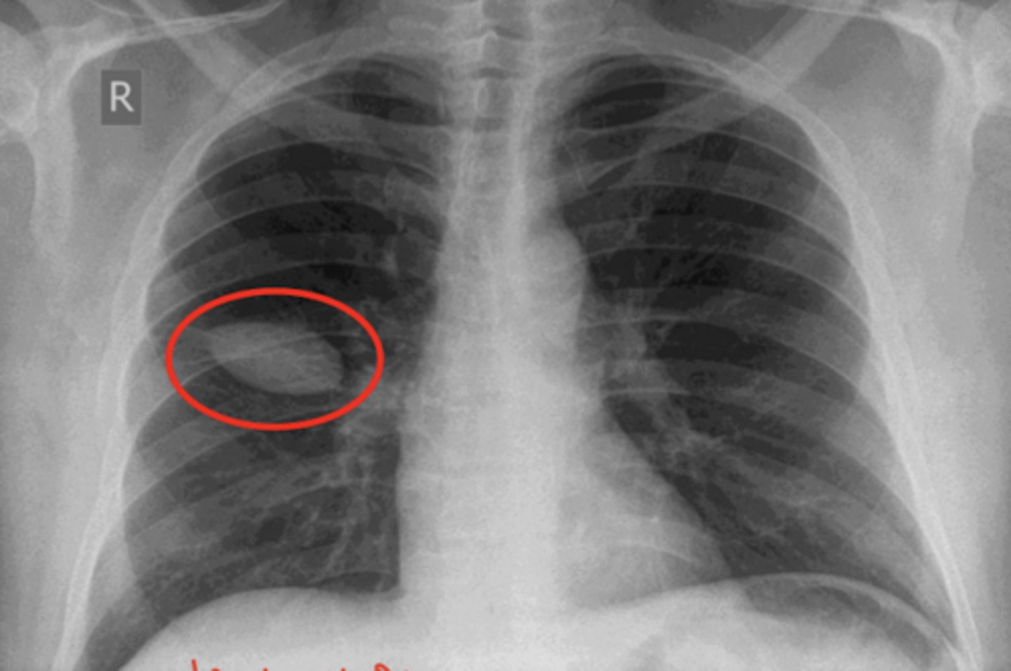
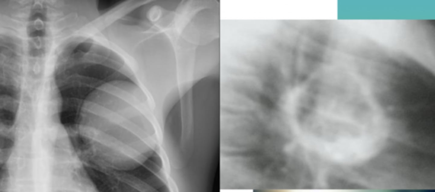
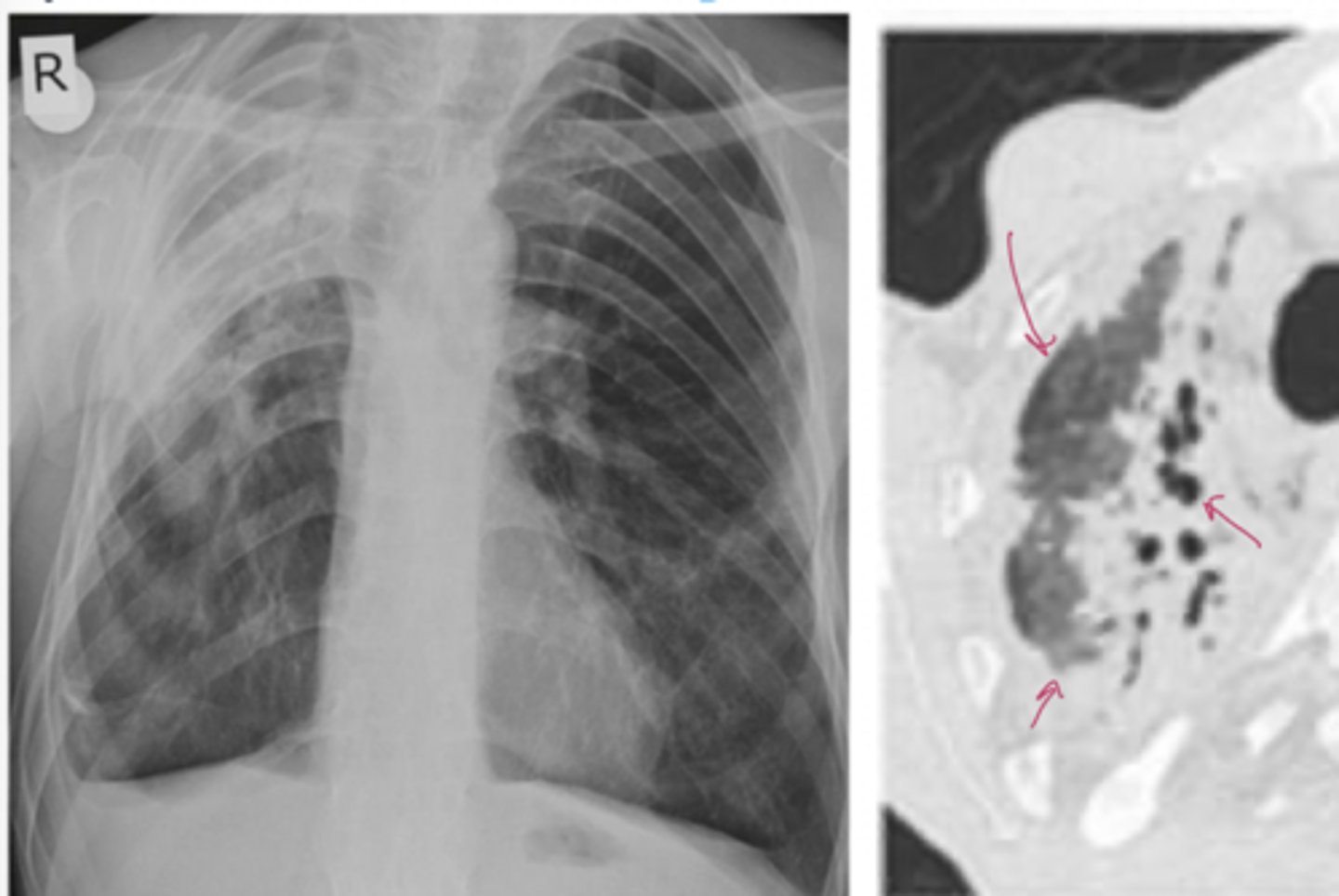
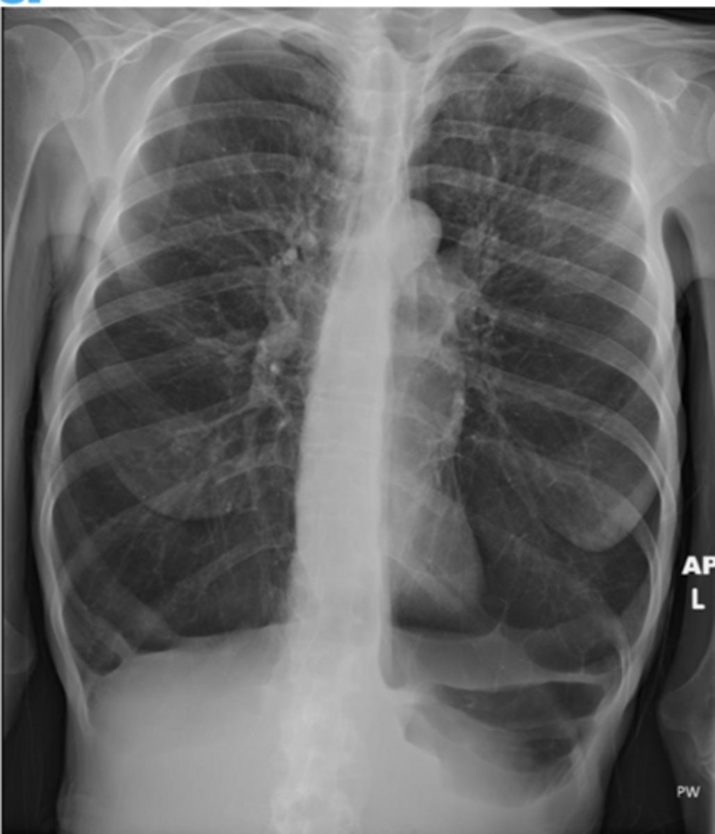
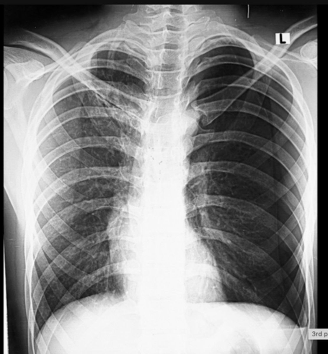
azygous fissure
What normal variant is present here?
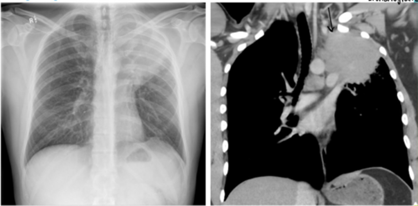
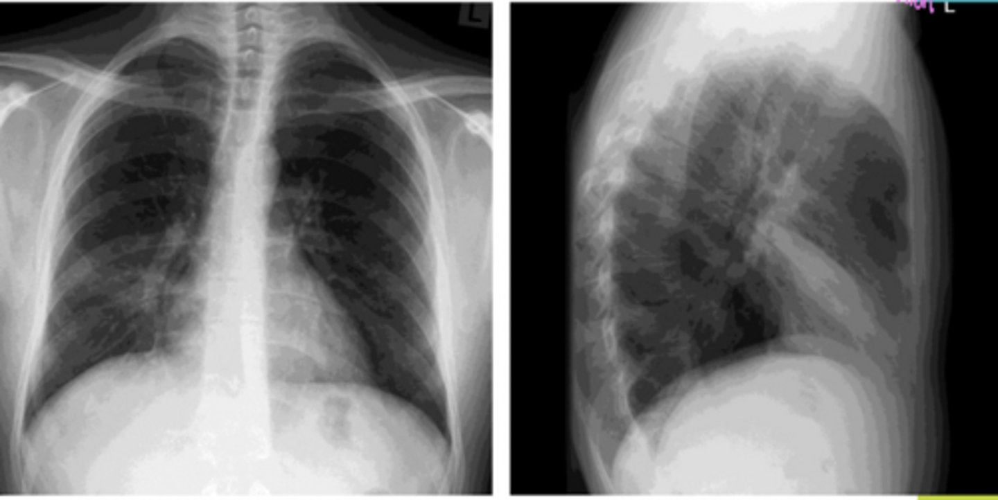
Carcinoid tumor
A 35-year-old woman presents with a 1-year history of chronic cough, intermittent wheezing, and recurrent pneumonia localized to the right middle lobe. She is a non-smoker and reports that her symptoms have not improved with inhalers for presumed asthma.
Over the past 4 months, she has developed:
Weight gain with central obesity
Purple abdominal striae
Proximal muscle weakness
Easy bruising
Physical exam reveals mild unilateral wheezing over the right lung.
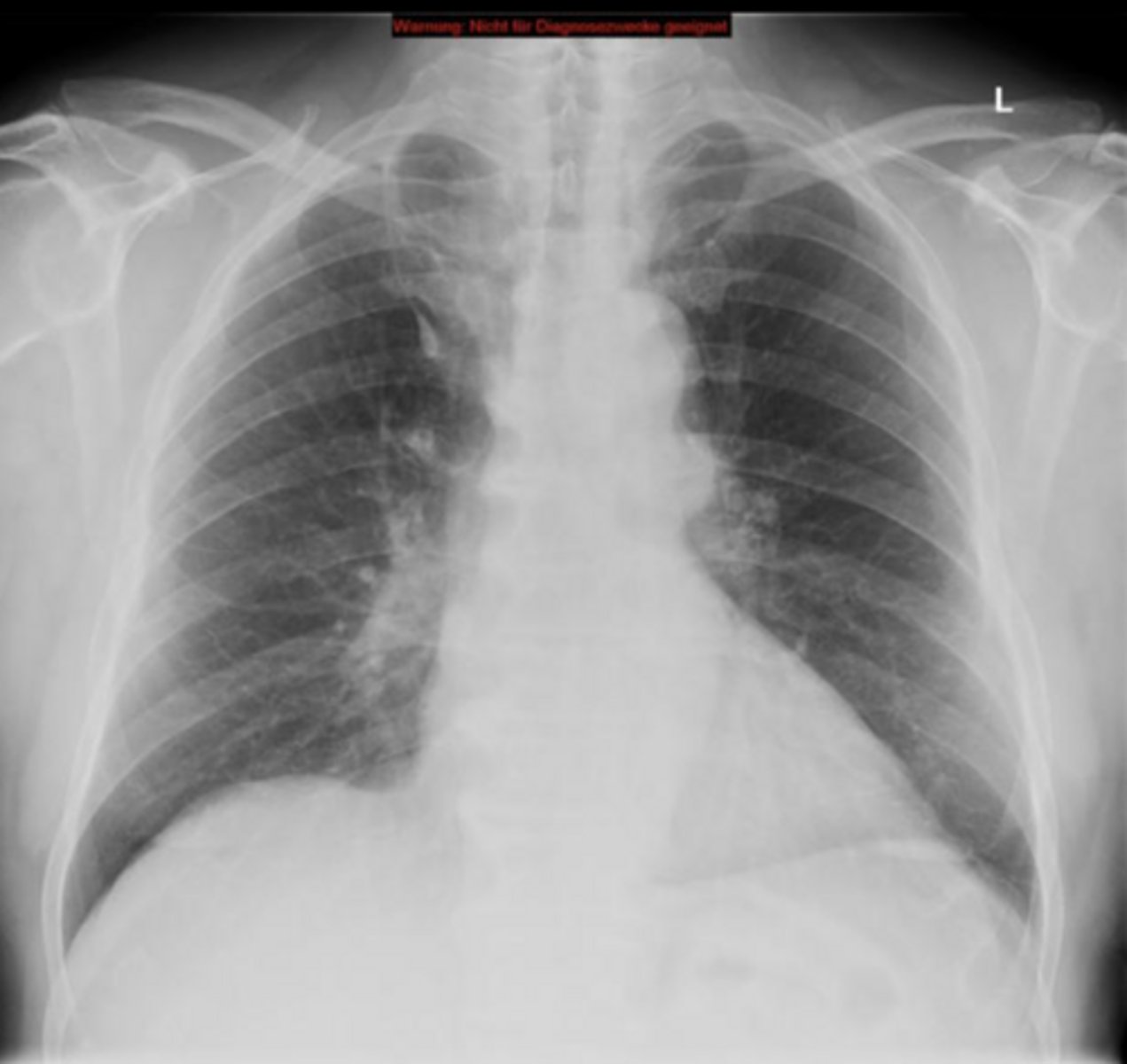
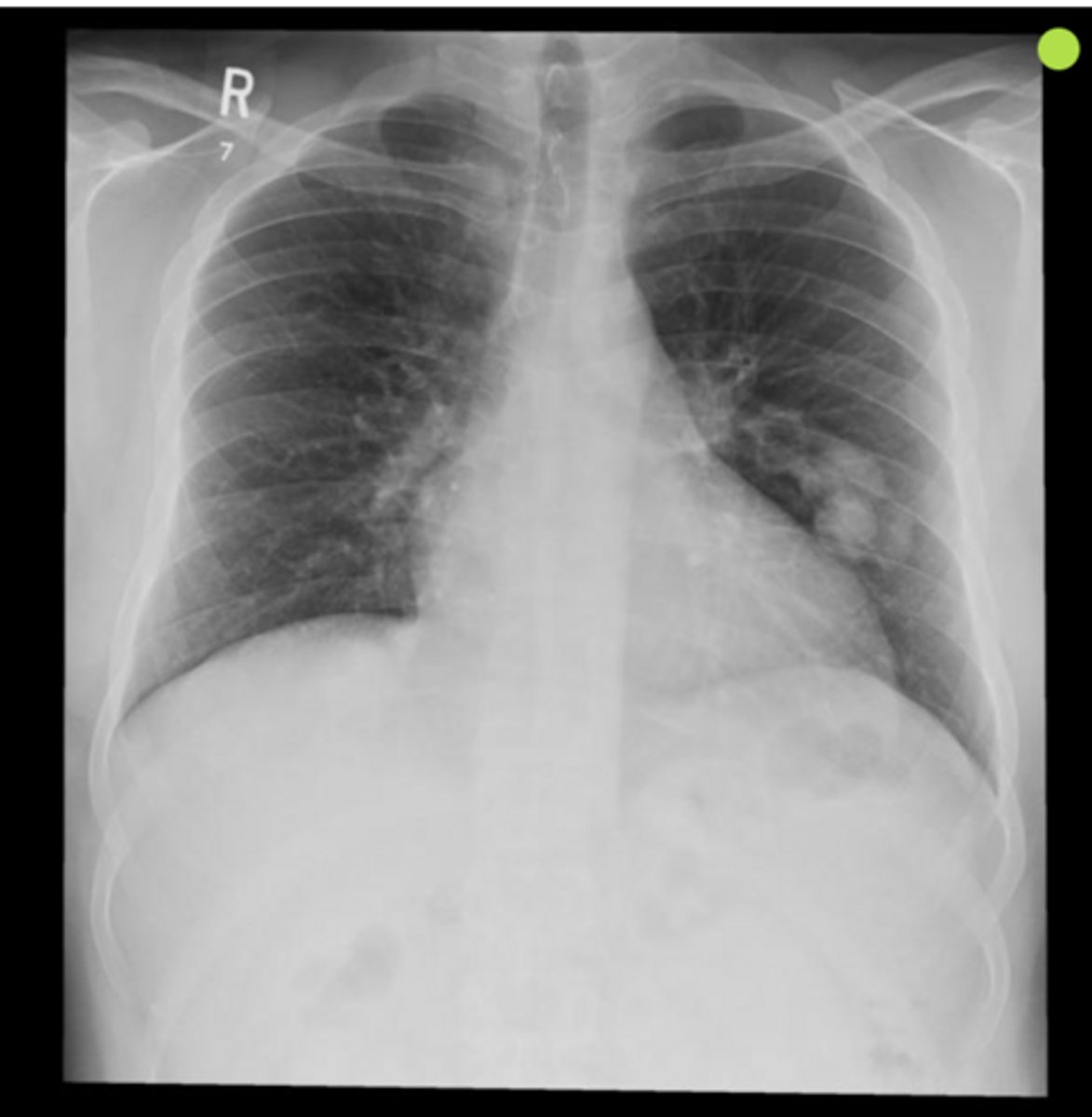
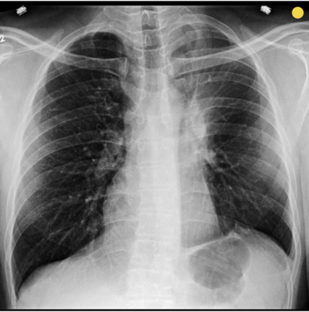
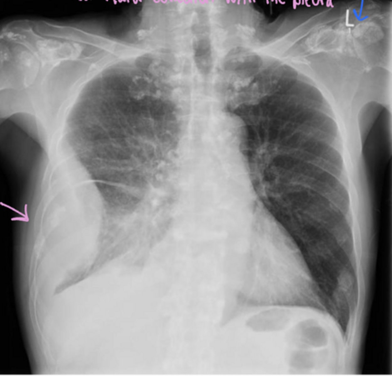
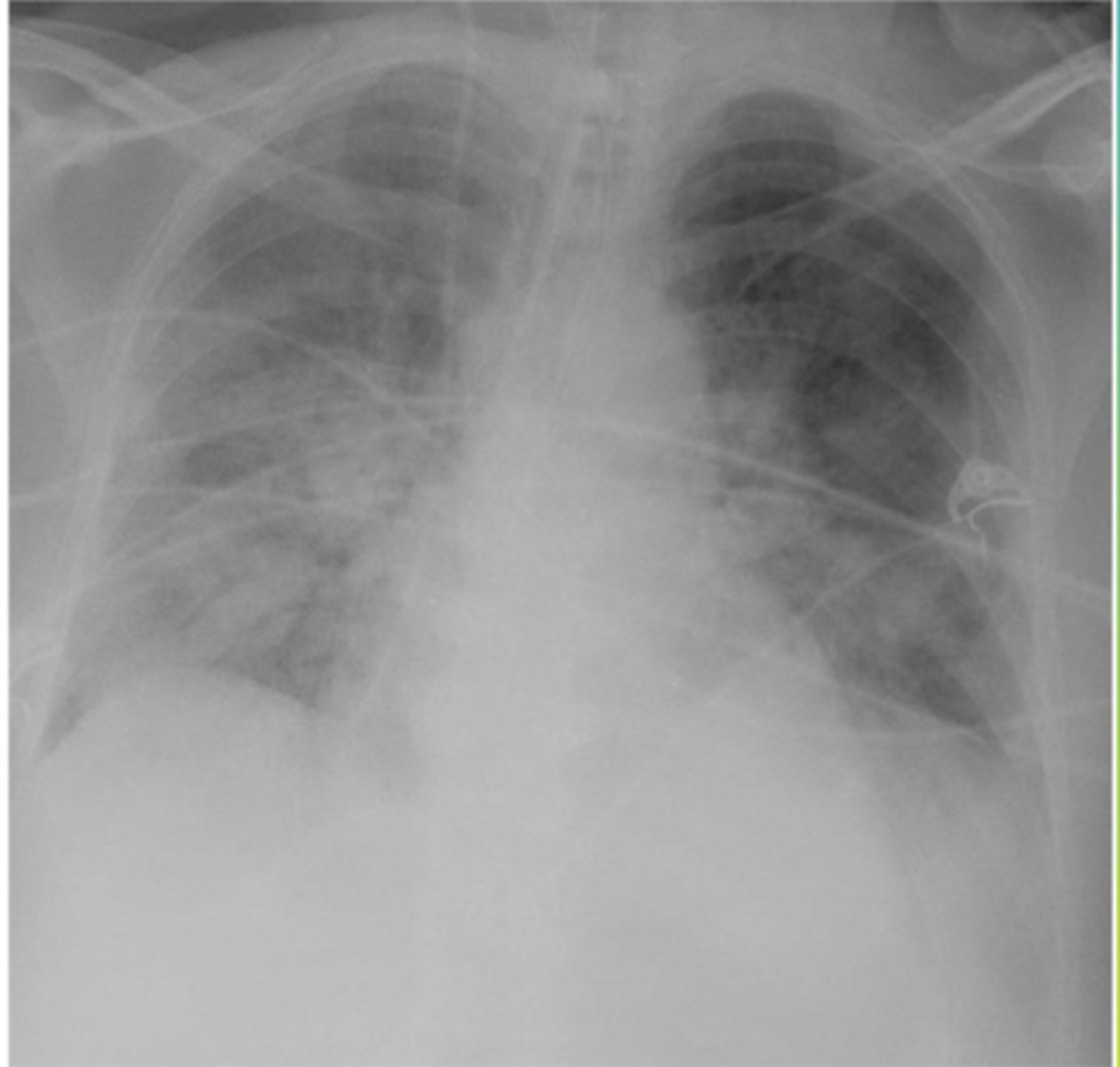
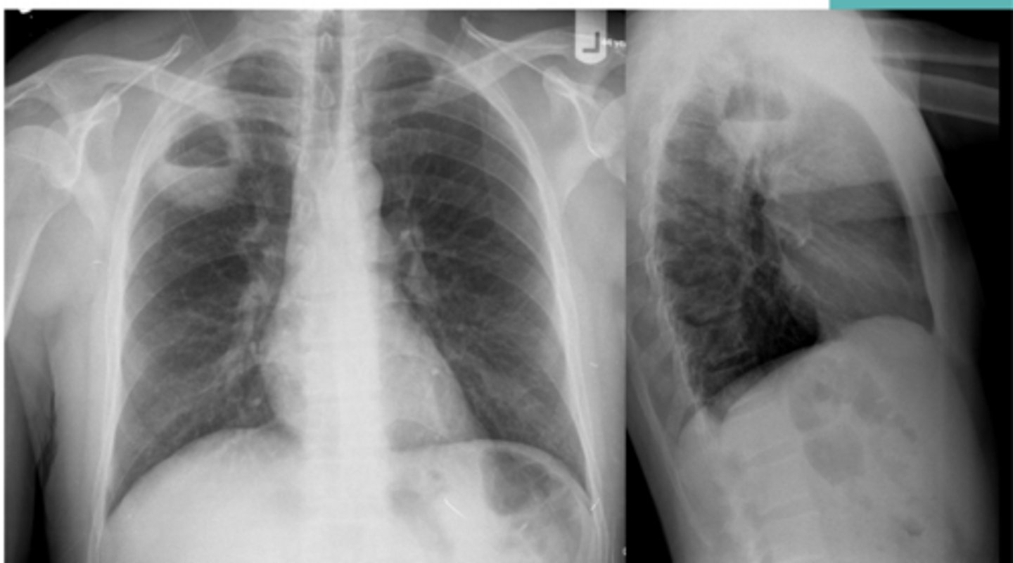
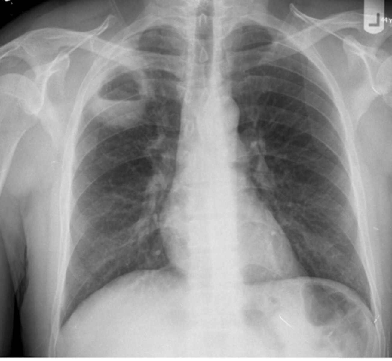
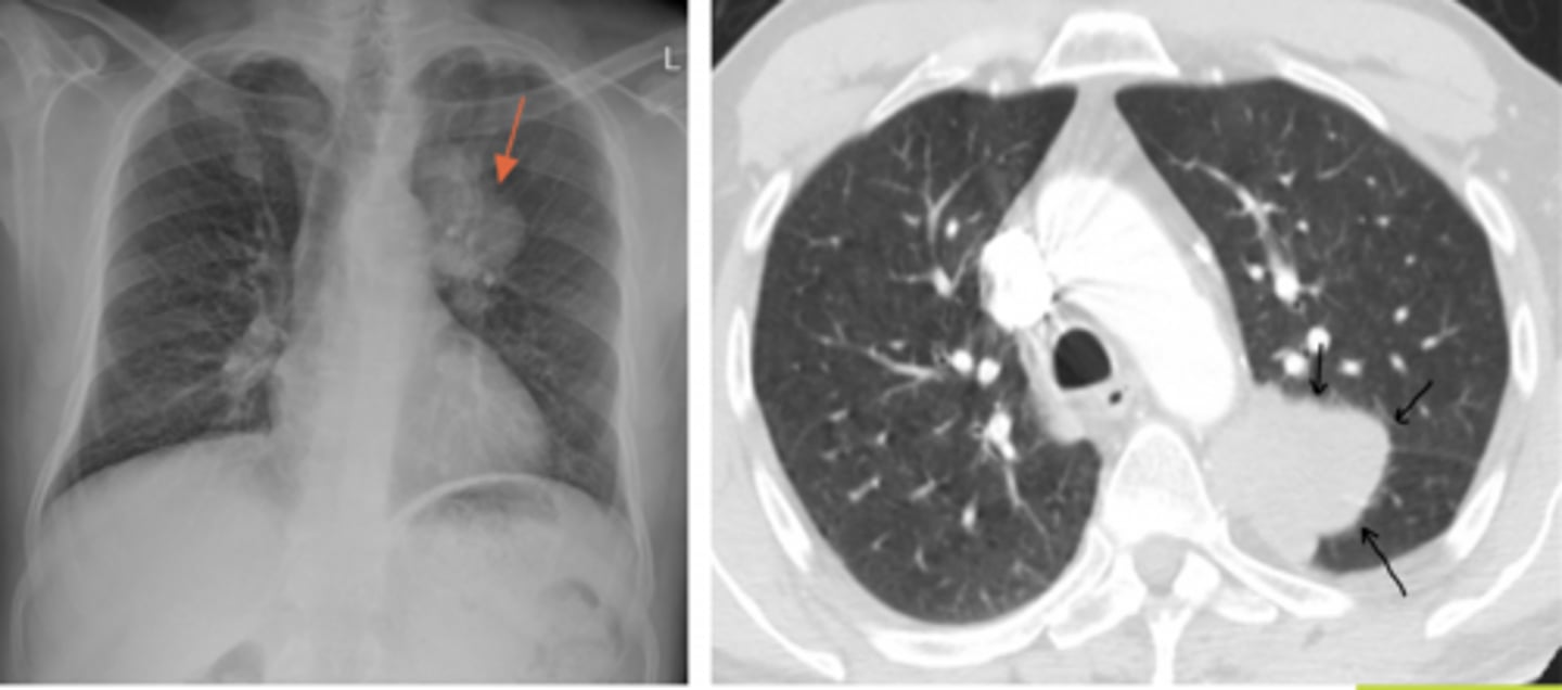
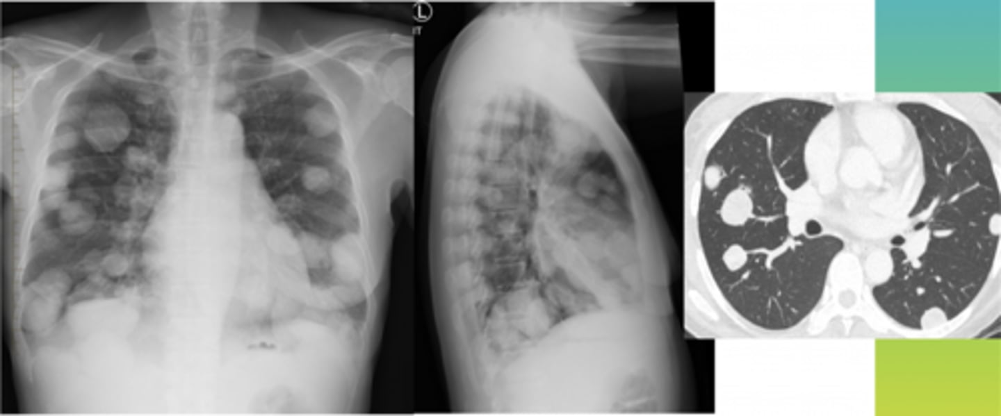
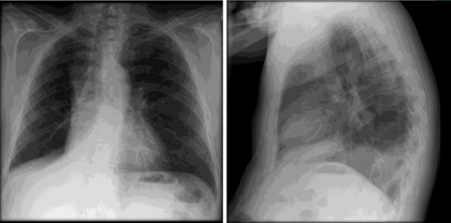
Pulmonary metastasis (cannonball pattern)
Patient has a previous history of pancreatic cancer
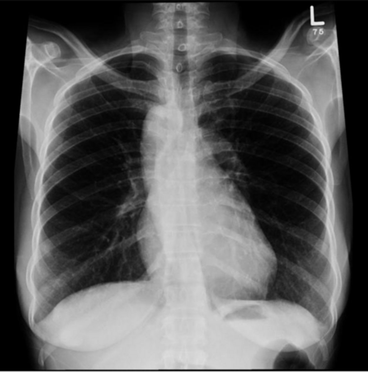
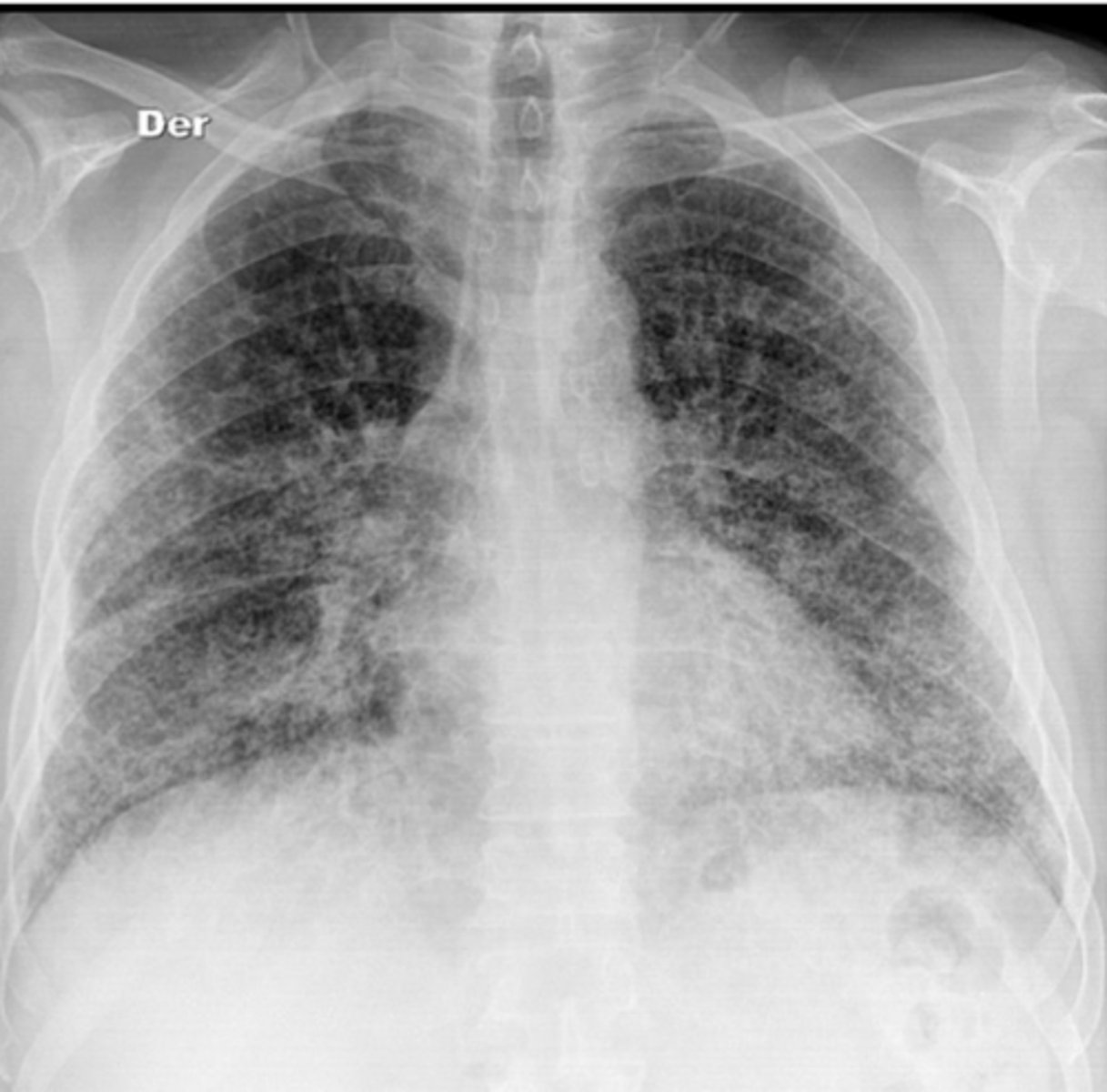
Pulmonary metastasis (snowstorm pattern)
Patient has a previous history of thyroid cancer
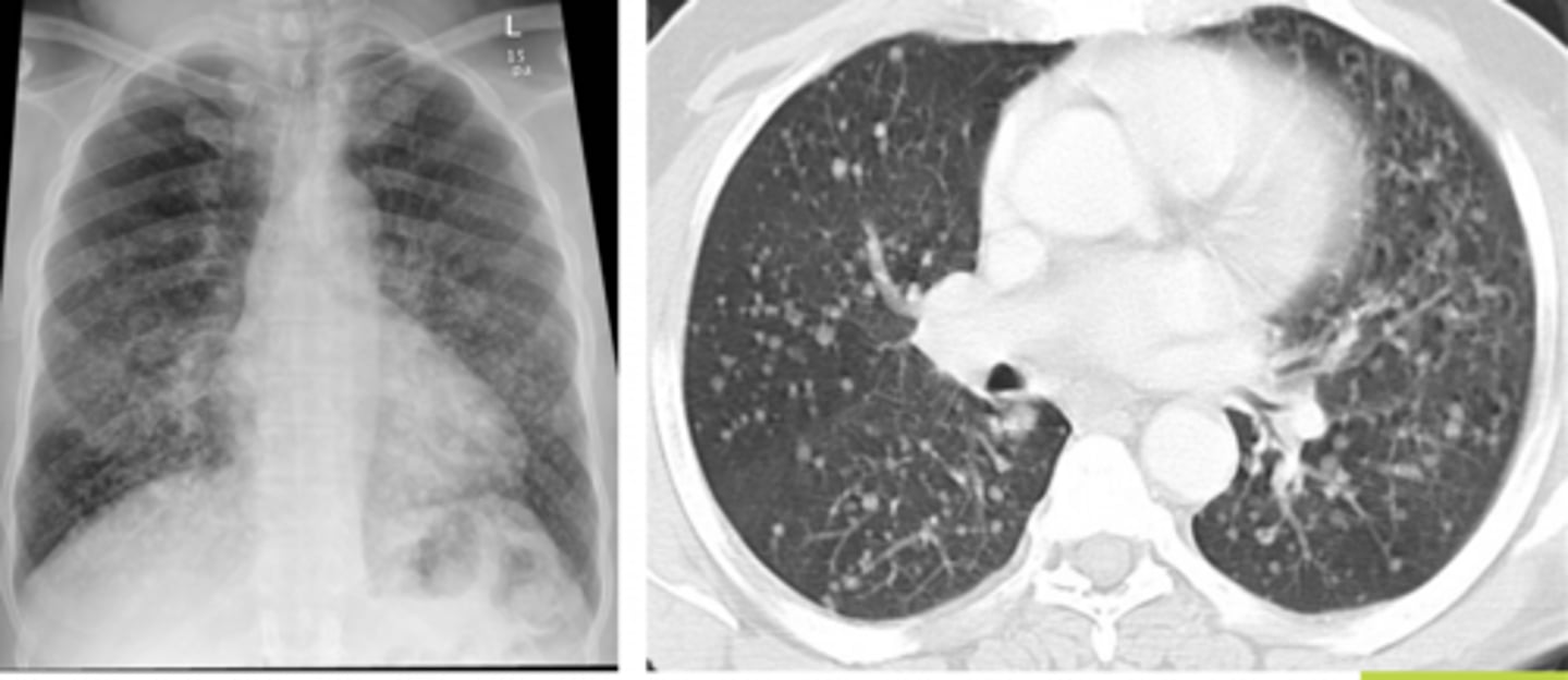
2 primary carcinomas that are most common with the snowstorm pattern of pulmonary METS
renal cell + thyroid
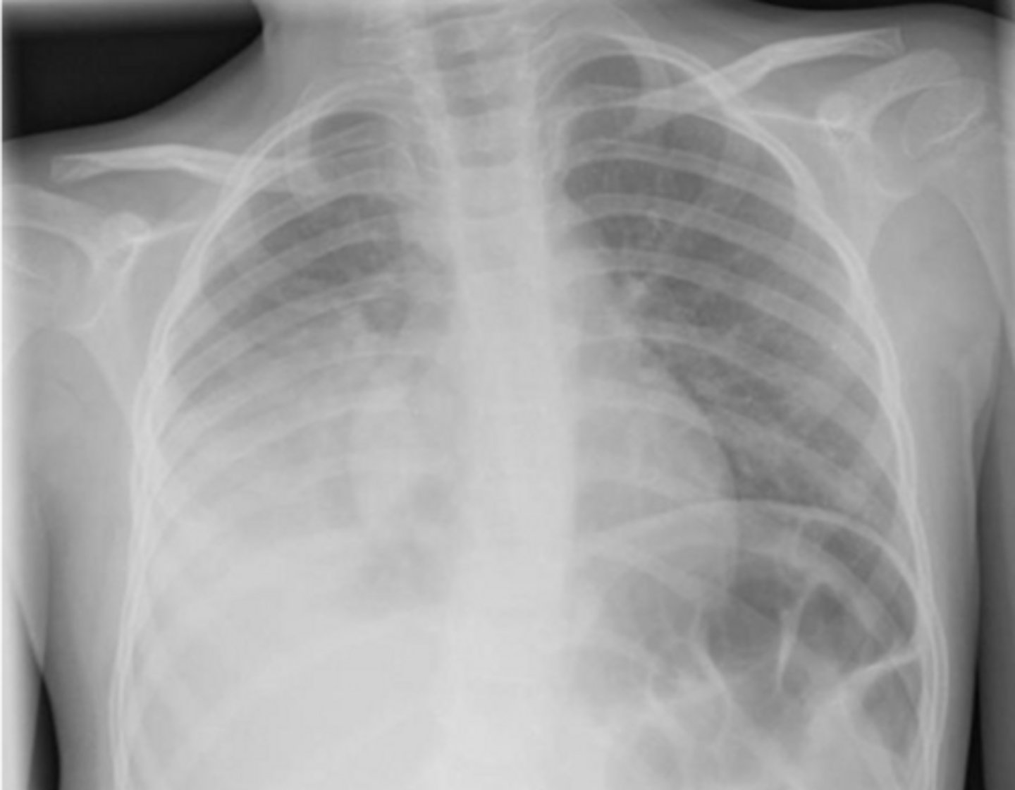
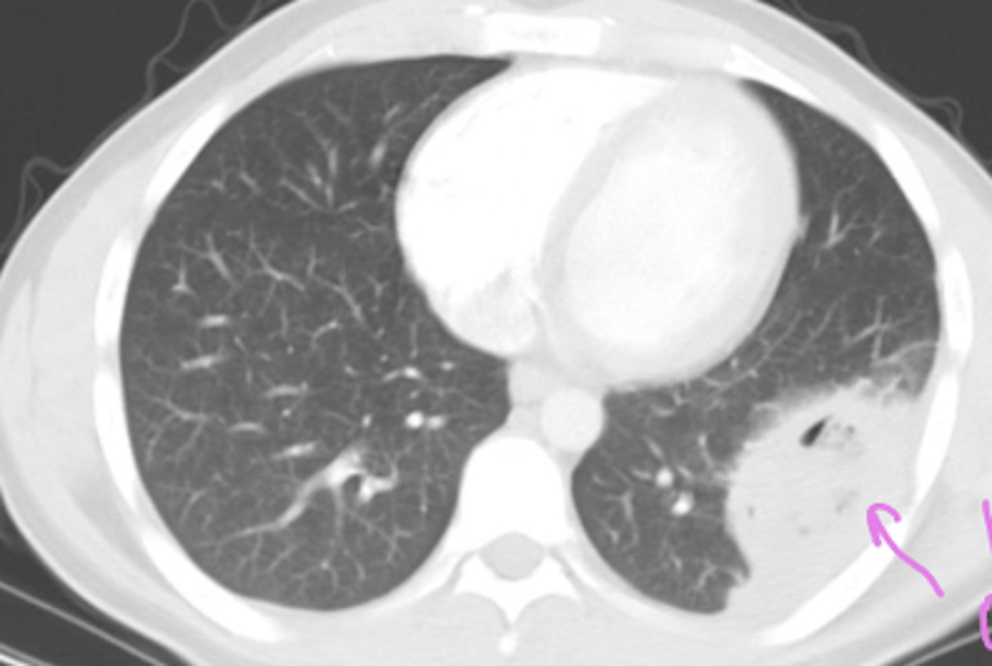
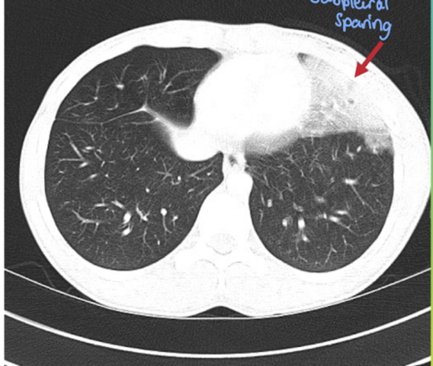
What causes the meniscus sign?
pleural effusion
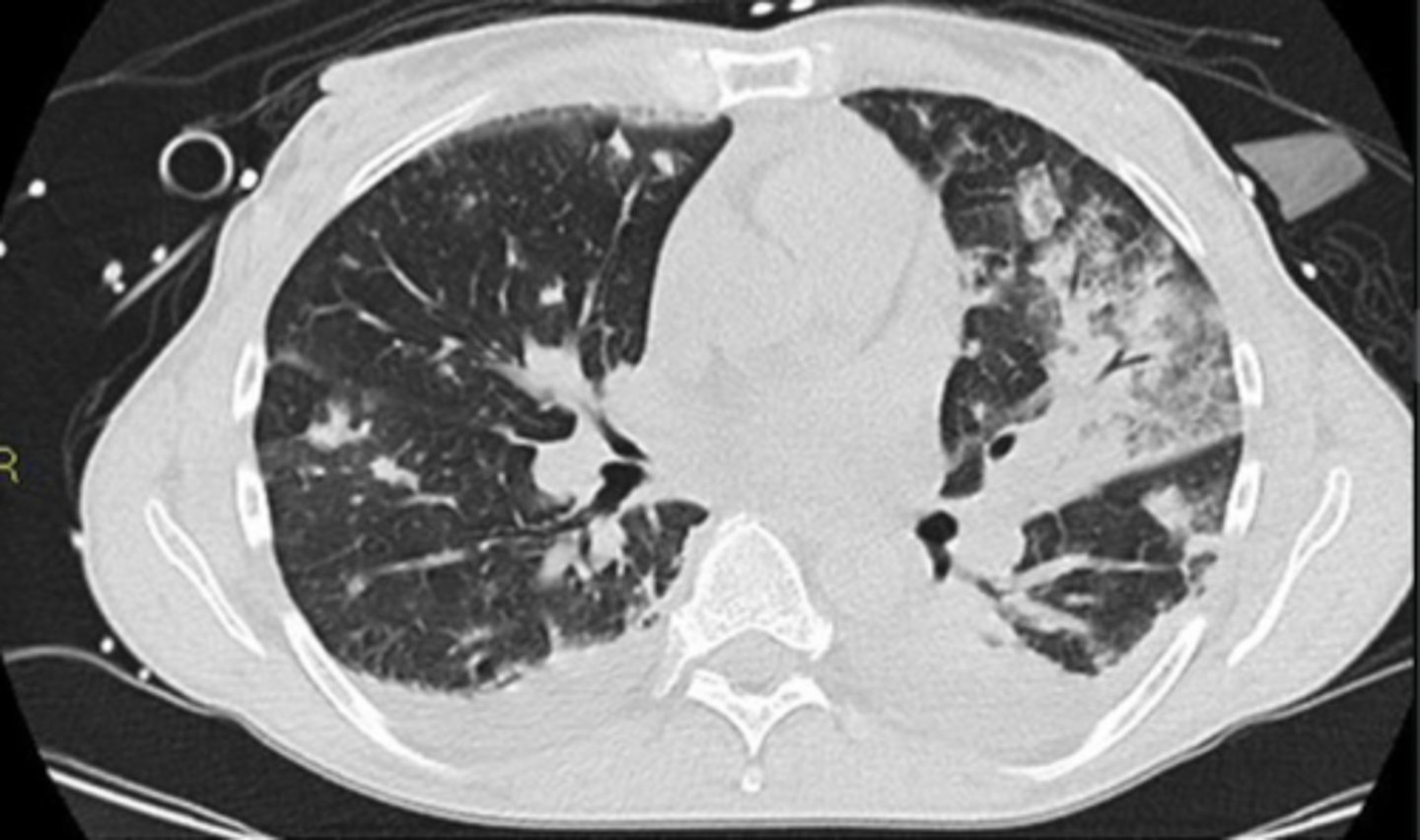
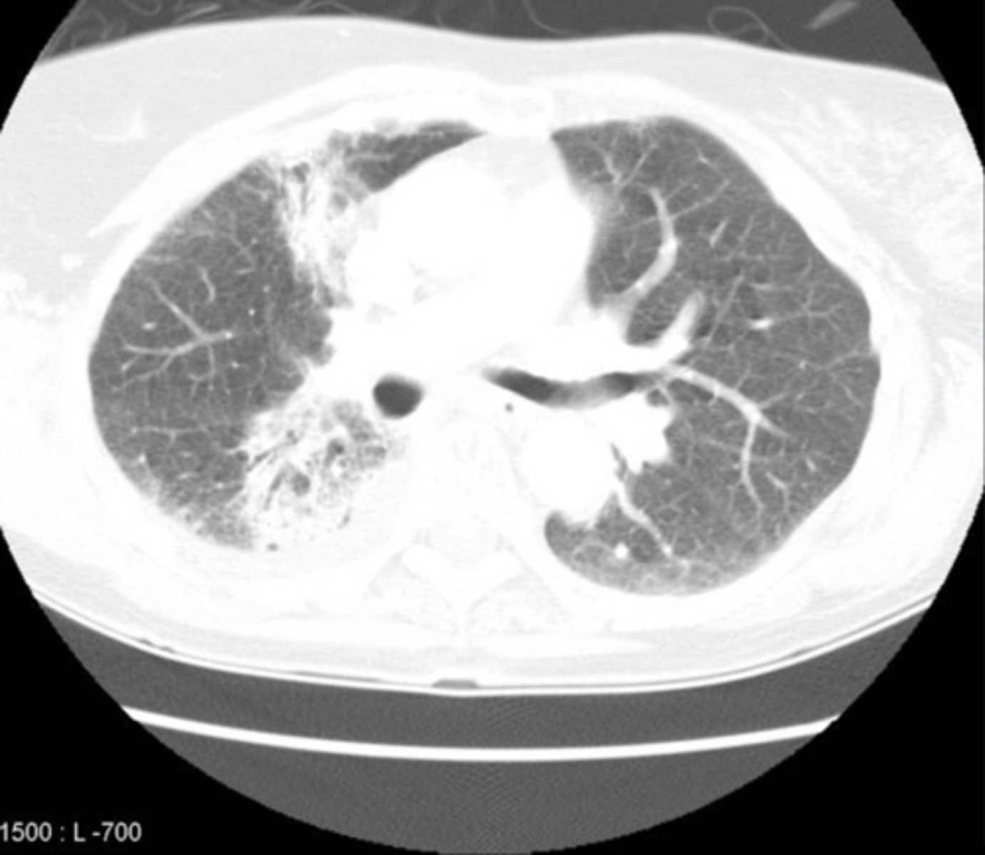
Hypersensitivity pneumonitis
A 46-year-old man presents with progressively worsening shortness of breath and dry cough over the past 6 months. He also reports fatigue, intermittent low-grade fevers, chills, and unintentional weight loss. Symptoms seem worse after work and improve slightly on weekends.
He works on a farm handling moldy hay and grain.
Physical examination reveals:
Mild tachypnea
Fine inspiratory “squeaks” (crackles) at the lung bases
Early digital clubbing
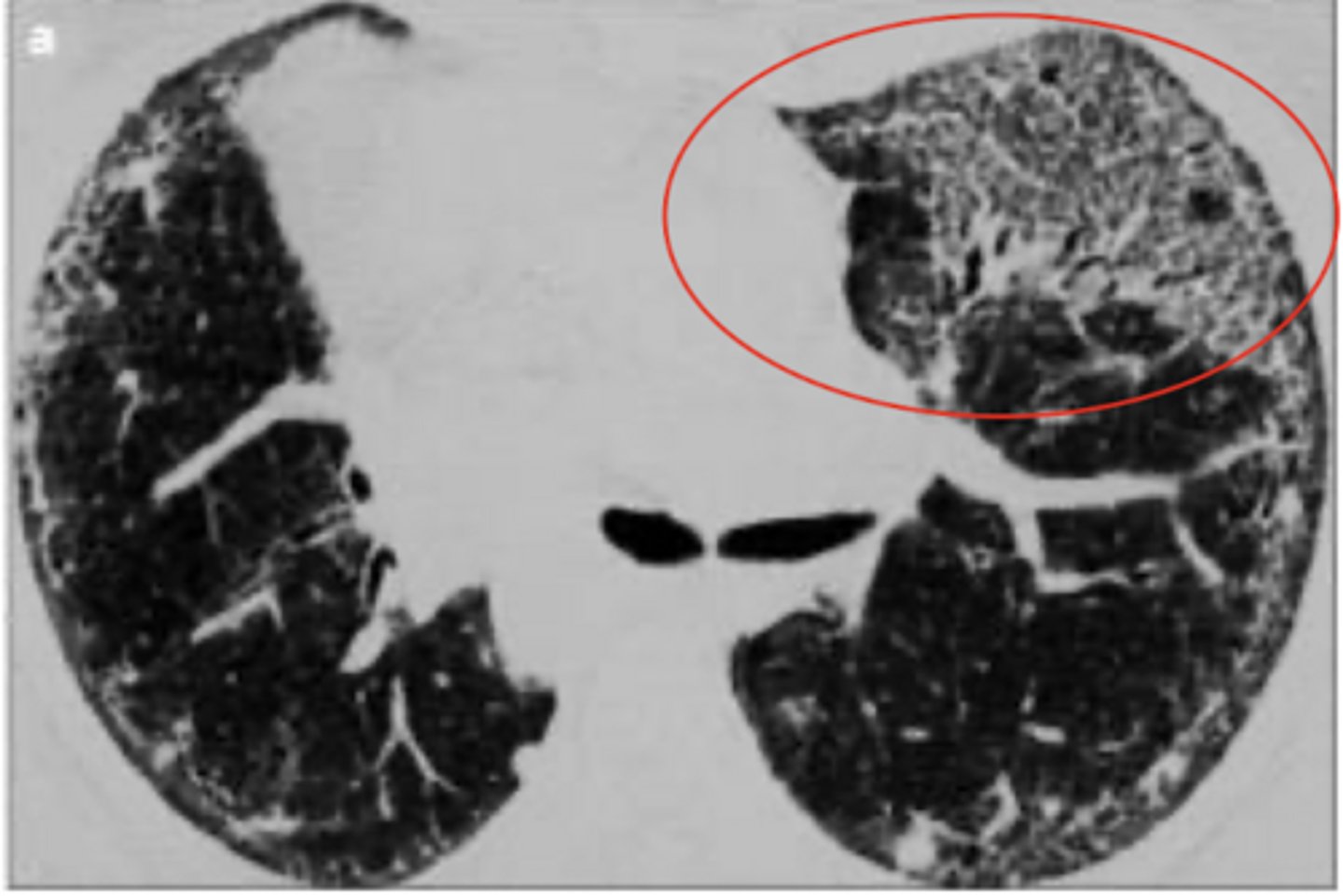
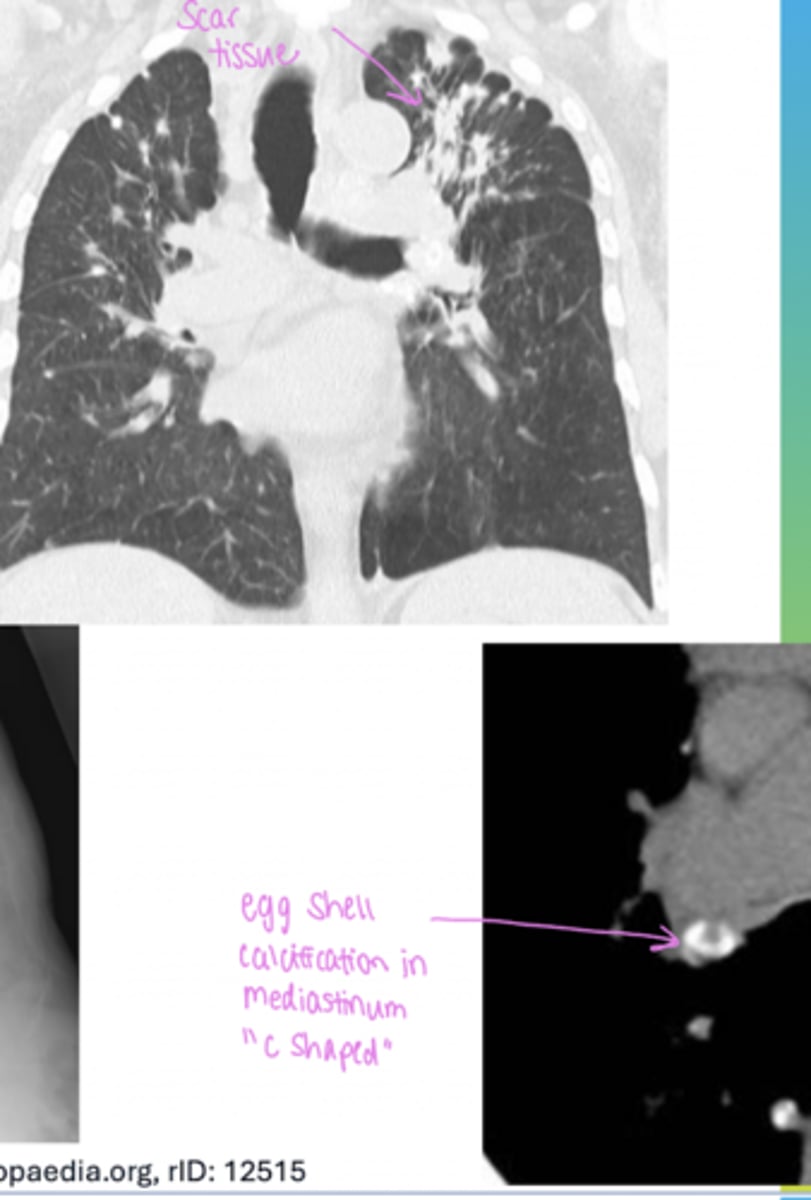
silicosis that lead to Progressive Massive Fibrosis
Patient's occupation was mining and quarrying (worked in that field for 70+ years)
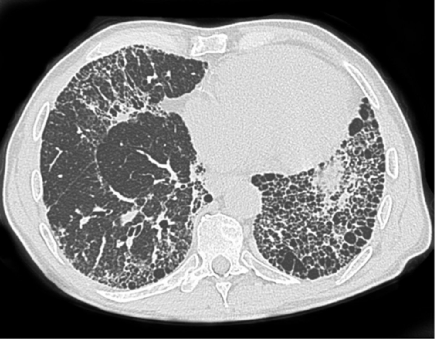
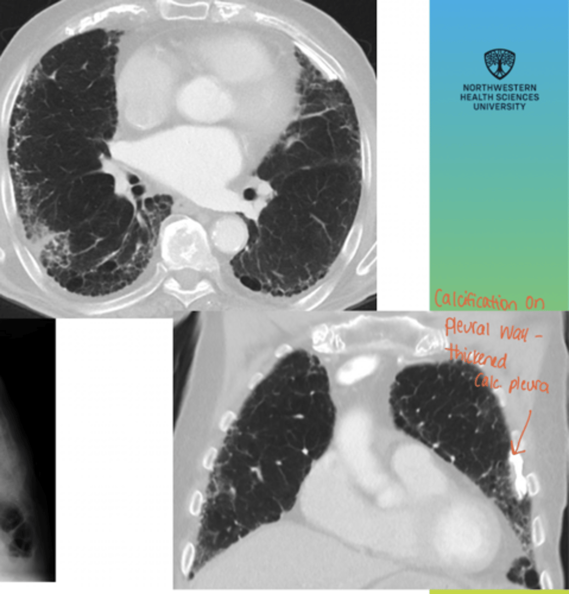
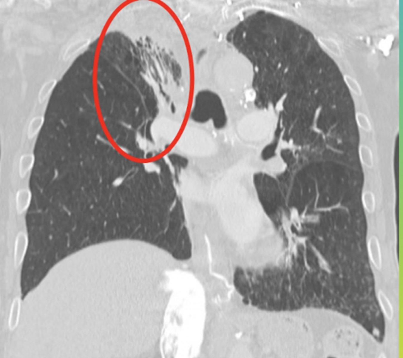
asbestosis
This patient worked in the ship/automotive industry his whole life

asbestosis
This patient worked in the ship/automotive industry his whole life
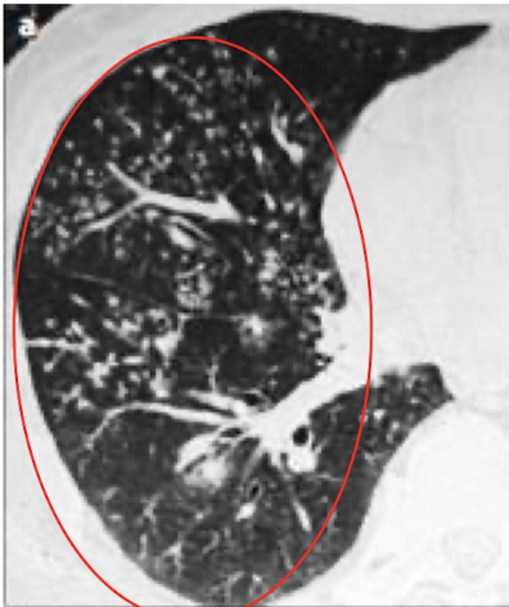
silicosis
This patient worked in the tunneling industry his whole life
silicosis
This patient worked in the tunneling industry his whole life
Radiation-Induced Lung Disease
A 62-year-old woman presents with progressive shortness of breath and a persistent dry cough over the past several months. She denies fever, sputum production, or chest pain. She notes that her symptoms have been slowly worsening and are now affecting her ability to climb stairs.
Her history is significant for left-sided breast cancer treated with radiation therapy, completed 10 months ago. She did well immediately after treatment with no acute complications.
Radiation-Induced Lung Disease
Recently received radiation treatment for breast cancer
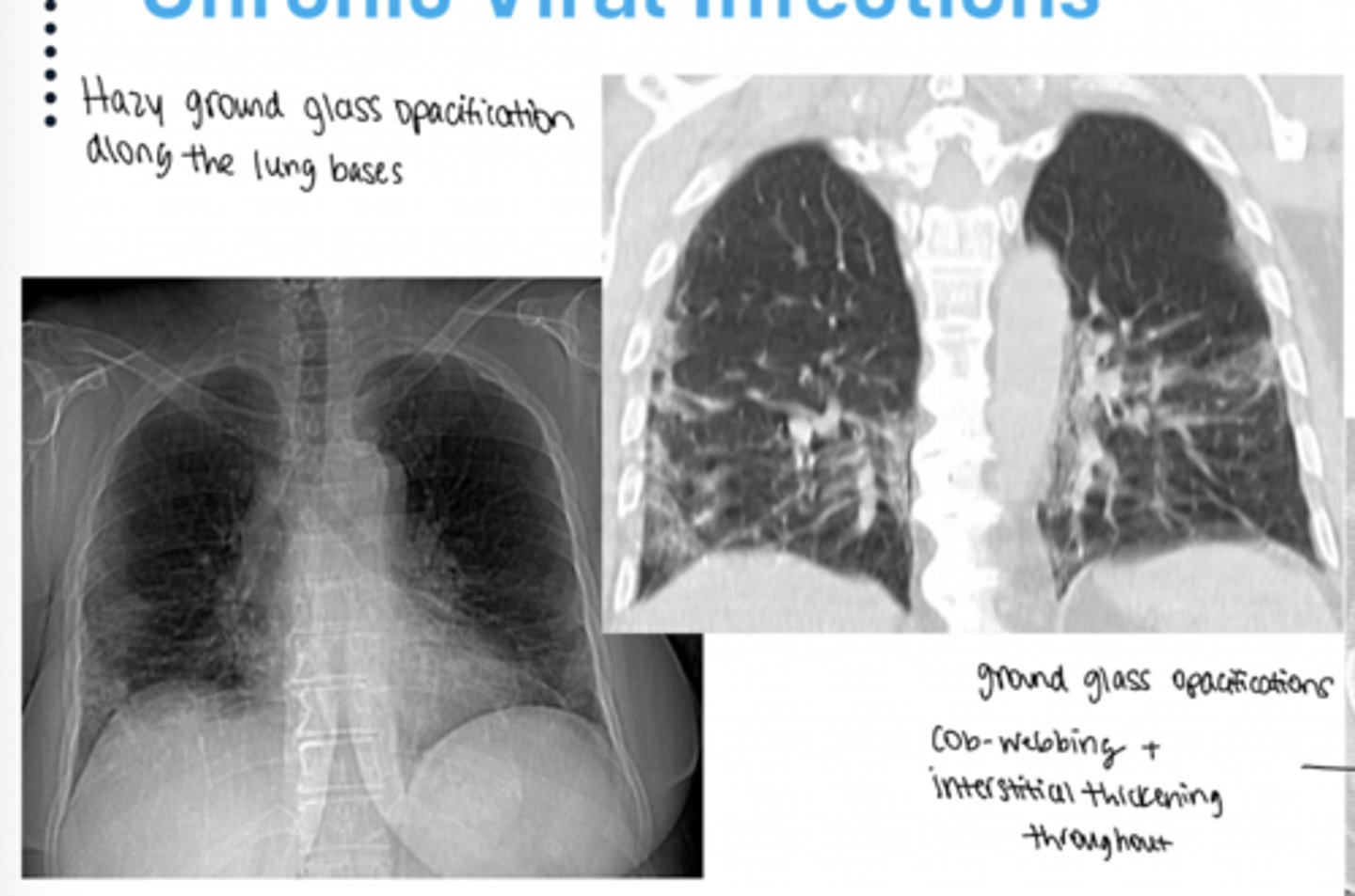
chronic viral infection lung disease
This patient was immunocompromised, got COVID-19, and never received any treatment
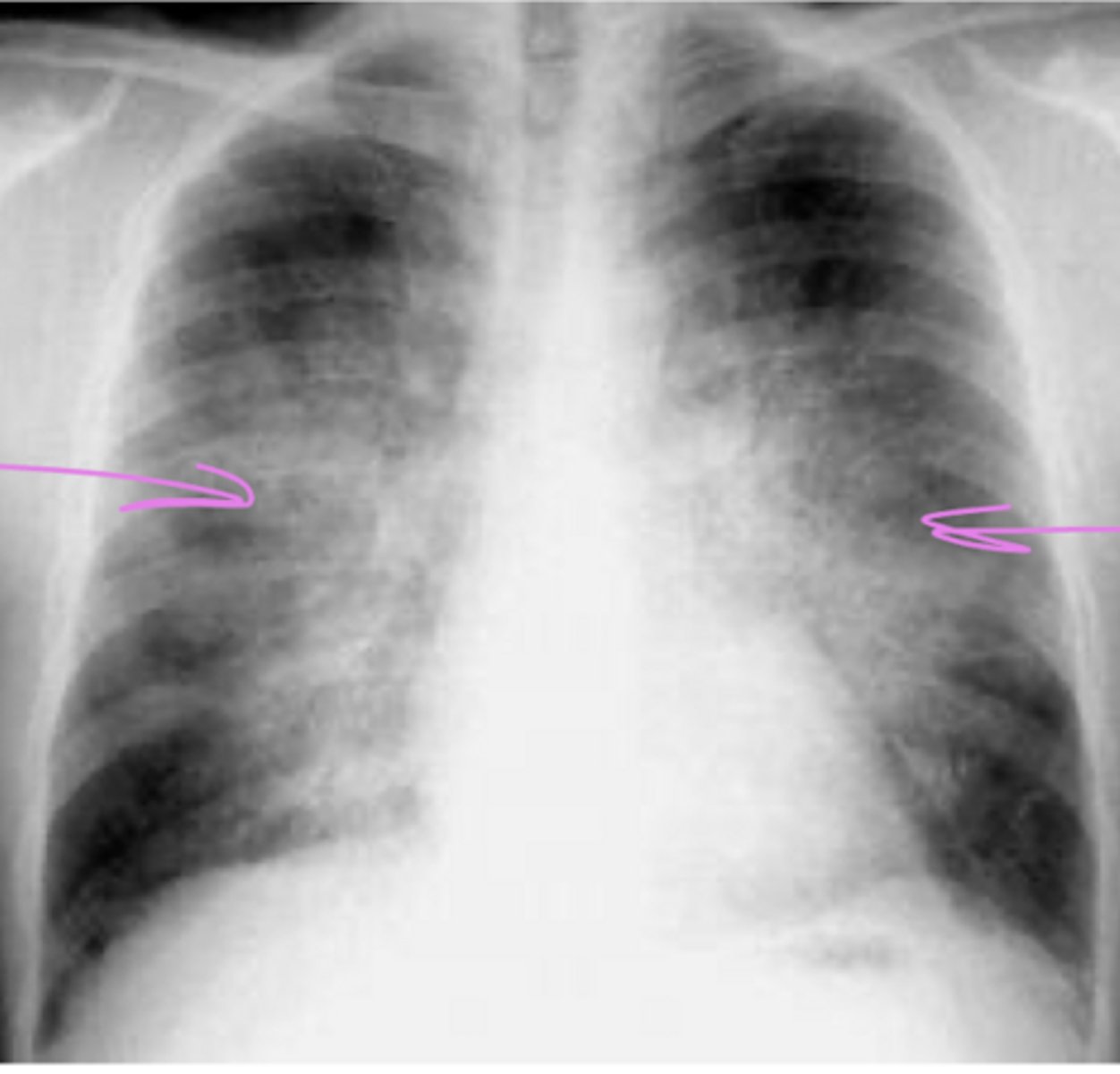
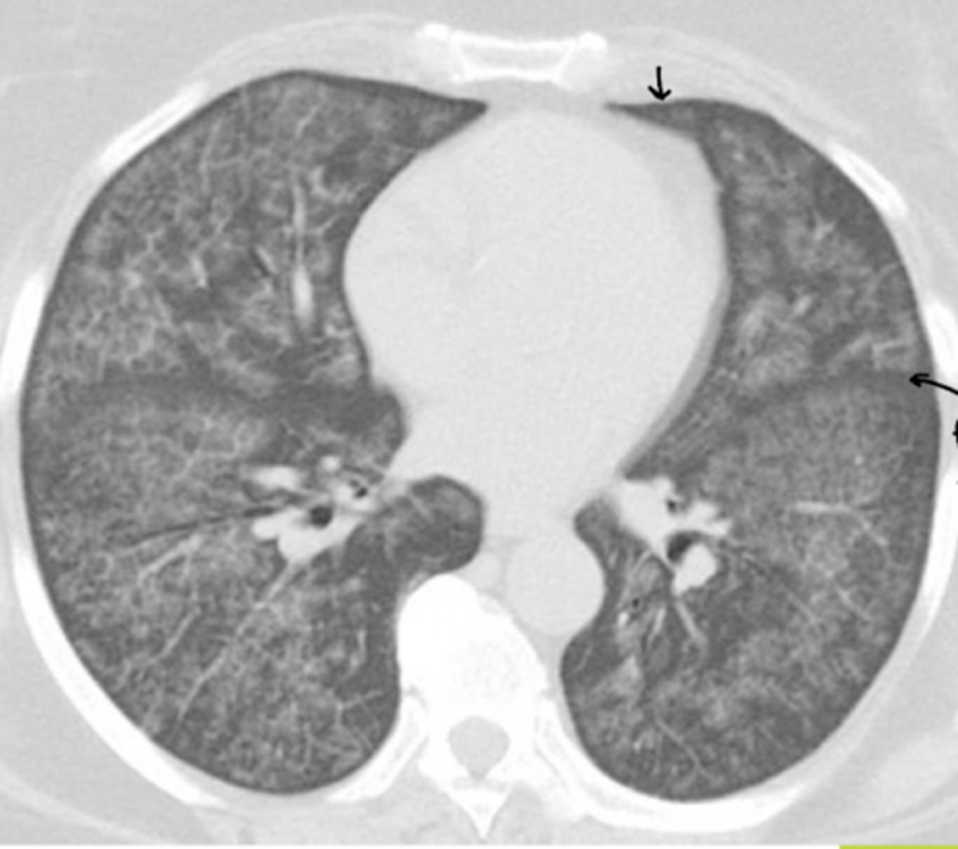
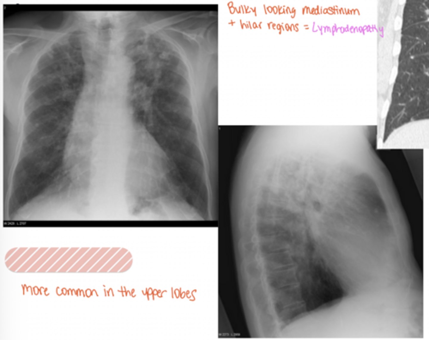
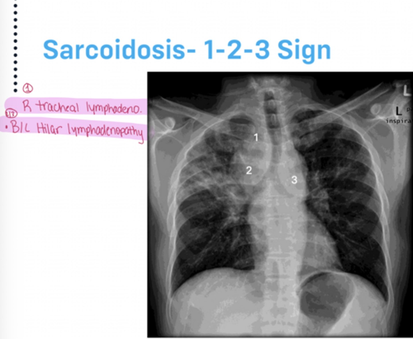
Sarcoidosis
1-2-3 sign
African American female presents with cough, dyspnea, and a skin finding called erythema nodosum
exam: restrictive pattern with reduced lung volumes and reduced diffusion capacity on LFTs
What sign is present?
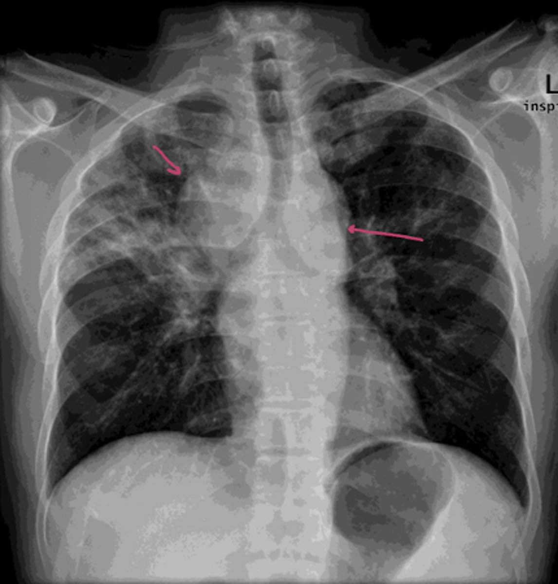
Sarcoidosis
Mediastinal / bilateral hilar lymphadenopathy
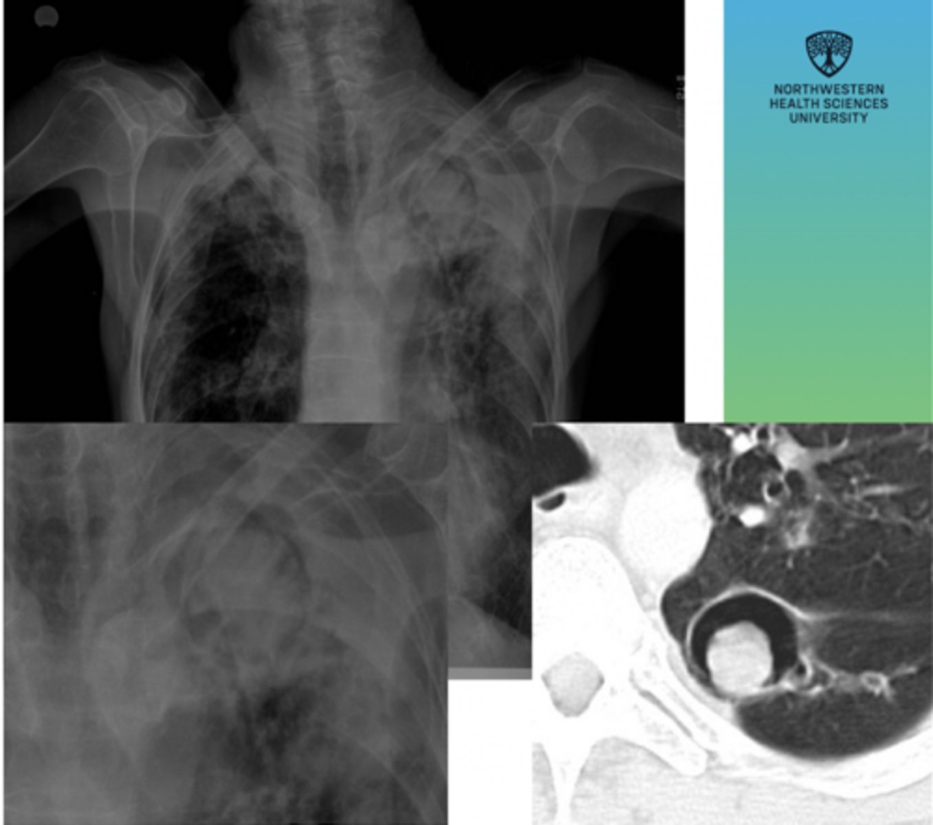
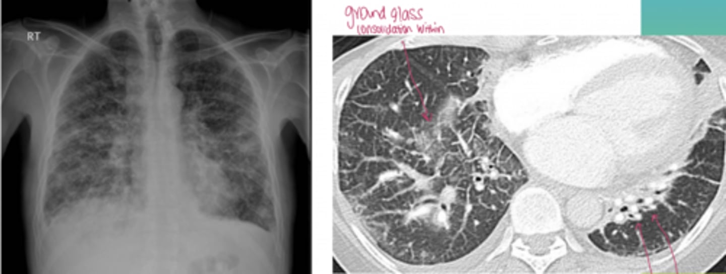
Lymphangitis carcinomatosis
Patient has a history of recent breast cancer, has been experiencing some dyspnea and has abnormal lung function tests, got an x-ray a few months ago but it showed nothing. Got another x-ray and this is what shows- but overall lung architecture is preserved
SLE
ankylosing spondylitis
A 41-year-old man presents with progressive shortness of breath and chronic dry cough over the past year. He denies fever or sputum production. Symptoms have been slowly worsening and are now limiting his exercise tolerance.
His history is notable for long-standing low back pain and morning stiffness since his 20s, which improves with activity. He also reports reduced spinal mobility over time.
Rheumatoid arthritis
Her medical history is notable for a 10-year history of symmetric pain, swelling, and prolonged morning stiffness affecting the wrists, hands, and feet. She reports that stiffness lasts more than an hour each morning and improves with movement throughout the day. Over time, she has noticed increasing difficulty with grip strength and fine motor tasks such as buttoning clothing and opening jars.
COPD risk factors
Patient presents with dyspnea upon exertion, wheezing, productive cough, pursed-lip breathing, use of accessory respiratory muscles
Emphysema
-flattened hemidiaphragm
-blunted costophrenic angle
-large hyperinflated lungs w/ lack of bronchovascular markings
Patient presents with tachypnea, absence of cyanosis, pursed-lip breathing, tripod position, barrel chest
Exam:
reduced breath sounds, hyper-resonant to percussion
panlobular
-lower lobes w/ uniform distribution
What type of emphysema is this?
centrilobular
-upper zones of lobes w/ patchy distribution
-"white dot" representing bronchovascular bundle
What type of emphysema is this?
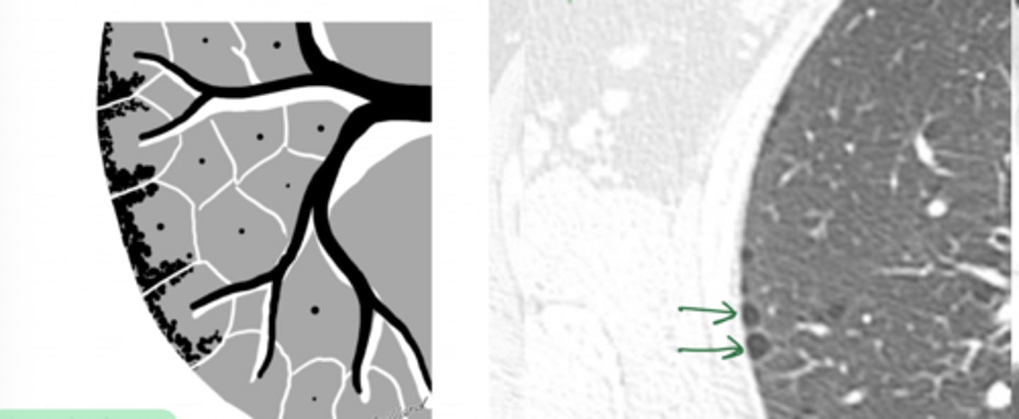
paraseptal
-small focal lucencies peripheral distribution
What type of emphysema is this?
asthma
Patient presents with wheezing, SOB, chest tightness, cough, episodic symptoms worse at night or early in the morning, known atopic disorder
Exam:
widespread wheezing on auscultation
blood test: serum eosinophil count, IgE, allergen skin prick tests
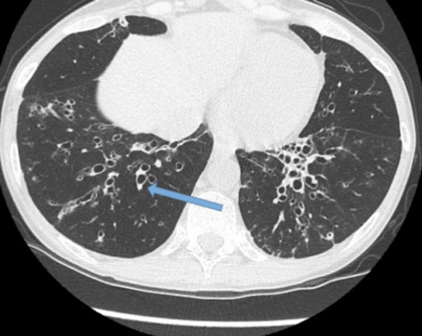
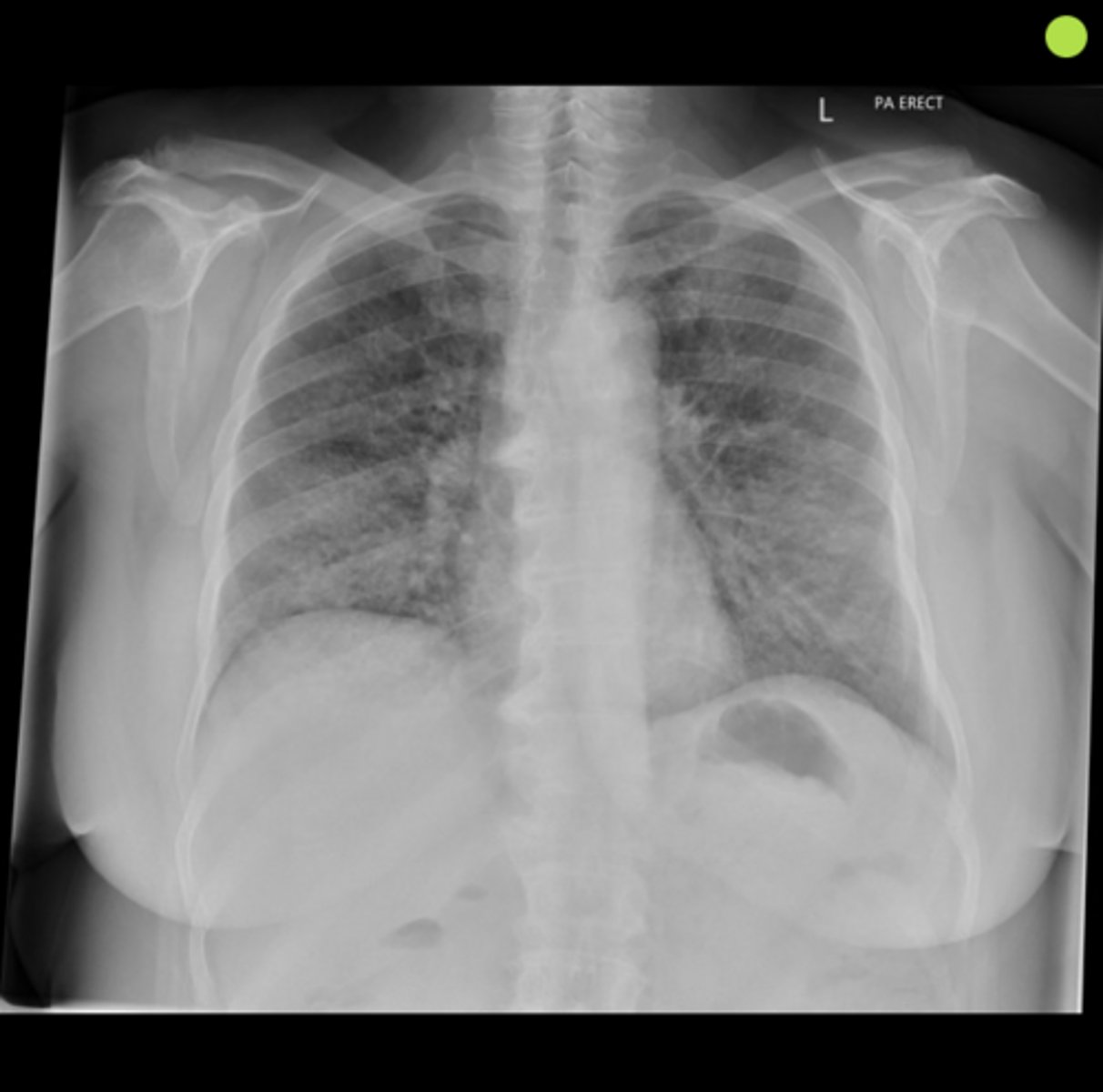
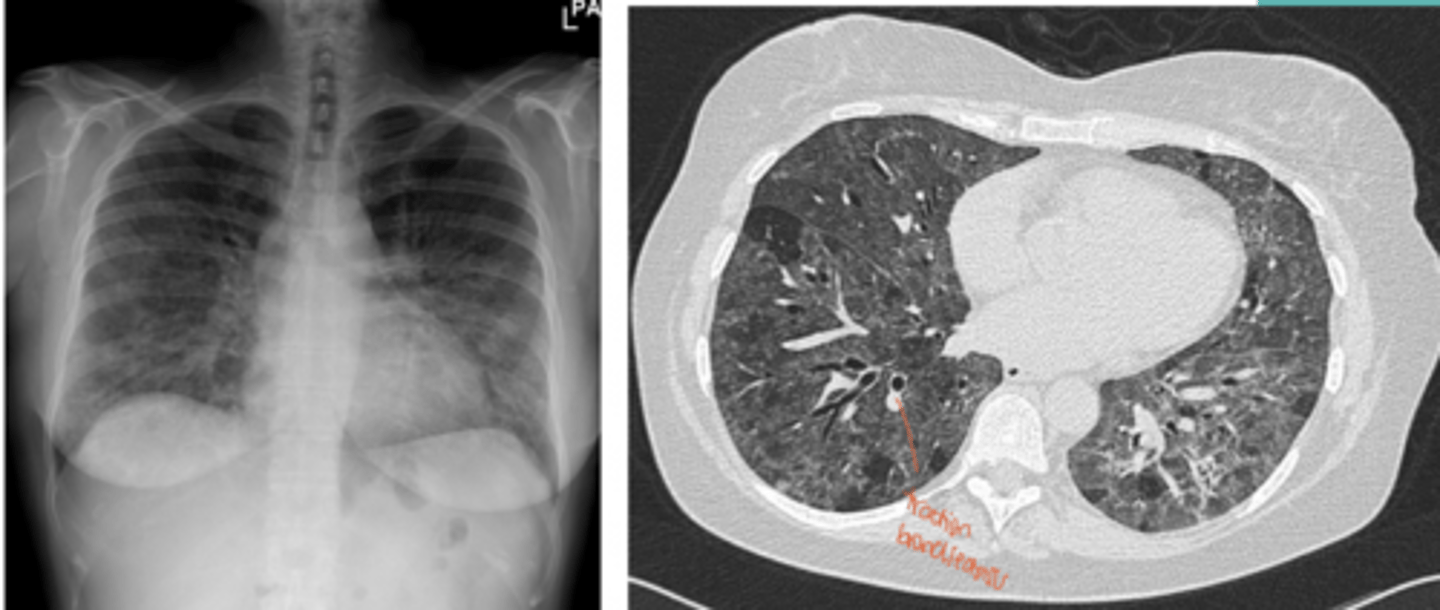
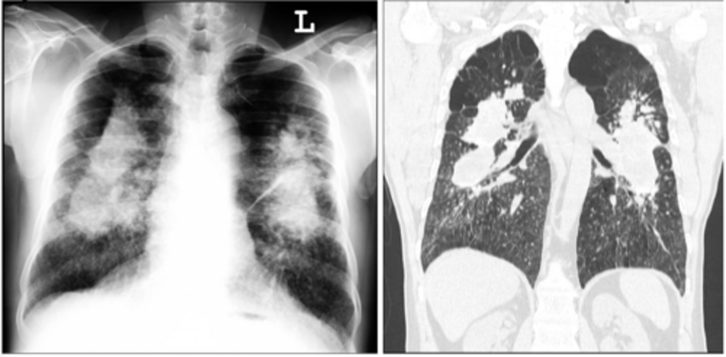
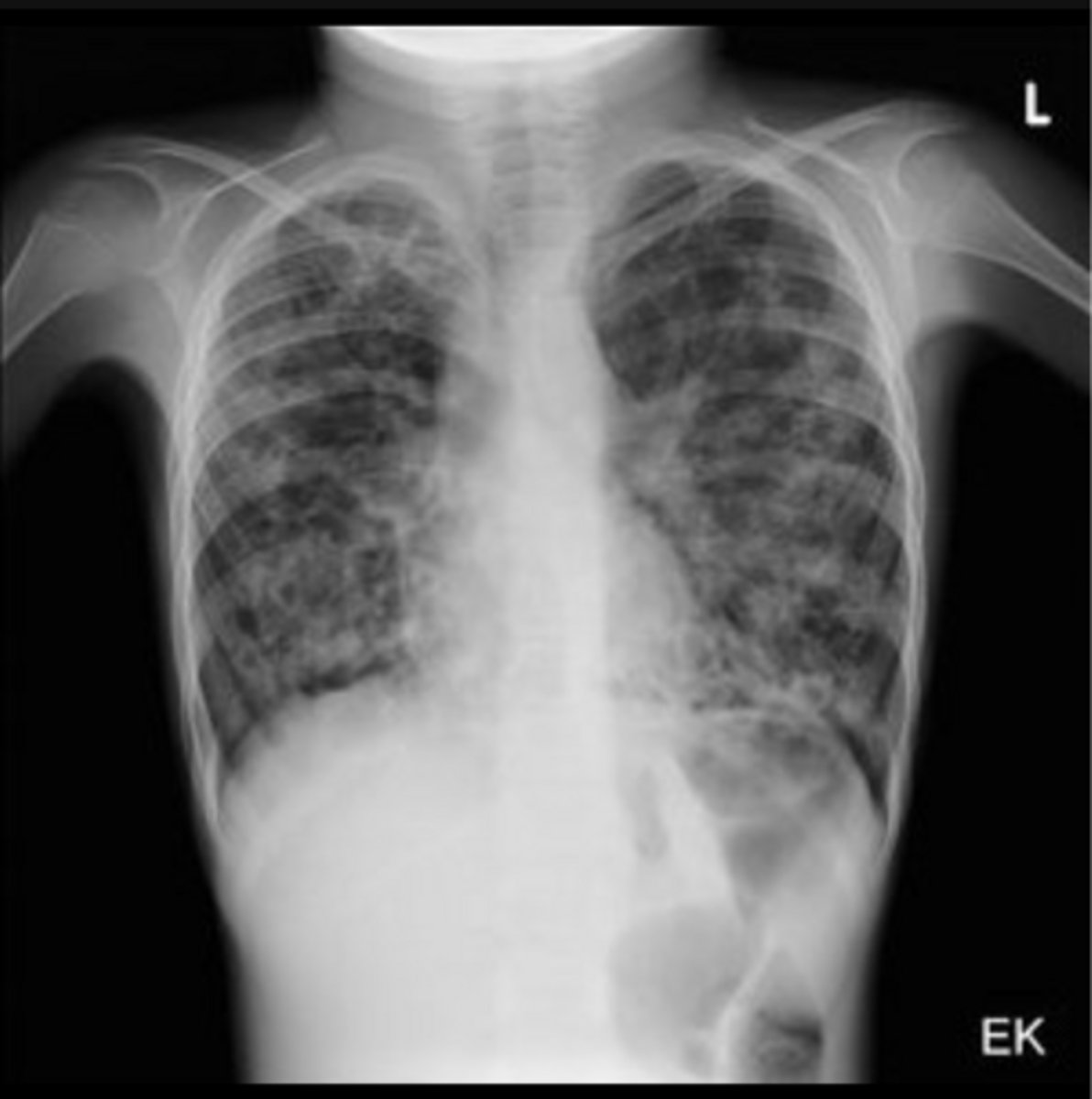
bronchiectasis
Patient presents with chronic productive cough with thick, foul-smelling sputum, recurrent chest infections, hemoptysis, SOB, wheezing
cystic fibrosis
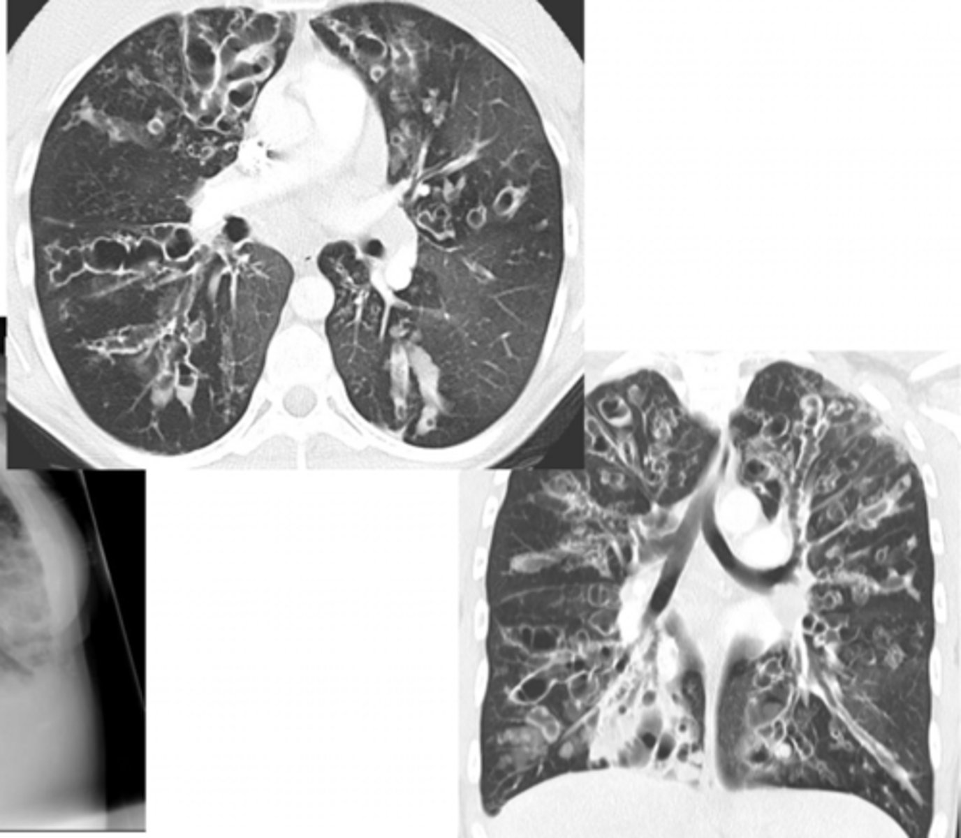
Child presents with recurrent pulmonary infections, hemoptysis, poor weight gain, steatorrhea, copious amounts of sputum, frequent blood-stained and containing mucus plugs
cystic fibrosis w/ signs of bronchiectasis
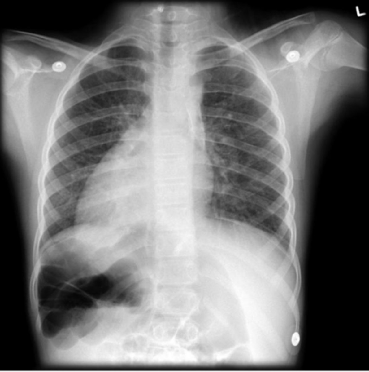
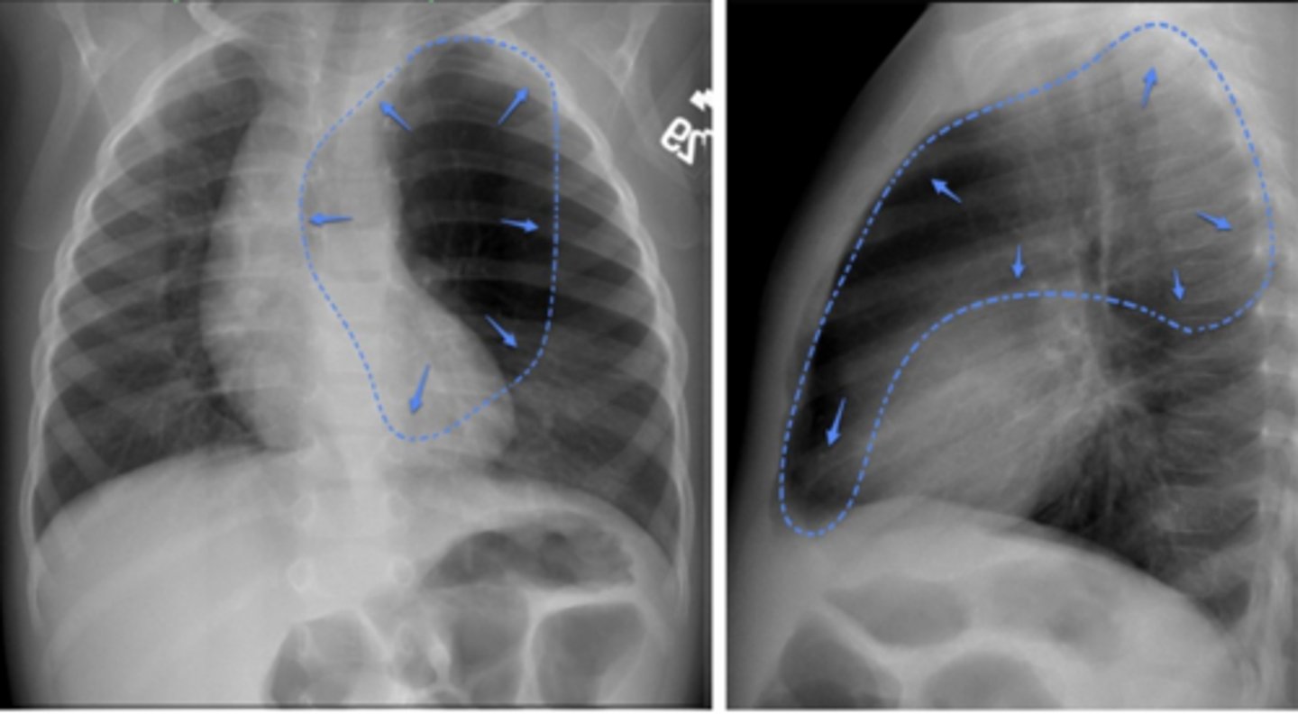
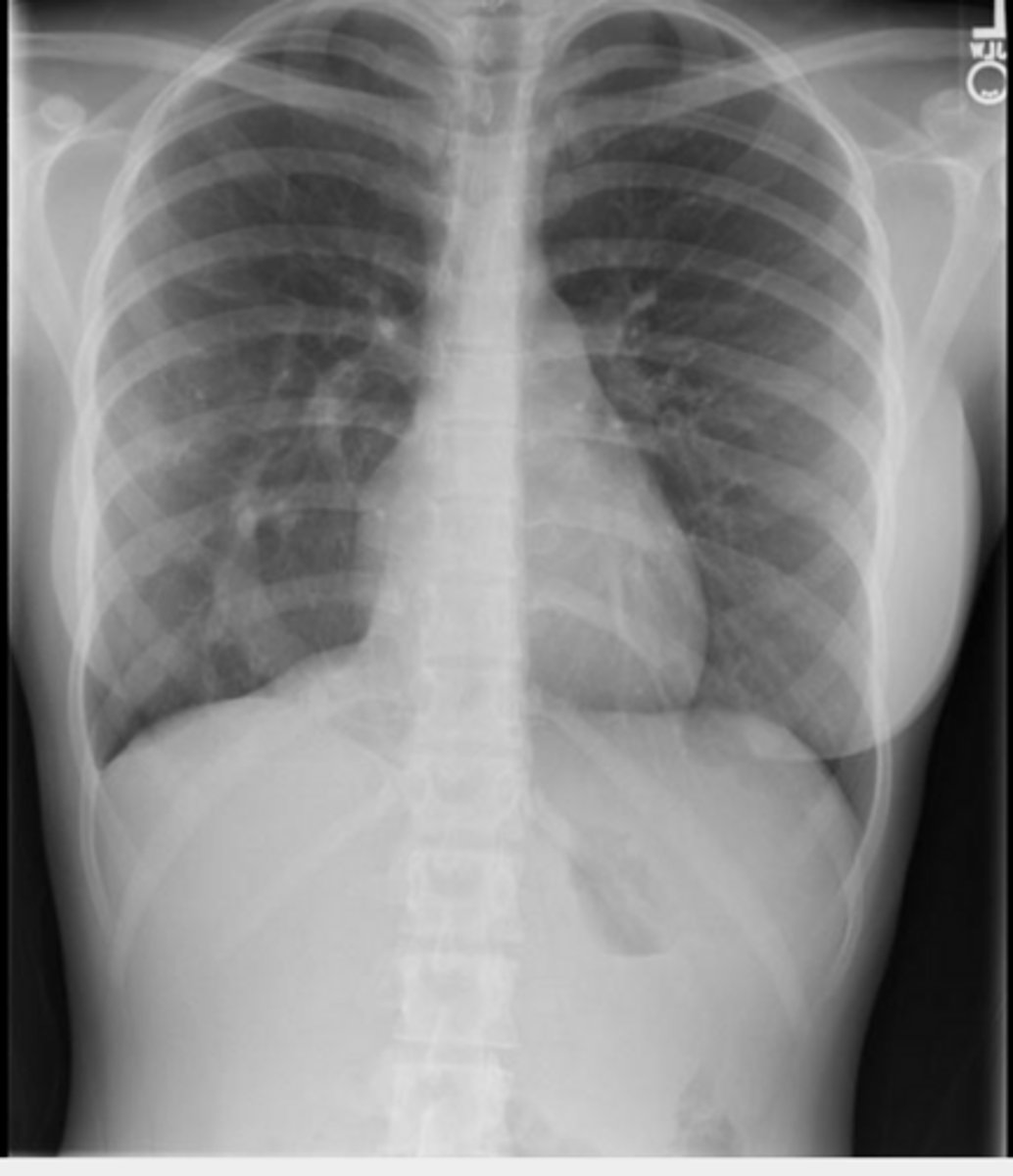
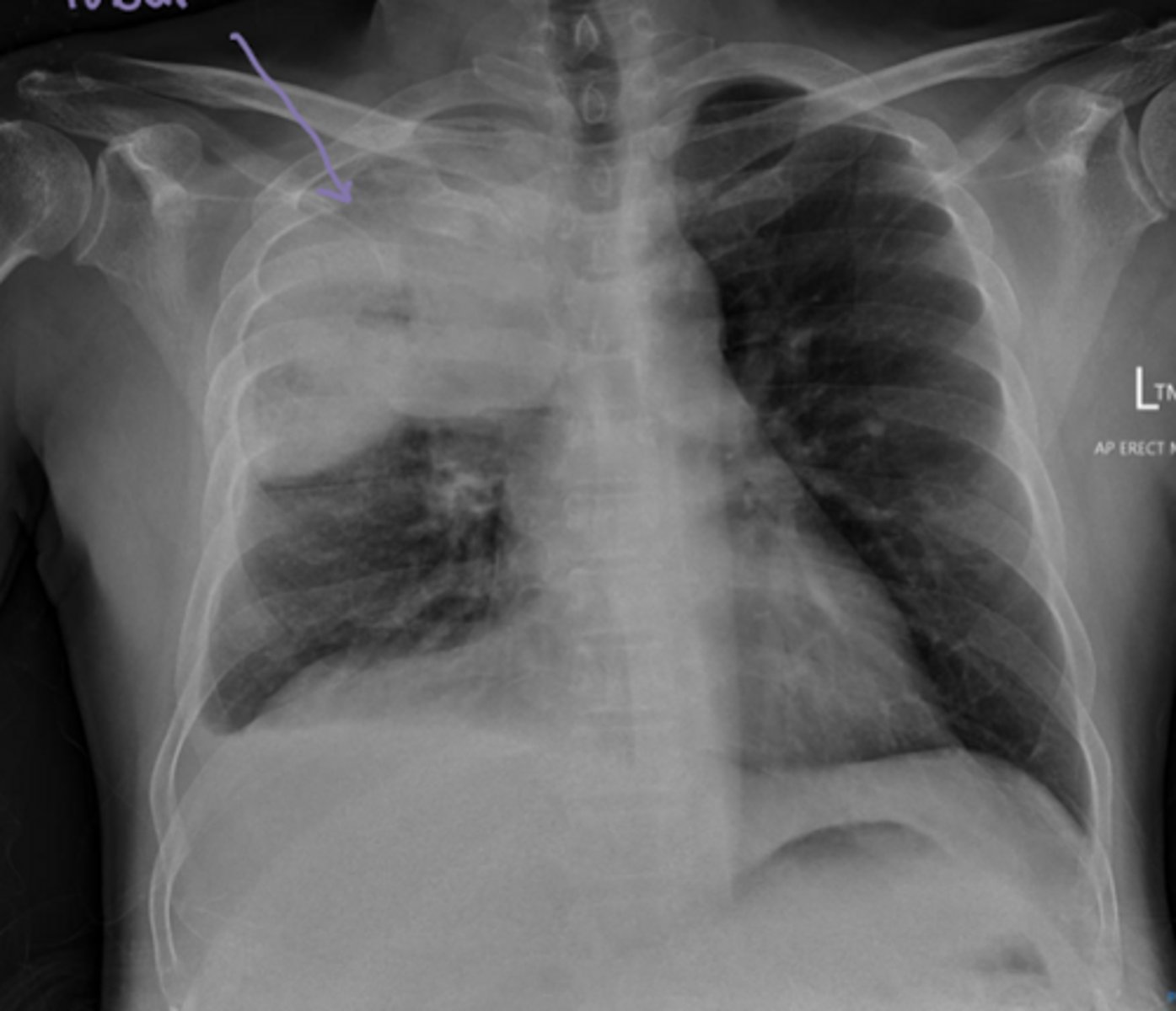
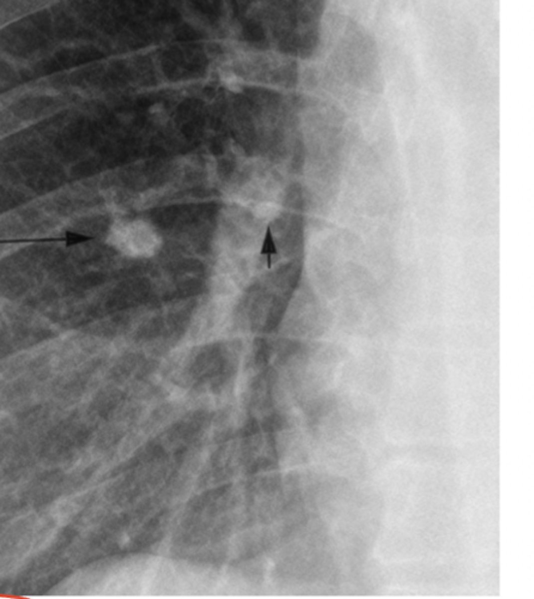
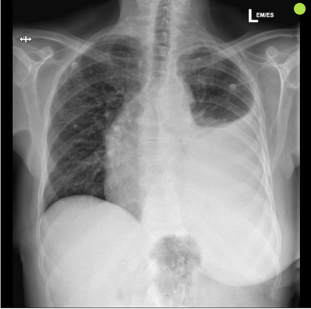
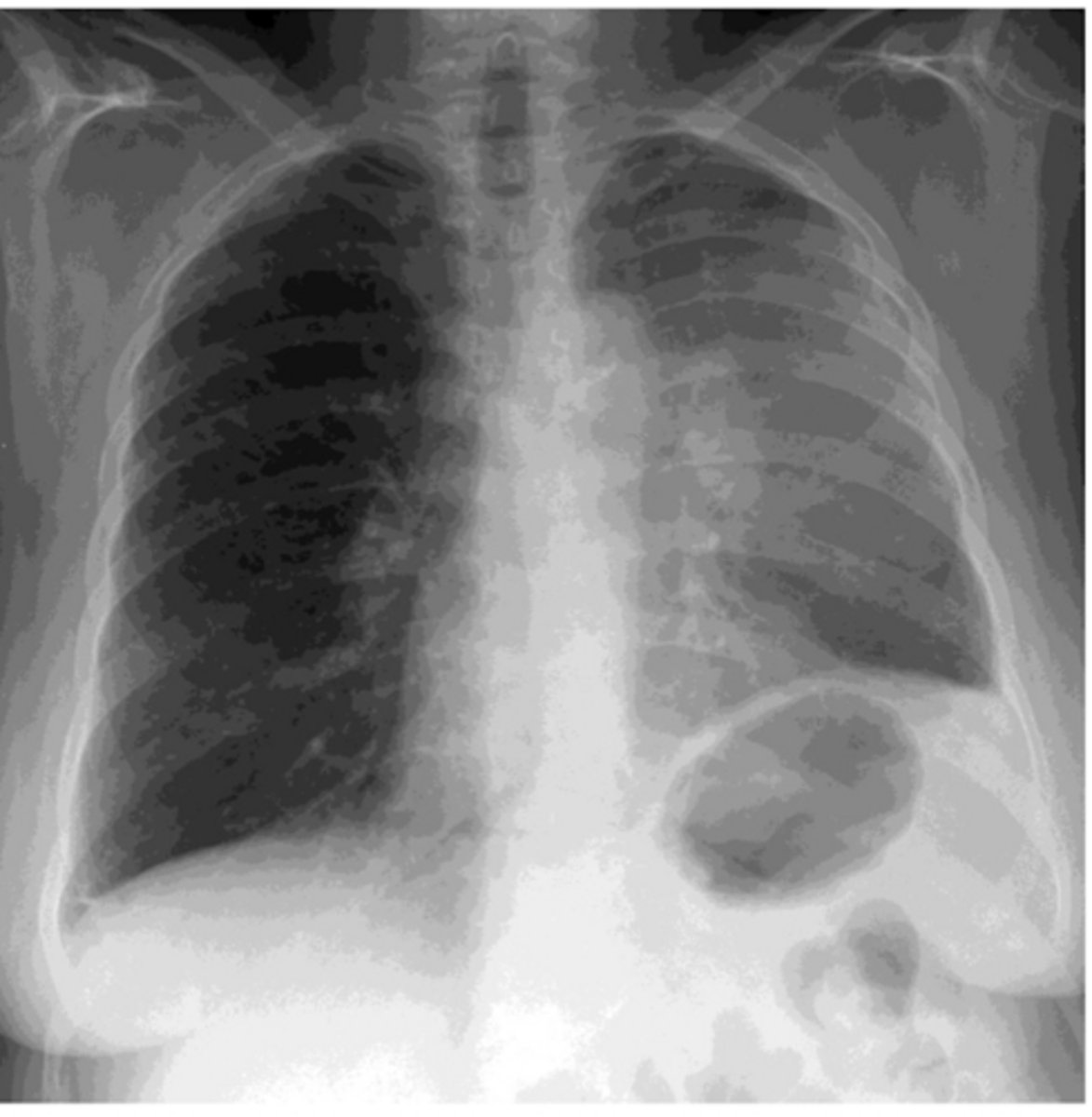
Atelectasis of the right upper lobe
Golden's sign is present
What is the diagnosis? What sign is present?
atelectasis of the right middle lobe
What diagnosis is present?
Atelectasis of the right lower lobe
What is the diagnosis?
Atelectasis of the left upper lobe
What is the diagnosis?
Atelectasis of the left lower lobe
What is the diagnosis?
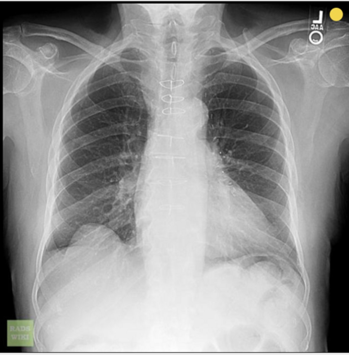
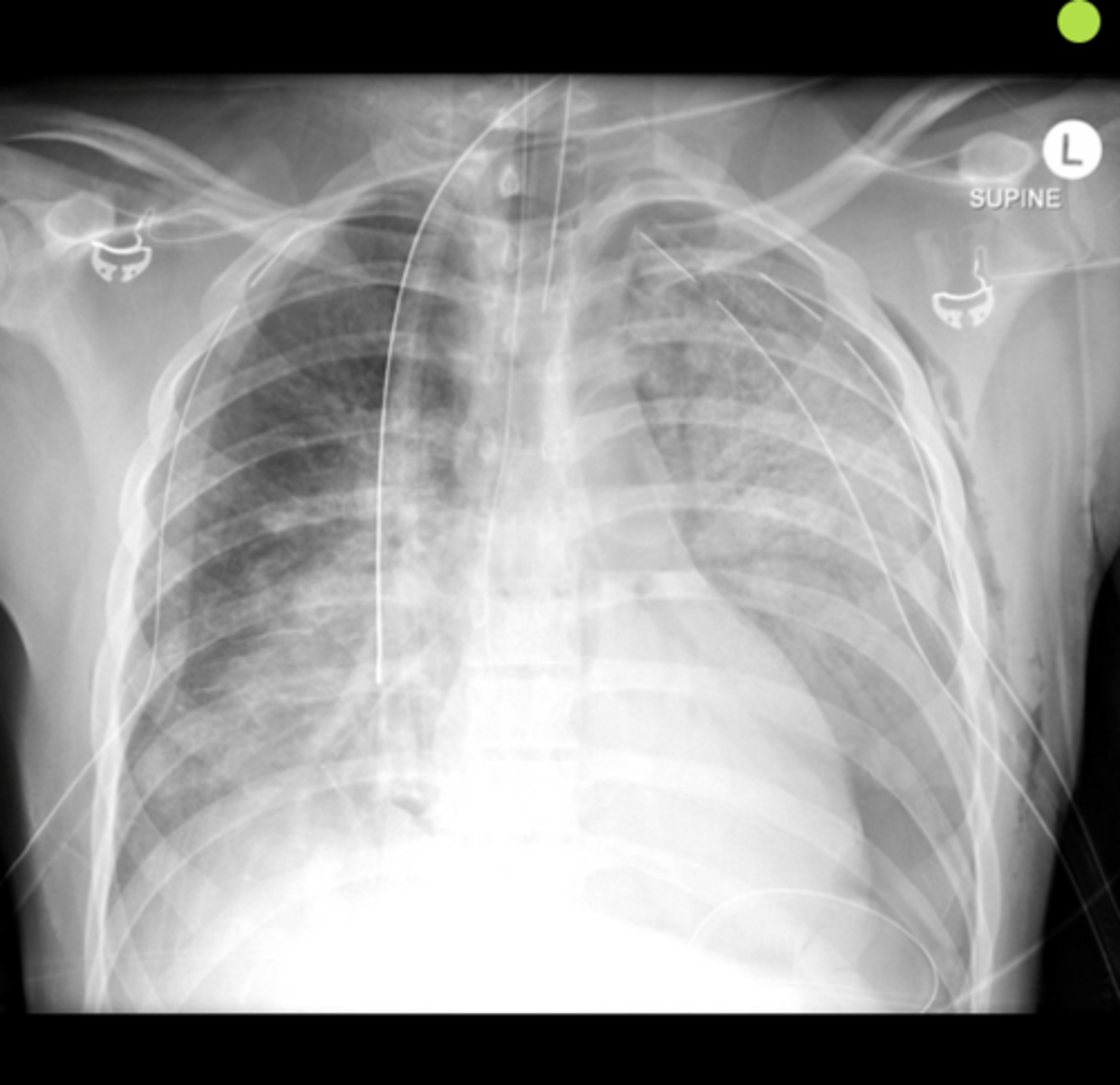
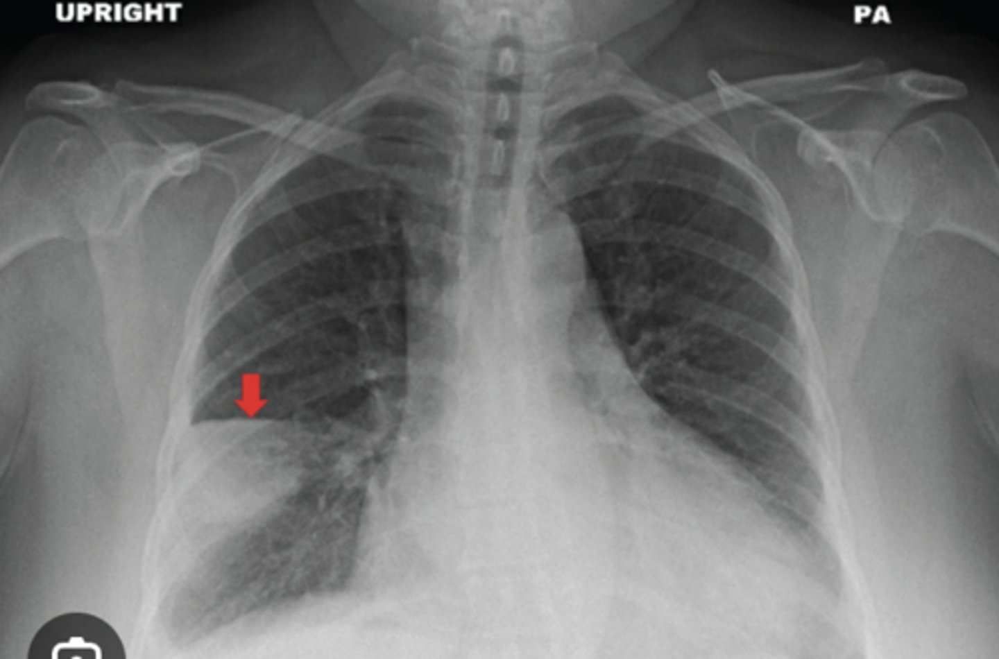
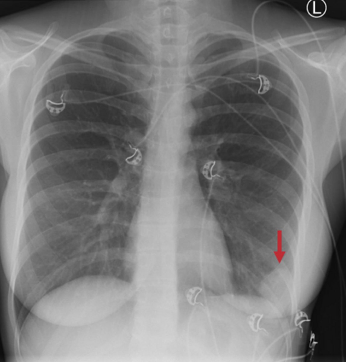
white pleural line sign
dx: pneumothorax
What sign is present on this x-ray? What is the diagnosis based on this sign?
pneumothorax (white line sign present)
What is the diagnosis?