43 - Development of the Eye
1/47
There's no tags or description
Looks like no tags are added yet.
Name | Mastery | Learn | Test | Matching | Spaced |
|---|
No study sessions yet.
48 Terms
Where does eye development begin in the embryo?
From a single, midline eye field at the rostral end of the neural plate.


What splits the single eye field into right and left fields?
Migrating cells from the prechordal plate.

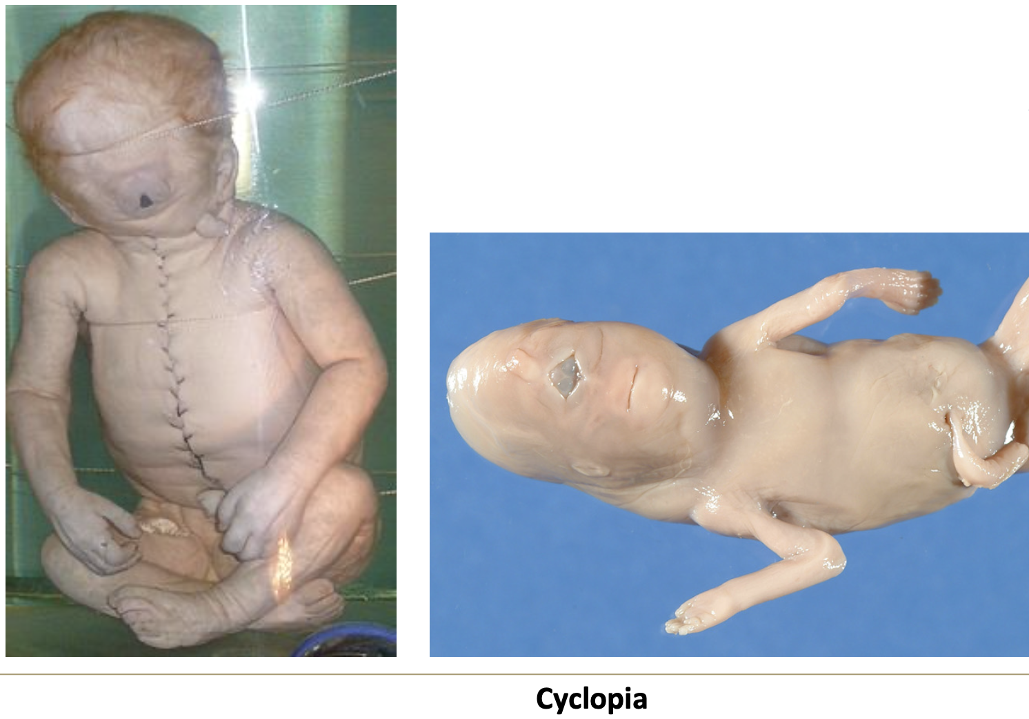
What condition results from failure to split the midline eye field?
Cyclopia — one central eye, due to teratogens or mutations in developmental genes.

When does eye development begin?
4th week of development.
What structures appear before closure of the cranial neuropore?
Optic grooves.

What forms when optic grooves evaginate laterally from the forebrain?
Optic vesicles.

What is the retinal disk?
The distal face of the optic vesicle, which becomes the retina.

What is the optic stalk?
The narrowed portion connecting the optic vesicle to the forebrain.

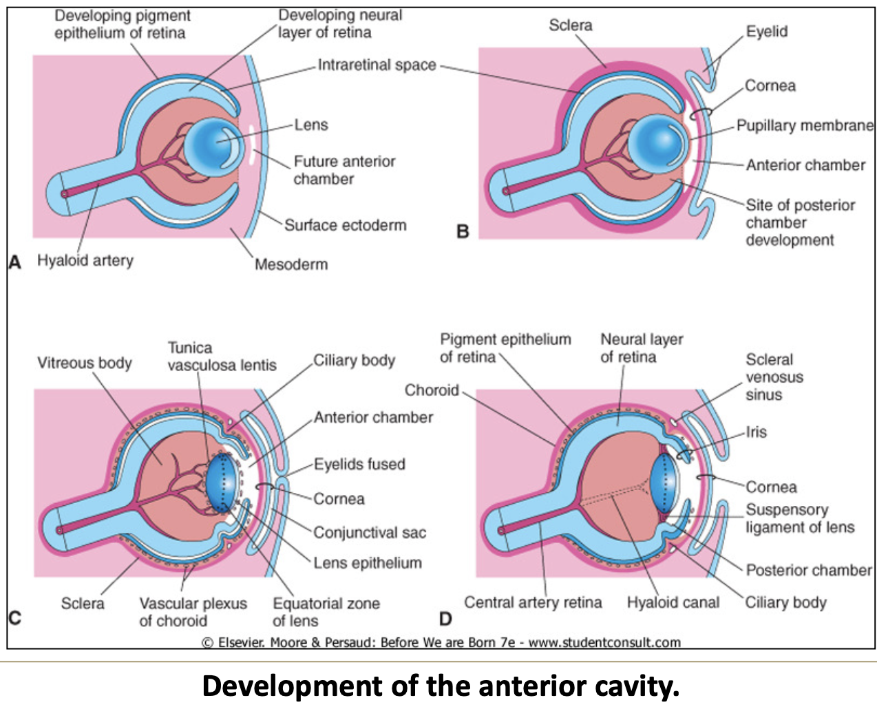
How does the optic cup form?
By invagination of the retinal disk.
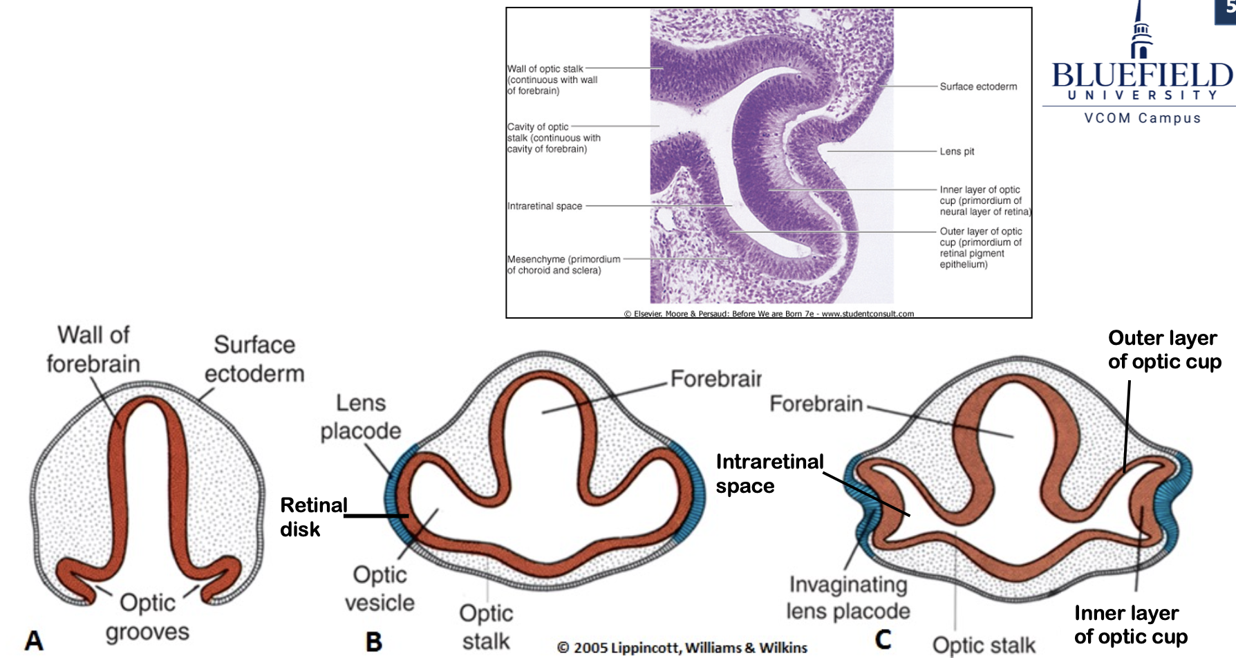
What layers does the optic cup consist of?
Inner and outer layers with an intraretinal space in between.

What is the intraretinal space?
A space between the optic cup layers, continuous with the lumen of the optic stalk and cavity of the forebrain.

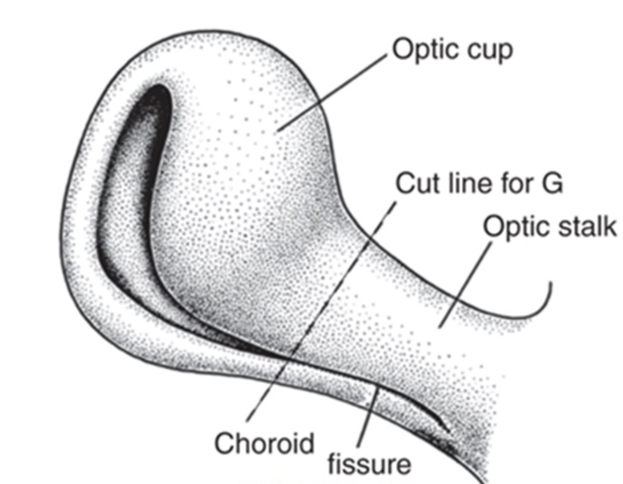
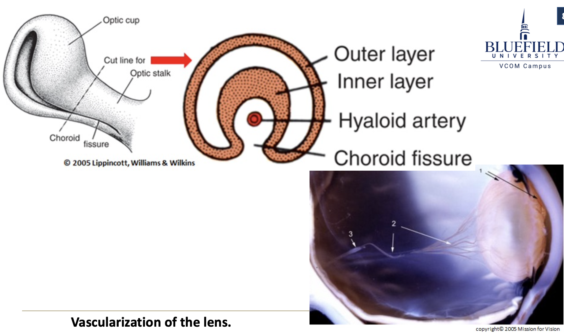
What is the optic fissure?
A longitudinal groove on the ventral optic cup and stalk, allowing vasculature to access the eye.

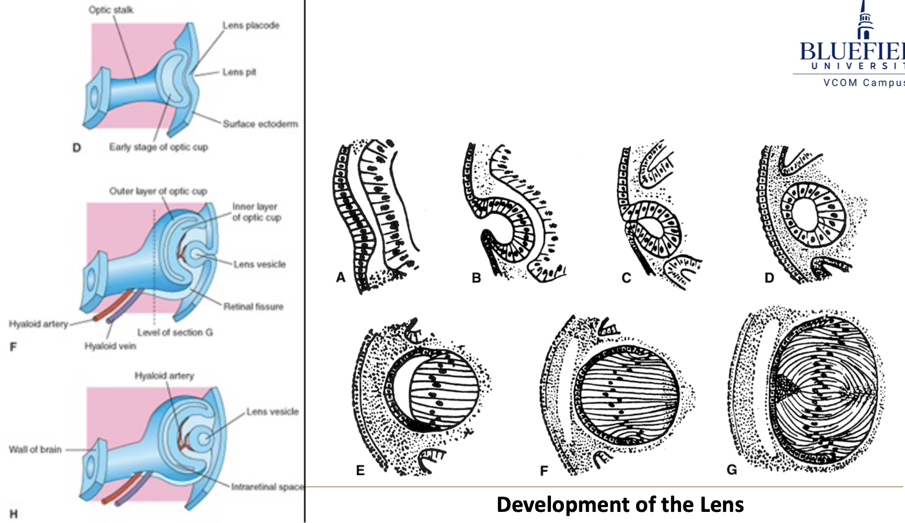
What is the lens placode?
A thickening of the surface ectoderm induced by the retinal disk.

What is the lens pit?
An invagination of the lens placode, mirroring the optic cup.


What happens to the optic vesicle as it detaches from surface ectoderm?
It becomes enclosed within the optic cup and surrounded by mesenchymal capsule.

What are primary lens fibers?
Cells from the posterior lens wall that elongate and obliterate the lens cavity.
What are secondary lens fibers?
Fibers that develop from epithelial cells at the equator and continue to grow until around age 20.
What supplies blood to the developing lens?
The hyaloid artery, a branch of the ophthalmic artery.

When do the lips of the optic fissure fuse?
Around day 37 of development.
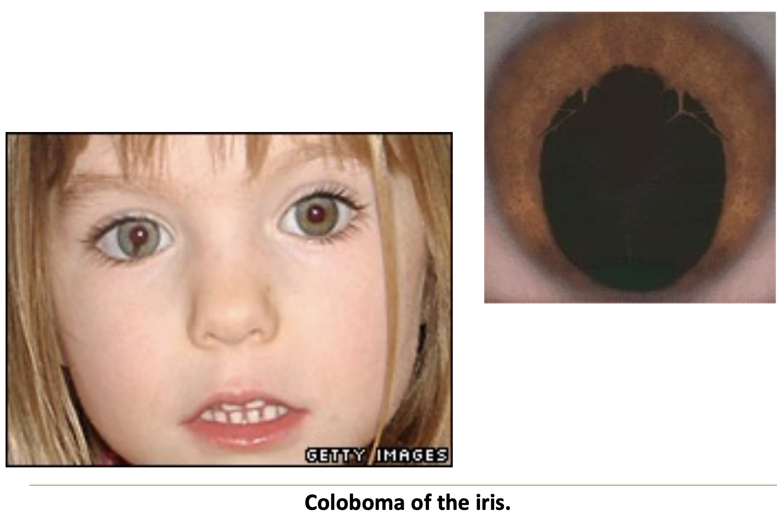
What is coloboma of the iris?
A defect from failure of optic fissure to close; can be genetic and affect the retina, sclera, and ciliary body.

What happens to the hyaloid artery in the fetal period?
The distal vessels degenerate, leaving the hyaloid canal in the vitreous.
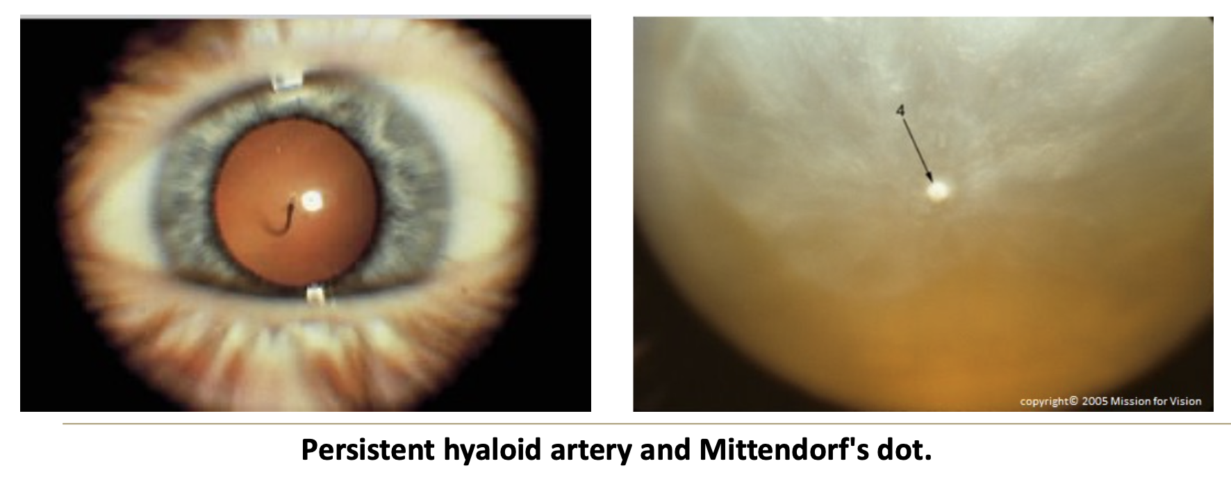
What is a persistent hyaloid artery?
A rare condition where hyaloid vessels remain intact after birth.

What is Mittendorf’s dot?
An opaque dot on the lens indicating the former attachment of the hyaloid artery.

What are short arteriovenous loops from the retina a sign of?
Persistent hyaloid vessels, which usually degenerate with age.
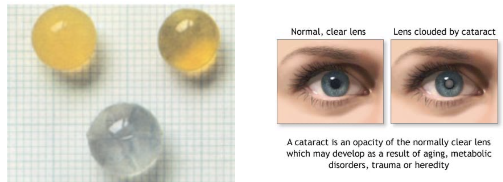
What are congenital cataracts?
An opaque lens caused by genetics or teratogens like rubella or radiation.

What does the outer layer of the optic cup form?
The pigmented layer of the retina.

What does the inner layer of the optic cup form?
The neural retina, including photoreceptors, bipolar cells, ganglion cells, and support cells.
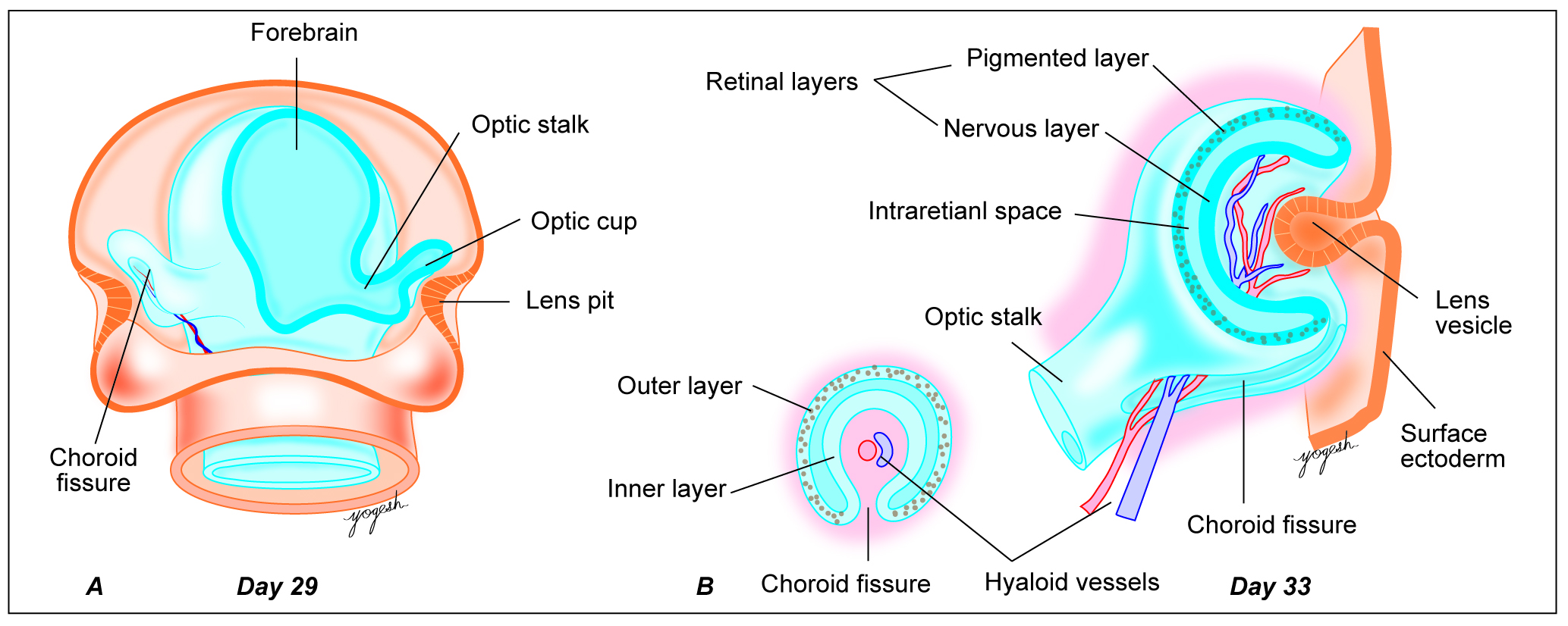
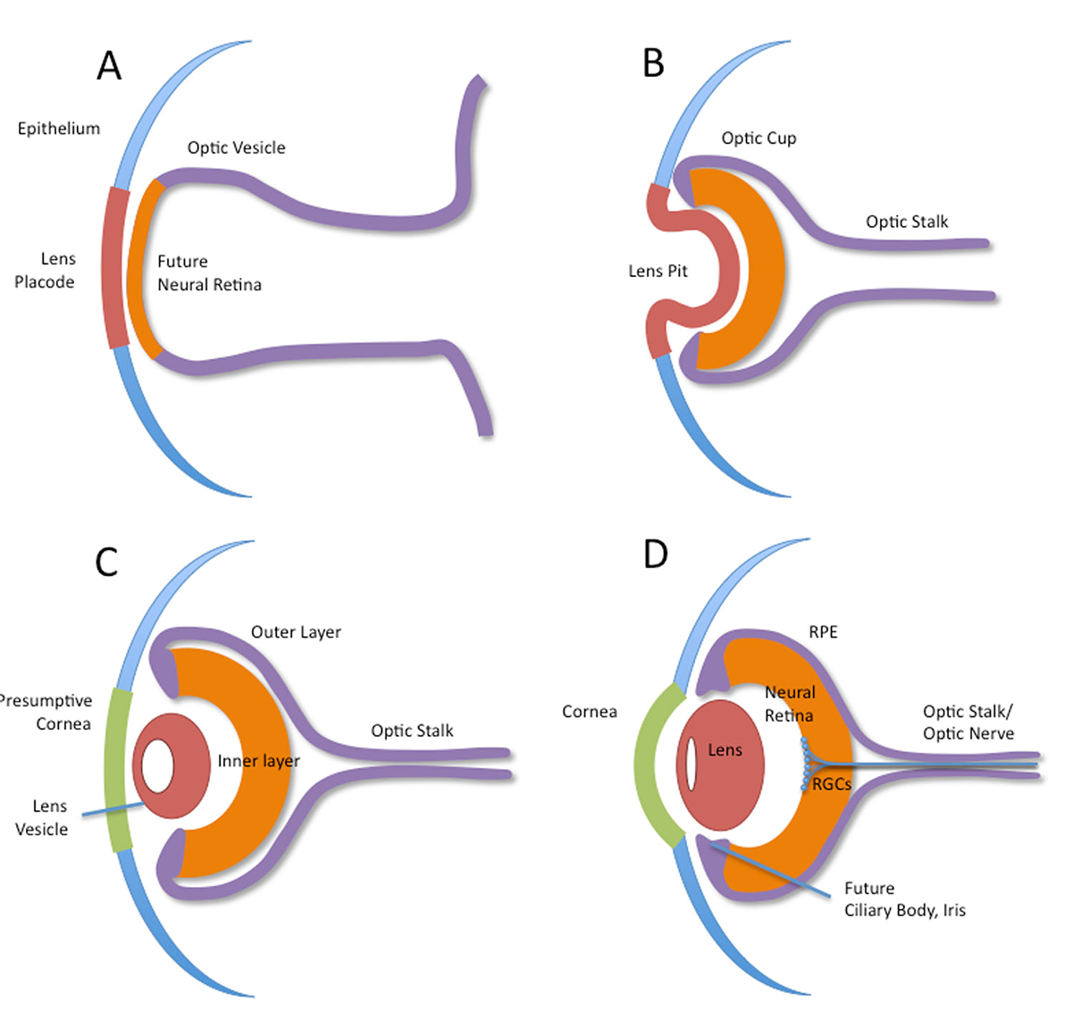
What forms the optic nerve?
Axons of ganglion cells that pass through the optic stalk.

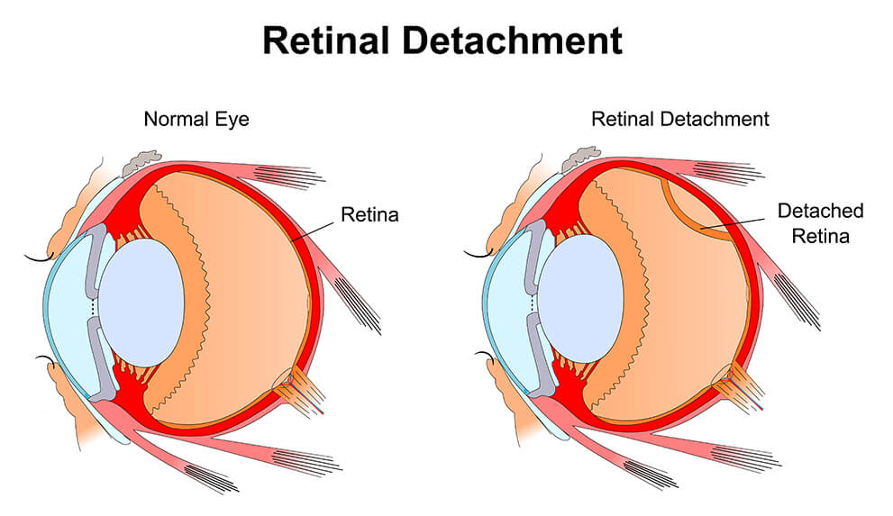
What causes congenital retinal detachment?
Failure of fusion between the pigmented and neural layers of the retina.

What is non-congenital retinal detachment?
A separation between the neural retina and pigmented epithelium after birth.
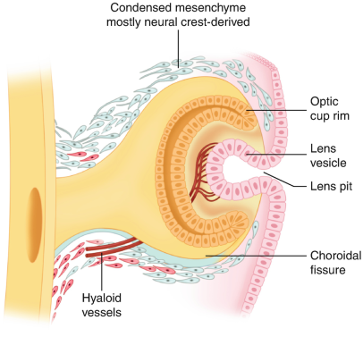
What does the mesenchymal sheath around the optic cup become?
The inner part becomes the choroid (vascular), and the outer part becomes the sclera (fibrous).

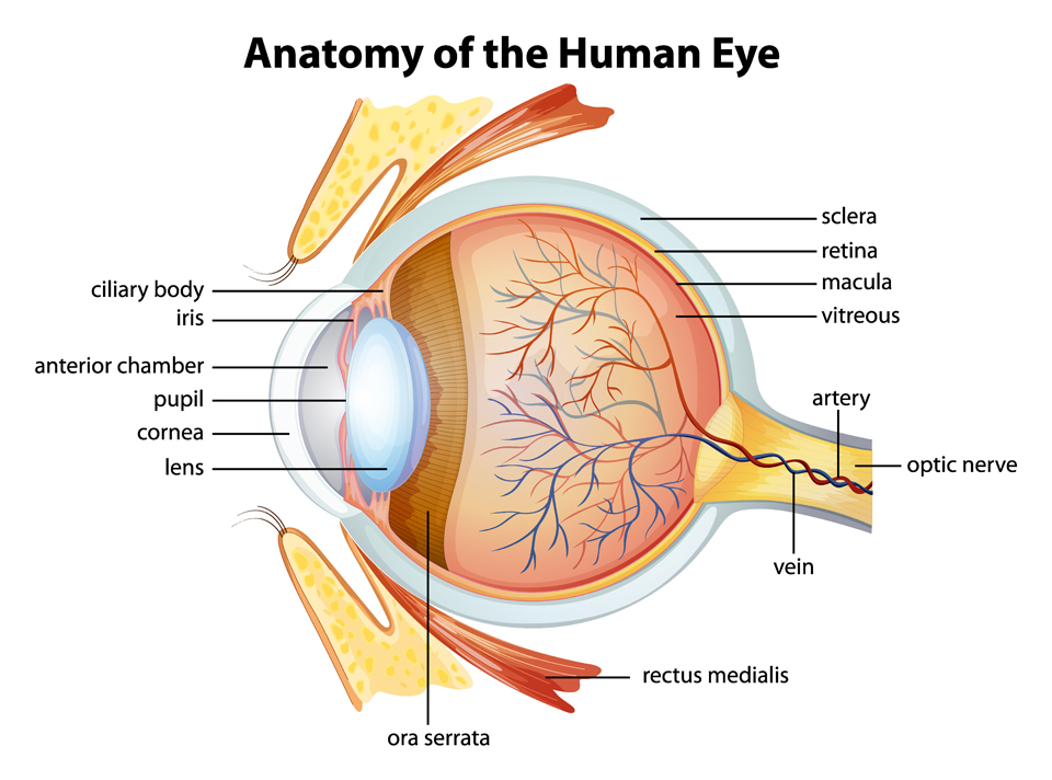
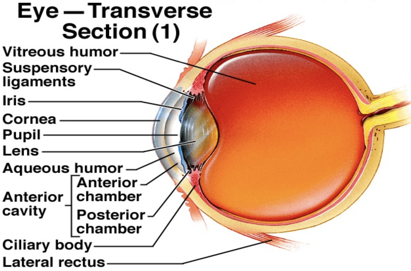
What forms the anterior chamber of the eye?
Mesenchymal cells that migrate between the lens and surface ectoderm, splitting into two layers.
What does the outer layer of anterior mesenchyme become?
The endothelium and stroma of the cornea.
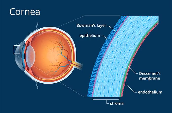

What does the corneal epithelium derive from?
Surface ectoderm.

What does the inner mesenchymal layer of the anterior eye become?
The pupillary membrane.

How is the posterior chamber formed?
By vacuolization of the deep layer of the pupillary membrane.
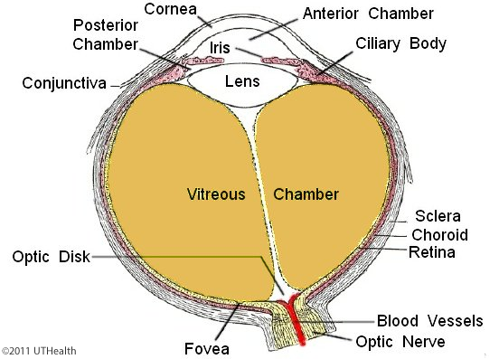
What allows communication between anterior and posterior chambers?
Breakdown of the thin pupillary membrane during early fetal life.

What forms the epithelium of the ciliary body and iris?
The fused inner and outer layers of the ventral optic cup (stratified cuboidal).
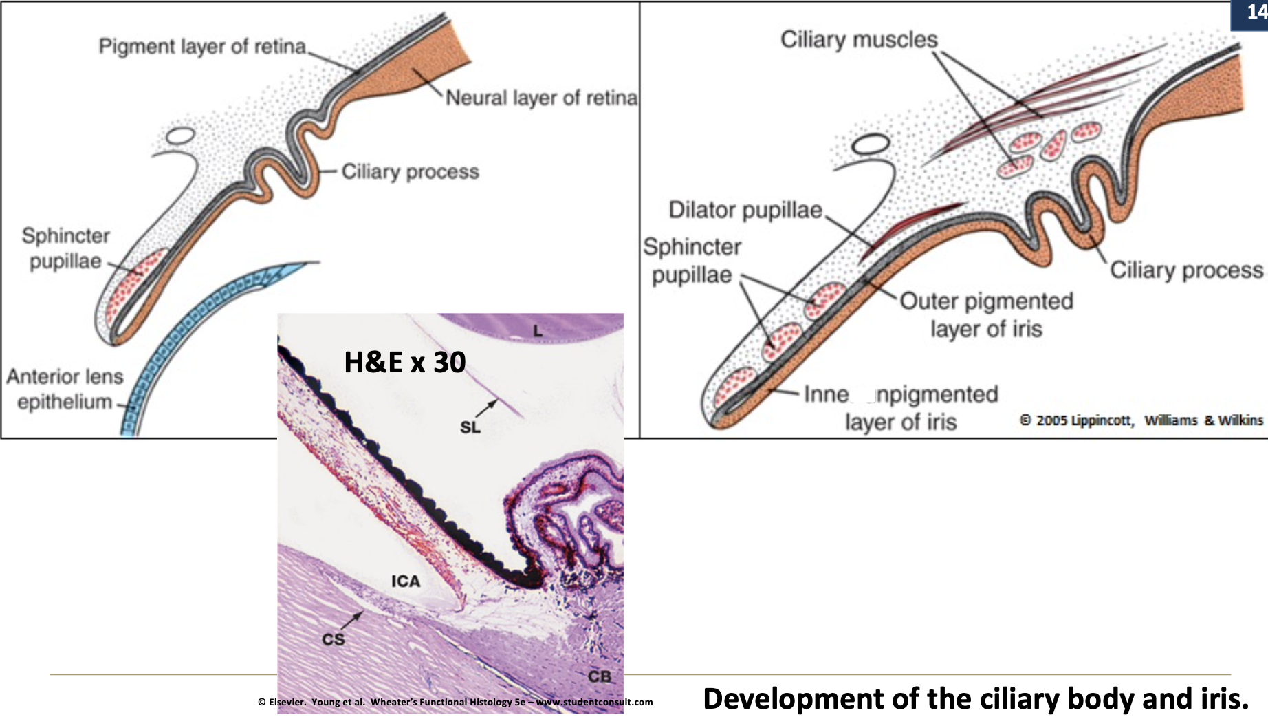
Where do the vascular and muscular components of the ciliary body and iris stroma come from?
Migration of mesenchymal sheath cells.
Why are many newborn irises blue or gray?
Pigmentation occurs after birth.

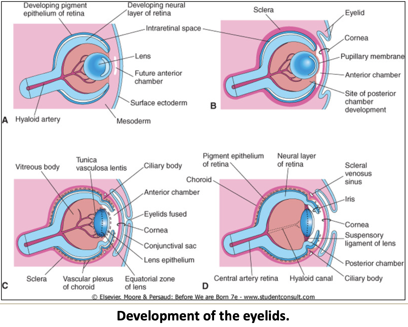
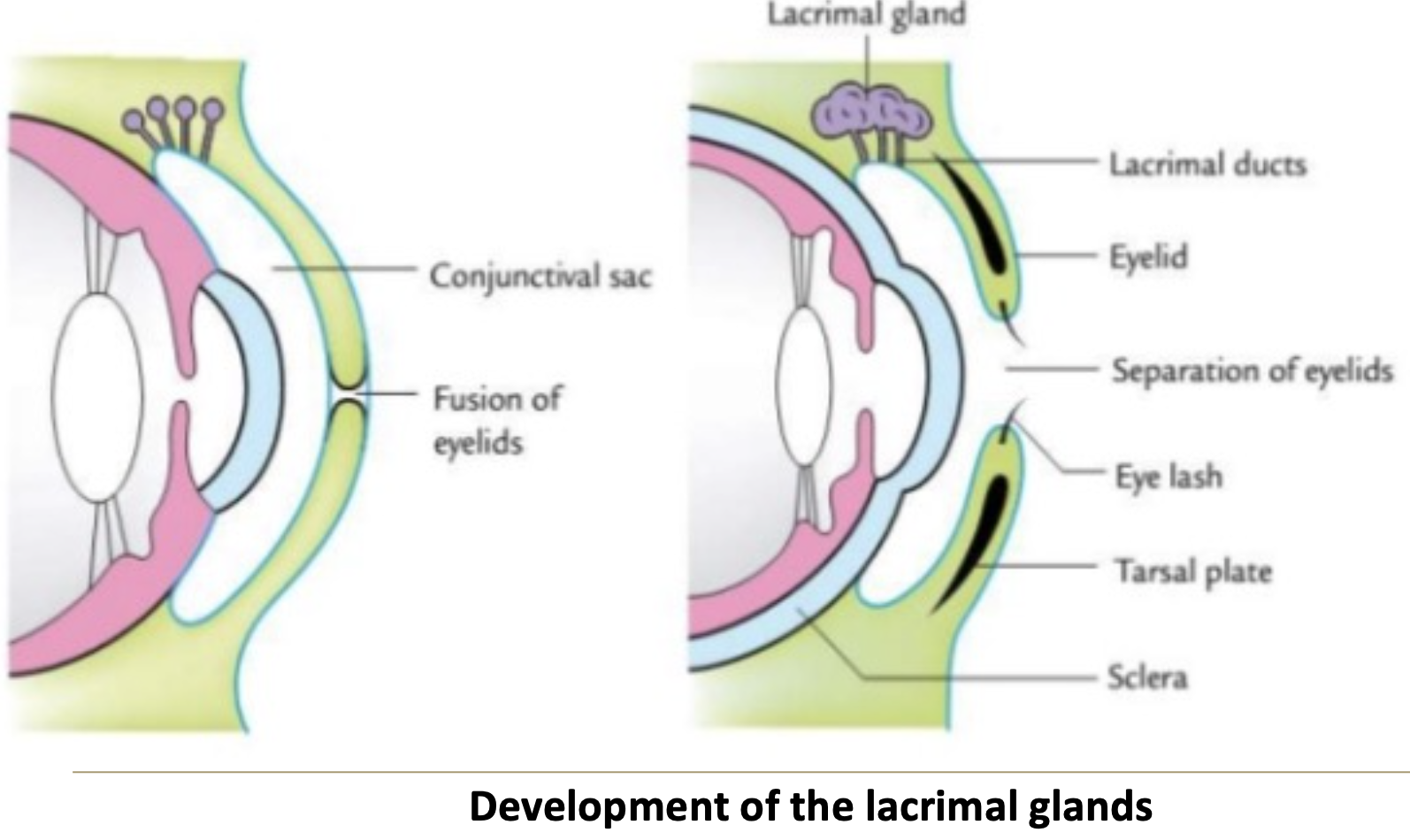
How do the eyelids form?
From superior and inferior ectodermal folds with a mesenchymal core.

When do the eyelids fuse and separate?
Fuse by 8th week, separate by 27th week.


What is the conjunctival sac?
The space between fused eyelids and the cornea, lined with ectodermal epithelium.

What does the conjunctival epithelium become?
The conjunctiva.

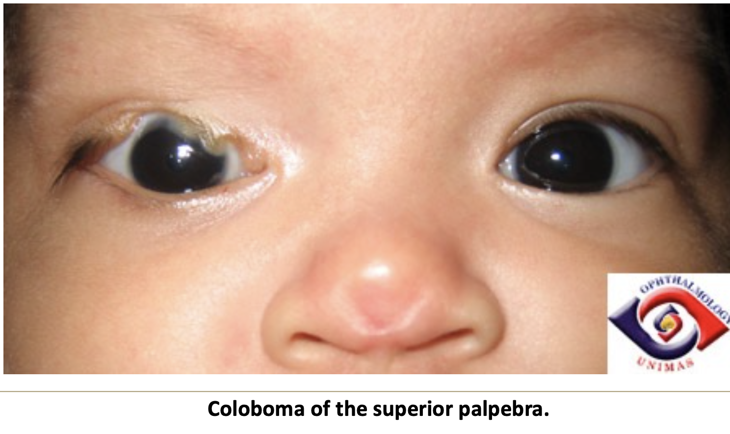
What is coloboma of the eyelid?
A notch-like defect usually affecting the superior palpebra.

How do the lacrimal glands develop?
As epithelial invaginations at the superolateral conjunctival sac angles.


When do lacrimal glands become functional?
About 6 weeks after birth.
When are lacrimal glands fully mature?
By 3 to 4 years of age.