Bones and Joints of the Distal Limb (Week 2, Mod 7)
1/26
Earn XP
Description and Tags
Name | Mastery | Learn | Test | Matching | Spaced |
|---|
No study sessions yet.
27 Terms
What are the two MAIN components of the carpus?
1) Proximal row
2) Distal row
What are the 3 main bones of the proximal row? List and describe them from medial to lateral:
Radial carpal bone / intermediate carpal bone
Is one big, fused bone… largest bone of the carpus
Ulnar carpal bone
Accessory carpal bone
Tucked CAUDALLY behind ulnar carpal bone (see image)
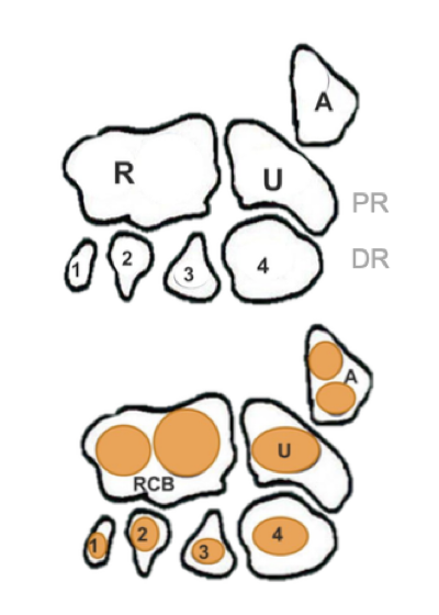
What are the 4 main carpal bones in the DISTAL row of the carpus? List them medially to laterally.
1st carpal bone
2nd carpal bone
3rd carpal bone
4th carpal bone
Which carpal bones only have 1 center of ossification? Which have 2?
All carpal bones have 1 center of ossification, EXCEPT for the radial / intermediate carpal bone and the accessory carpal bone
Species differences: How is the carpus of the pig different from the dog?
The radial and intermediate carpal bones are SEPARATE… has a complete set of proximal and distal rows
Species differences: How is the carpus of the horse different from the dog? How is this significant?
Radial and intermediate bones are separate as well, BUT:
1st carpal bone is MISSING
2nd and 3rd carpal bones are FUSED
Interacts the MOST with the 3rd metacarpal, which is weight bearing
Is fused for support
What are the 3 joints that can be found within the carpus?
1) Antebrachio-carpal joint
2) Middle carpal joint
3) Carpo-metacarpal joint
** also has intercarpal joints keeping the individual bones next to each other; are fibrous, and have no flexibility
Describe the antebrachio-carpal joint… where is it found in the carpus? How flexible is it?
Is found between the antebrachium and the proximal row of the carpus
Facilitates the MOST movement of the carpus
Describe the middle carpal joint… where is it found in the carpus? How flexible is it?
Found between the proximal and distal rows of the carpus
Allows for some flexibility, but not as much as the antebrachio-carpal joint
Describe the carpo-metacarpal joint… where is it found in the carpus? How flexible is it?
Is found between the distal row of the carpus and the metacarpal bones
Has the LEAST amount of flexion here of the other carpal joints
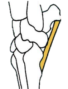
What is the importance of the accessory carpal bone?? What ligament in particular attaches here, and what is its function?
Tendons of the foreleg muscles attach to the accessory carpal bone, allowing the bone to act as a sort of lever for FLEXION of the carpus
However, when animals are standing, the legs are in extension
To prevent hyperextension, the PALMAR LIGAMENT attaches to the distal portion of the accessory carpal bone, inserting at the METACARPALS to stabilize it
What are 3 features that contribute to stabilizing the carpal joint?
1) Collateral ligaments
Laterally: Styloid process of ulna to 5th metacarpal
Medially: Radius to 2nd metacarpal
2) CARTILAGE PLATE on the PALMAR aspect of the joint capsule
Keeps bones stacked and supports them
3) Retinaculum
A band of thickened connective tissue that surrounds the carpus, like a compression sock
How many digits to dogs and cats have? How many to ungulates have, and which ones did they keep? How many do horses have, and which one did they keep?
Dogs and cats:
Four (technically 5) digits… shed the 1st one (dew claw) for speed
Ungulates:
Have only 2 weight-bearing digits… 3rd and 4th digits remain
Termed artyrodactyla
Horses:
Only 1 weight bearing digit, kept the 3rd digit
Termed perissodactyla
What are the 3 different classifications of “stance”, and what are they based on? Which species is unique?
1) Plantigrade
Bears, humans
Bearing weight on the digits, metatarsals, AND tarsal bones
2) Digitigrade
Dogs, cats
Bear weight on the digits / phalanges; protected by foot pads
3) Unguligrade
Ruminants, horses
Bear weight on the distal phalanx ONLY; protected by hoof
*** RABBITS can be both 1 and 2… sit plantigrade, run digitigrade
How many centers of ossification to each metacarpal and phalanges have? What is the exception?
Each bone has 2 ossification centers, EXCEPT
Distal phalanx… only has 1
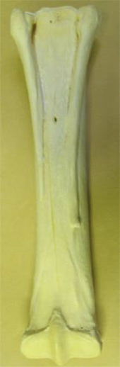
Describe the metacarpal bone of the equine… what is it also termed as? What is its function? What are 2 key features of the bone?
Is the 3rd metacarpal… also termed the canon bone
Is WEIGHT BEARING
2 key features:
Has a sagittal groove on the distal articular surface; interacts with the proximal phalanx
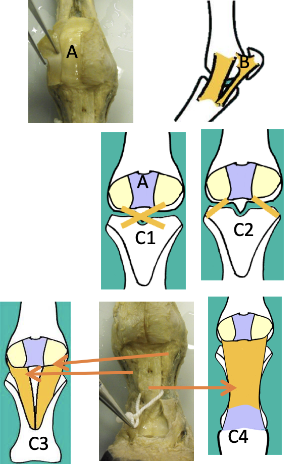
Has 2 collateral SPLINT BONES at the proximal, palmar end (see image)
Are vestigial and non-weight bearing; are palpable
Describe the proximal sesamoid bones… what species are they found in? What are their functions?
Paired proximal sesamoid bones are found in ALL species
Are palmar-distal to every metacarpal/ metatarsal
Will ALWAYS be paired
Are embedded in suspensory ligament
Function to PROTECT THE SUPERFICIAL DIGITAL FLEXOR TENDONS (SDFT) AND THE DEEP DIGITAL FLEXOR TENDON (DDFT)
Describe the distal sesamoid bones… what species are they found in? What are their functions?
Are ONLY found in the canine, and are not paired; single
Are found on the dorsal aspect of every metacarpal / metatarsal
Protects the EXTENSOR TENDONS
Describe the proximal phalanx of the horse… how is it unique compared to other species? How is it kept stable?
Is also called the “long pastern bone” / P1
Has a “v” shape on the palmar surface, which is where the oblique distal sesamoidian ligament attaches…
This provides support and stability to the fetlock by connecting the proximal sesamoid bones to the pastern bone
Has 2 sagittal grooves… one proximal, and one distal

Describe the equine metacarpo-phalangeal joint… what are its 3 main components? What kind of joint is it? What are 2 unique features of the joint that contribute to the protection of surrounding structures?
Also called the “fetlock”; composed of
3rd metacarpal
Proximal phalanx
Proximal sesamoids
Is a typical synovial joint, but has two out-pouchings:
Large palmar pouch - incorporates the proximal sesamoids
Large dorsal pouch - cushions exterior tendons
What is the suspensory ligament in the equine? What does it attach to, where is it located, and what is its function?
Origin - proximal MC3… runs palmar, fills entire space between the two splint bones
BRANCHES in 2, attaching to the ABAXIAL aspect of the proximal sesamoids and wrapping around the metacarpo-phalangeal joint
Passes around the DORSAL aspect of the limb and fuses with the common digital extensor tendon
Function
Acts like a sling; prevents prolonged hyperextension of the limb and returns the joint to a resting position
Relies on the STABILITY OF THE SESAMOIDS
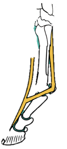
There are 3 sesamoidean ligaments…. what are they called, where are they found, and what are their functions?
1) Inter-sesamoidean ligament
Is a fibrocartilage plate BETWEEN the proximal sesamoid bones
Allows for the smooth passage of the DDFT and the SDFT
2) Collateral sesamoidean ligaments
Attach the sesamoids to the MC3 and the proximal phalanx (see image B)
3) Distal sesamoidean ligaments - there are 4
Cruciate and short -
labeled C1 and C2, stabilize the sesamoids to the proximal phalanx
Oblique -
Attach the sesamoids to the “v” shaped trough on the palmar aspect of the proximal phalanx, offers great stability
Straight -
Overlays entire palmar aspect of the proximal phalanx
Inserts with the SDFT
What is the suspensory apparatus and what structures is it made up of?
It is the set of ligaments and bones that stabilize the entire fetlock… very prone to injury, as every piece relies on the function of the other
Is composed of:
Suspensory ligament
Proximal sesamoids
Sesamoidean ligaments
Common digital extensor tendon
Describe the middle phalanx of the equine… what is it called? What is one unique feature that allows for the attachment of ligaments? Describe its associated proximal interphalangeal joint, and what is unique about it that provides protection…
Middle phalanx = short pastern bone
Has 2 sagittal grooves as well, 1 proximal and 1 distal
Has bony bumps along its surface that allow for better attachment of the collateral ligaments
The proximal interphalangeal joint (PIP) is a typical synovial joint:
Has an EXTENSIVE joint capsule; forms a DORSAL bursa to cushion the common digital extensor tendon
Describe the distal phalanx of the equine… has a “complex shape”, allowing for the attachment of multiple ligaments. What are these processes / aspects? (Hint: there are 4)
Extensor process -
Found on the proximal / dorsal aspect of the pedal bone (DP)
Allows for the attachment of the common digital extensor tendon (see image)
Palmar processes -
Are collateral processes at either end of the proximal DP… in the image, are proximal / palmar, kind of in the same line as the extensor process
Support the lateral cartilages
Solar surface -
Is the BOTTOM aspect of the DP
Deep digital flexor tendon attaches here
Dorsal surface -
Has striations on its surface for hoof attachment
Can see vascular channels for blood vessels

Describe the distal interphalangeal joint … What else is it called? What is it composed of? What is its function in terms of flexibility?
Called the “coffin”… is buried within the hoof
Composed of:
Middle / distal phalanges
Distal sesamoid (AKA NAVICULAR BONE)
Has an extensive joint capsule
ANOTHER dorsal bursa for cushioning of the extensor tendon
Is a typical synovial joint, stabilized by collateral ligaments
Function:
Flexion and extension
SOME ROTATION, unlike the rest of the interphalangeal joints
Accounts for uneven ground
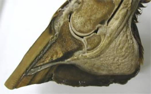
Describe the navicular bone… what other, protective structure is it associated with? What seems to separate it from the interphalangeal joint space?
The navicular bone has:
2 smooth articular surfaces
Is made of cancellous bone with vascular channels
Is associated with the podotrochlear bursa -
A fluid filled pocket between the navicular bone and the DDFT
CUSHIONS the DDFT
The navicular bone is separated from the joint space by the IMPAR LIGAMENT, which can be seen in the image
