Patho Lab - Hemodynamic Disorder
 Call Kai
 Learn
Learn Practice Test
 Spaced Repetition
 Match
Match Flashcards
 Knowt Play
1/53

There's no tags or description
Looks like no tags are added yet.
Name | Mastery | Learn | Test | Matching | Spaced | Call with Kai | Chat |
|---|
No analytics yet
Send a link to your students to track their progress
54 Terms
chronic passive congestion of lungs
gross findings: reddish in color due to congestion
endogenous, exogenous
In chronic passive congestion of lungs, hemosiderin are _________ and carbon pigments are __________.
T
T/F: In chronic passive congestion, CRACKLES are also heard during auscultation
hyperemia
Refers to the active process resulting from arteriolar dilation
congestion
Refers to the passive process resulting from impair outflow from tissue
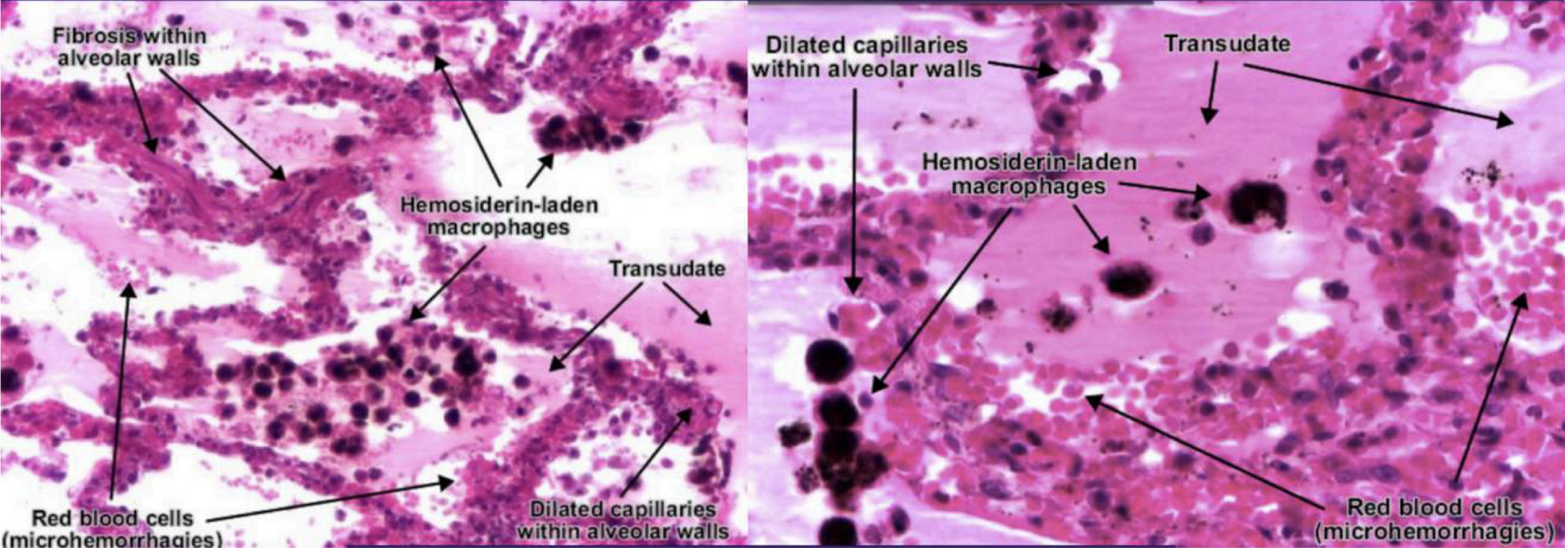
chronic passive congestion of lungs
5: transudate
diagnose & identify pointed structure
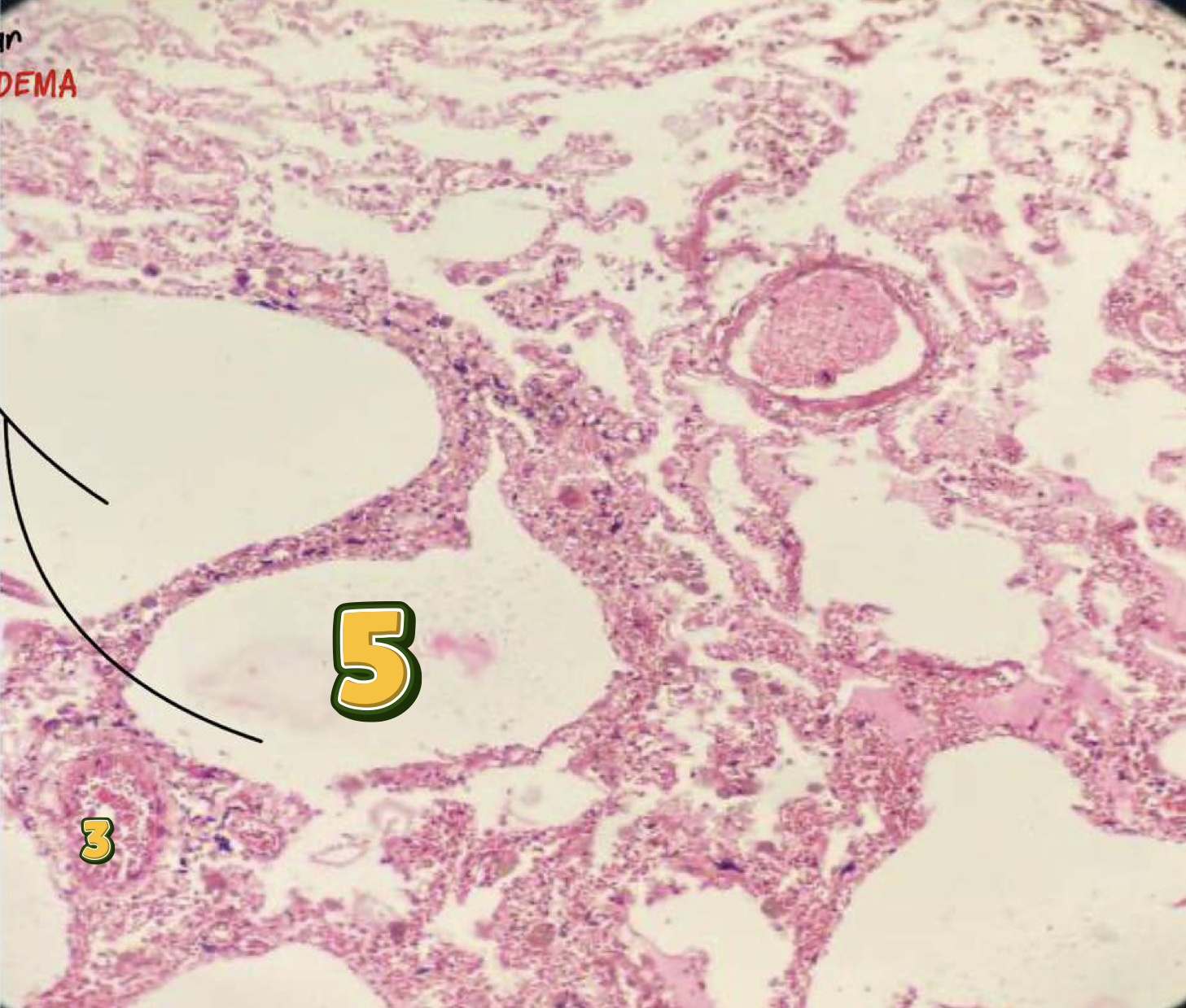
chronic passive congestion of lungs
4: hemosidering-laden macrophages
diagnose & identify pointed structure
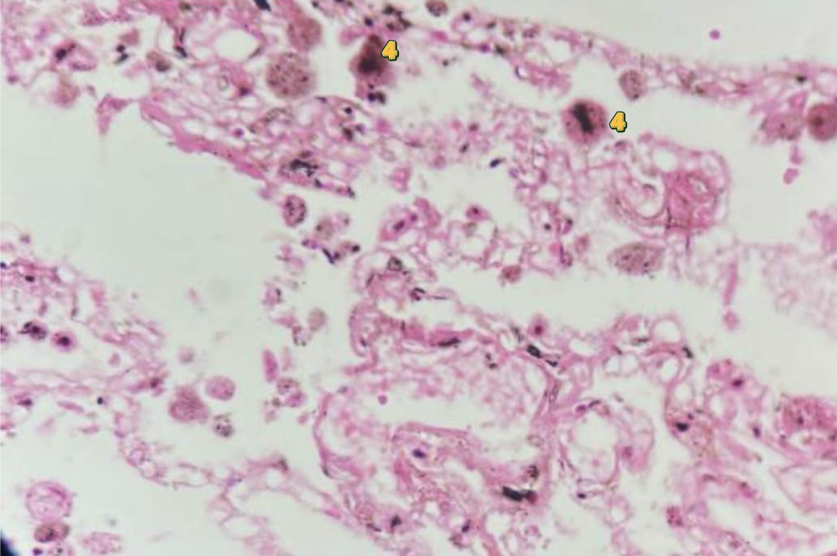
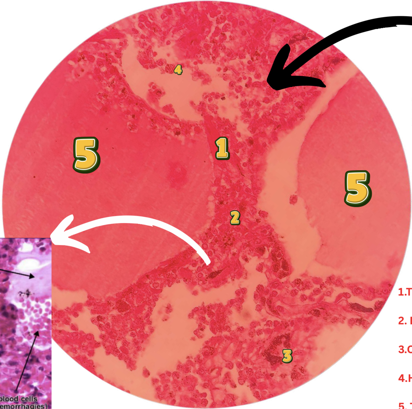
chronic passive congestion of lungs:
1: thickened fibrotic septa
2: dilated alveoli
3: congested blood vessels
4: hemosiderin laden macrophage
5: transudate
diagnose & identify pointed structure
Chronic passive congestion of lungs
Identify
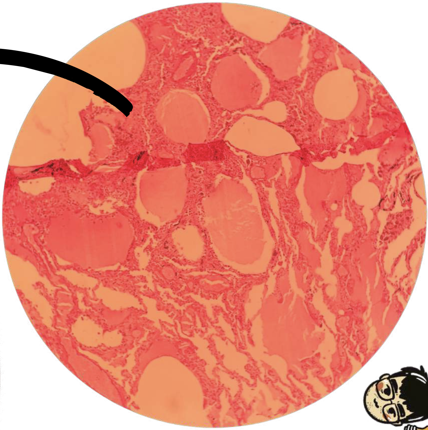
Chronic passive congestion of lungs
Identify
False
T/F: In chronic passive beharvior,
edema of lungs
Gross finding: distended and red lung due to congestion
increased hydrostatic pressure
decreased oncotic pressure
lymphatic obstruction
sodium and water retention
Causes of edema (4)
true
T/F: backflow of blood to the pulmonary circulation due to left sided heart failure causes pulmonary edema
false; alveolar walls, alveoli
T/F: In chronic passive congestion of lungs, transudate fluid is at the alveoli. While, in edema of lungs, transudate fluid is at the alveolar walls.
true
T/F: In chronic passive congestion of lungs, hemosiderin macrophages are referred to as heart failure cells. While, in edema of lungs, hemosiderin macrophages are not referred to as heart failure cells.
edema of lungs
Identify
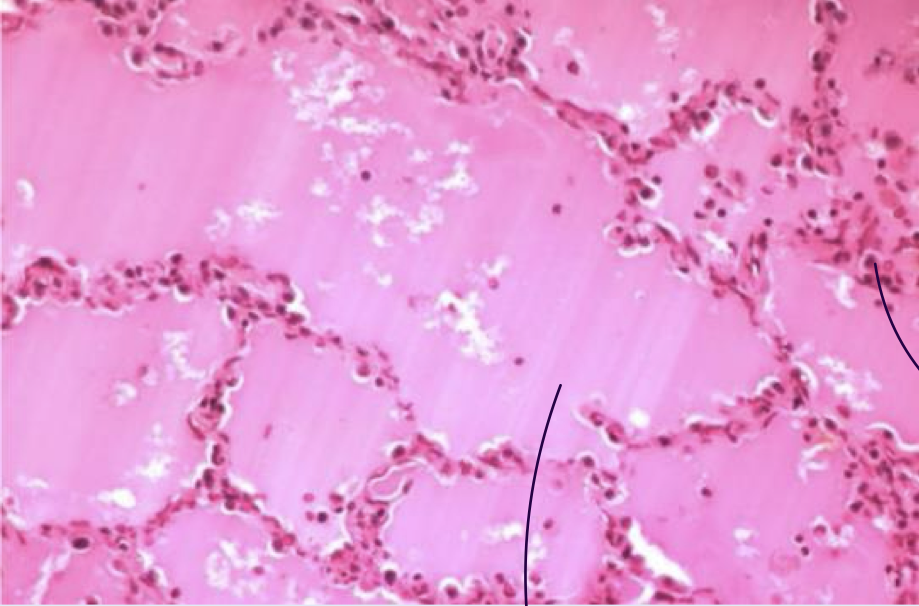
edema of lungs
Identify
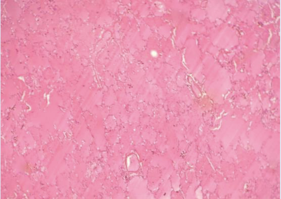
edema of lungs
Identify
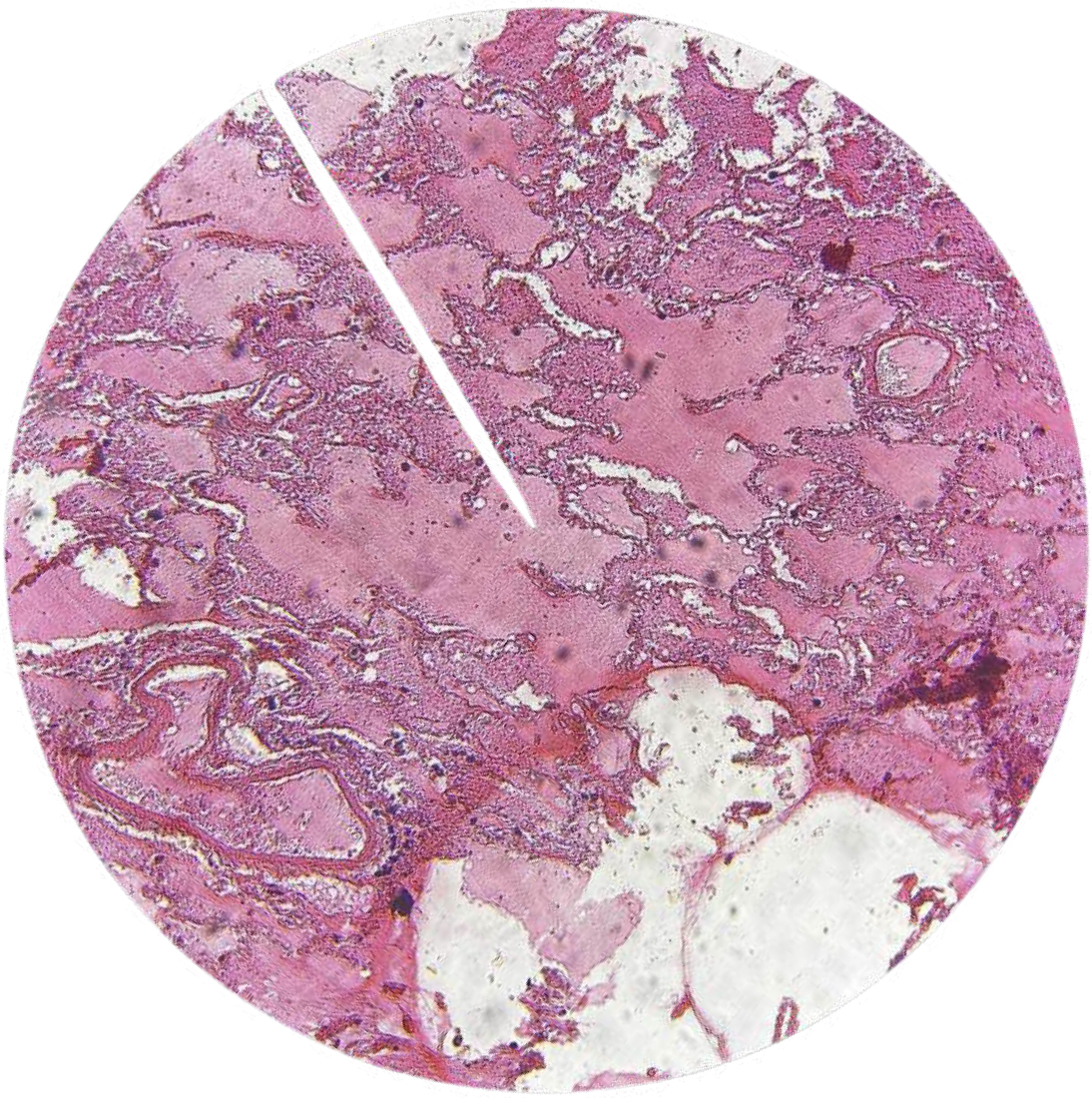
edema of lungs
Identify
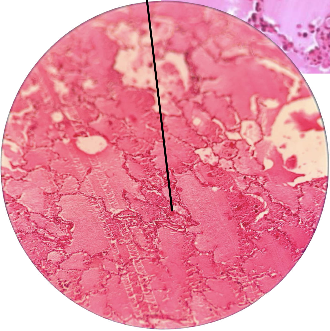
hemorrhoids with thrombus
Gross findings: edematous mass of pale connective tissue covered by gray-to-tan mucosa; cut surfacewith dilated vascular surfaces
columnar lining epithelium
Lining epithelium of internal hemorrhoids
squamous lining epithelium
Lining epithelium of external hemorrhoids
thrombus
Refers to blood clot formed and impeding blood flow
DOPE
dissolution
organization
propagation
embolization
Fate of thrombi
embolus
Refers to the detachment of thrombus and movement into other blood vessels
hemmorhoid
Refers to collateral vessels that develop secondary to persistently elevated venous pressure within hemorrhoidal plexus
stasis of blood flow
endothelial injury
hypercoaguability
Virchow triad
hemorrhoids with thrombus
Identify
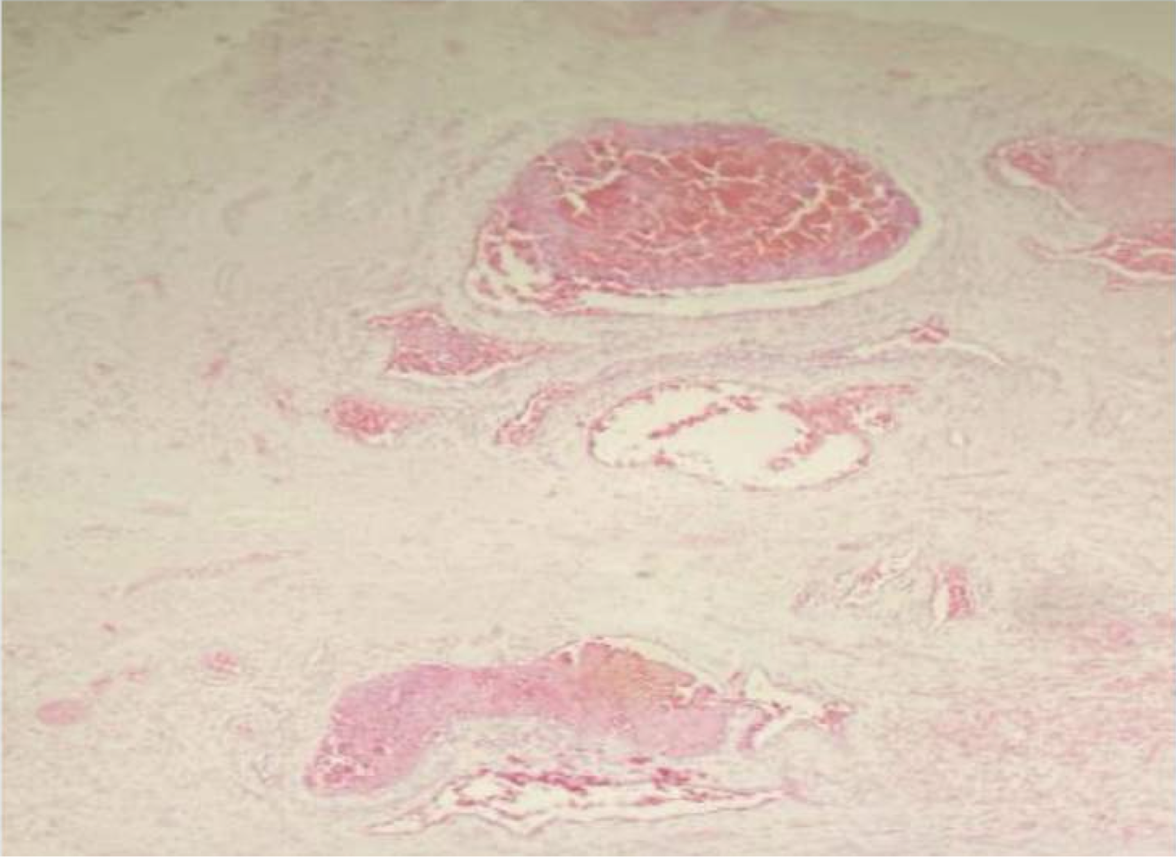
hemorrhoids with thrombus
A: dilated endothelial-lined vascular channels with RBC inside
B: squamous external lining
C: columnar internal lining
Identify structure and labeled areas
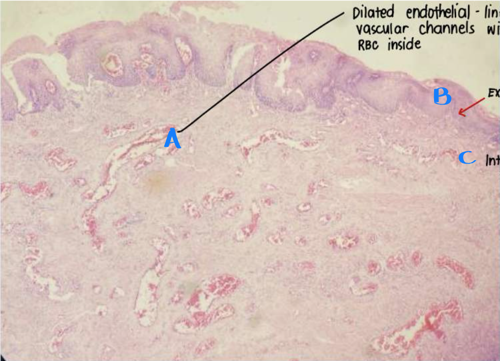
hemorrhoids with thrombus
Identify
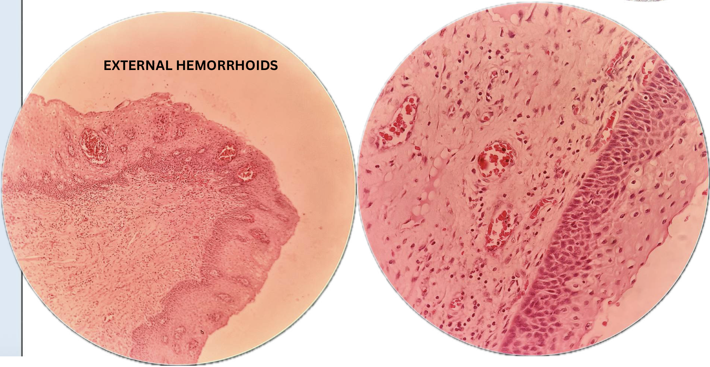
hemorrhoids with thrombus
Identify
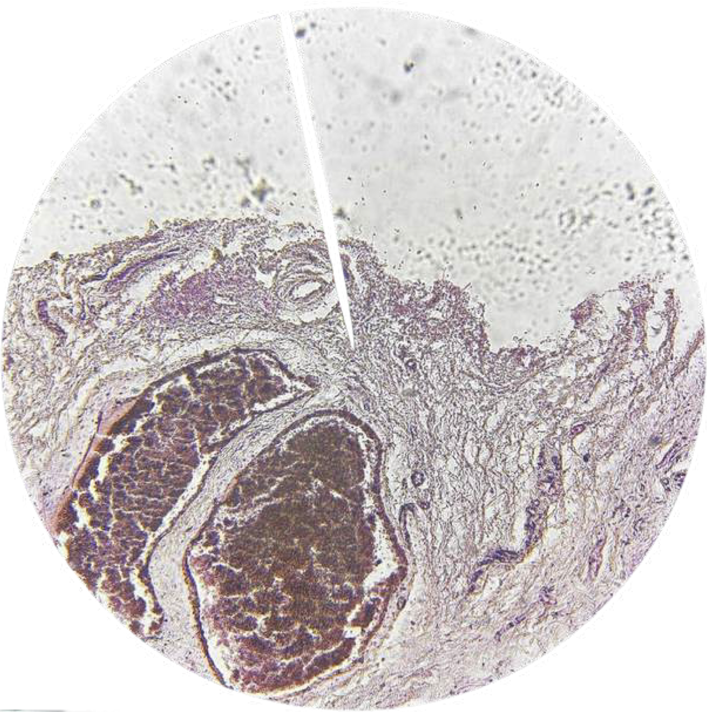
hemorrhoids with thrombus
Identify
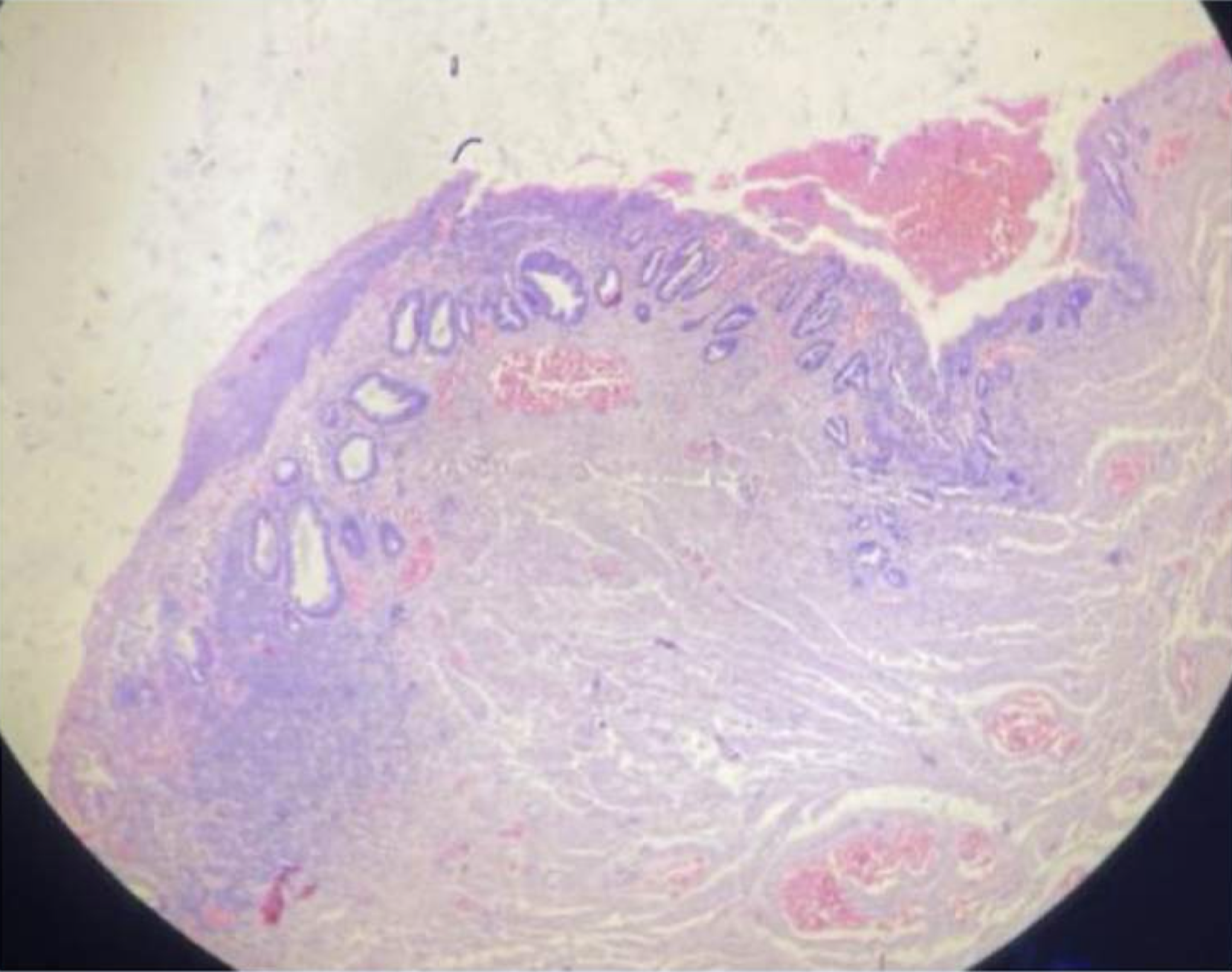
dissolution
Fate of thrombi: result of fibrinolysis, rapid shrinkage, and total disappearance
organization
Fate of thrombi: older thrombi become reorganized by ingrowth of endothelial cells
propagation
Fate of thrombi: thrombi accumulate additional platelets
embolization
Fate of thrombi: thrombi dislodge and travel to other sites into vasculature
myocardial infarction
Gross findings: yellowish center with necrosis and inflammation surrounded by a hyperemic border
infarct
A localized area of tissue damage or cell death that occurs due to inadequate blood supply, typically resulting from obstruction of blood flow to the area
white infarct
type of infarct referring to ARTERIES SOLID, WELL-STRUCTURED, ARTERIAL OCCLUSIONS
red infarct
type of infarct referring to VEINS, LOOSE, SPONGY TISSUE, VENOUS OCCLUSIONS
myocardial infarction
Identify
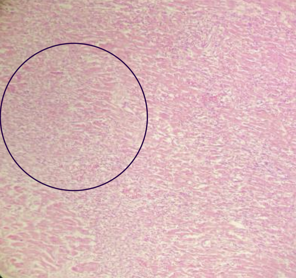
myocardial infarction
Identify
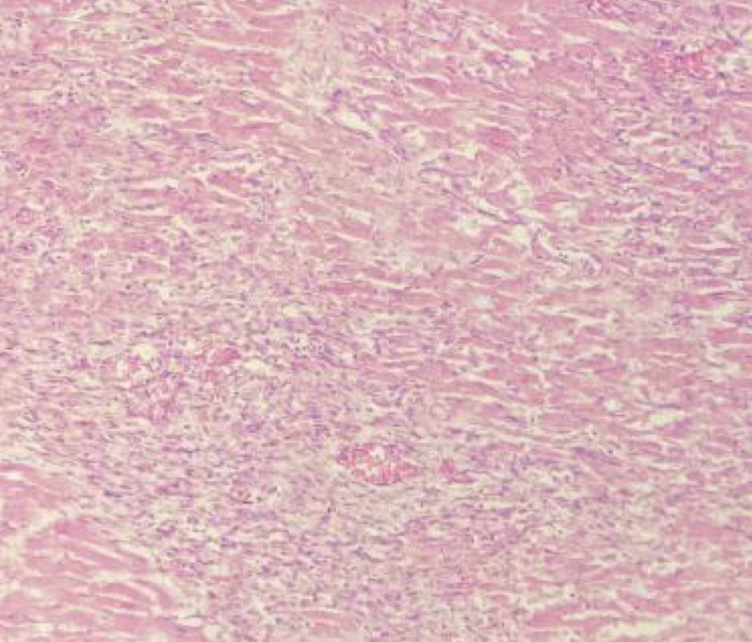
myocardial infarction
Identify
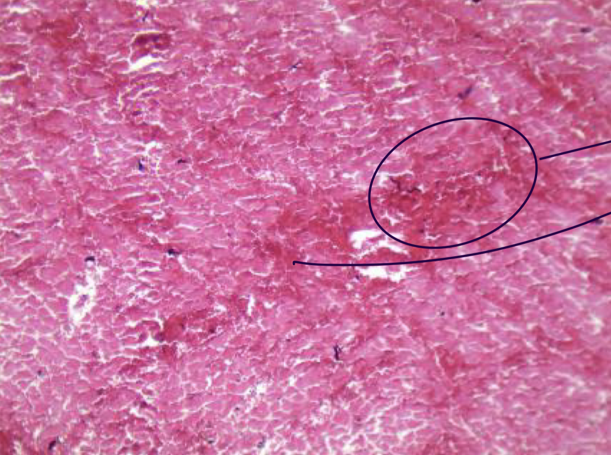
myocardia infarction; coagulation necrosis
Identify structure and type of necrosis
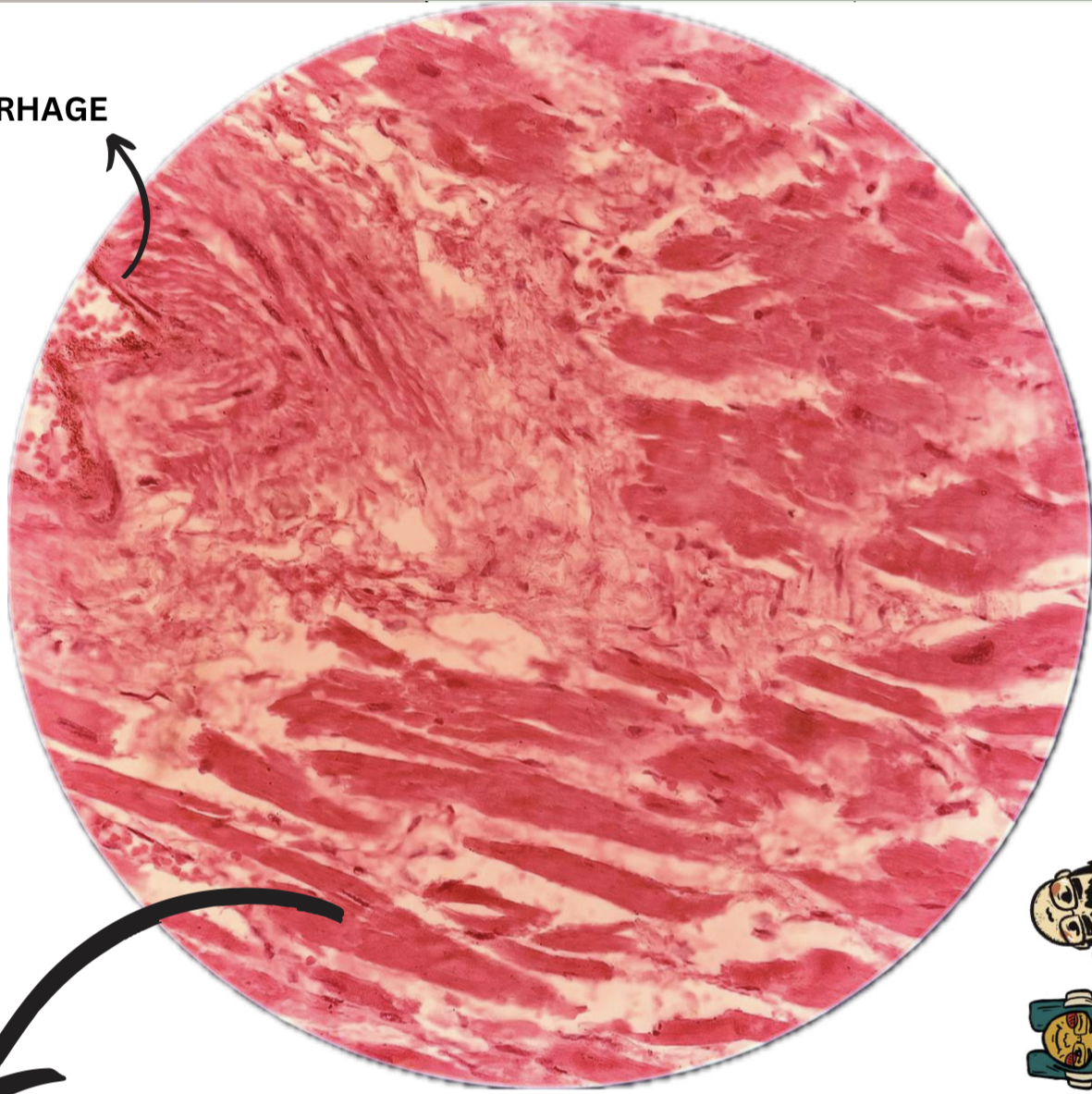
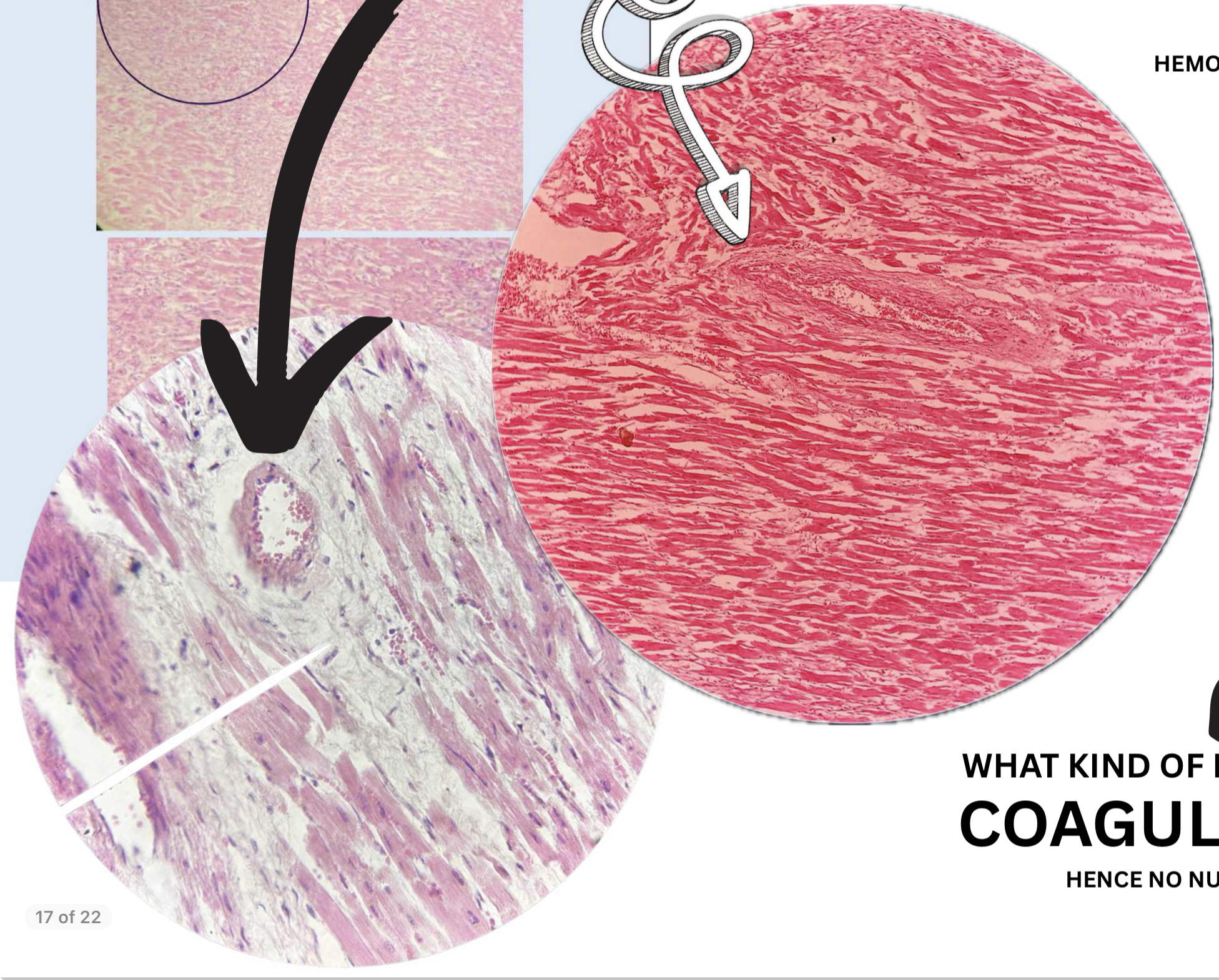
myocardial infarction
black arrow: white infarct
white arrow: red infarct
Identify structures
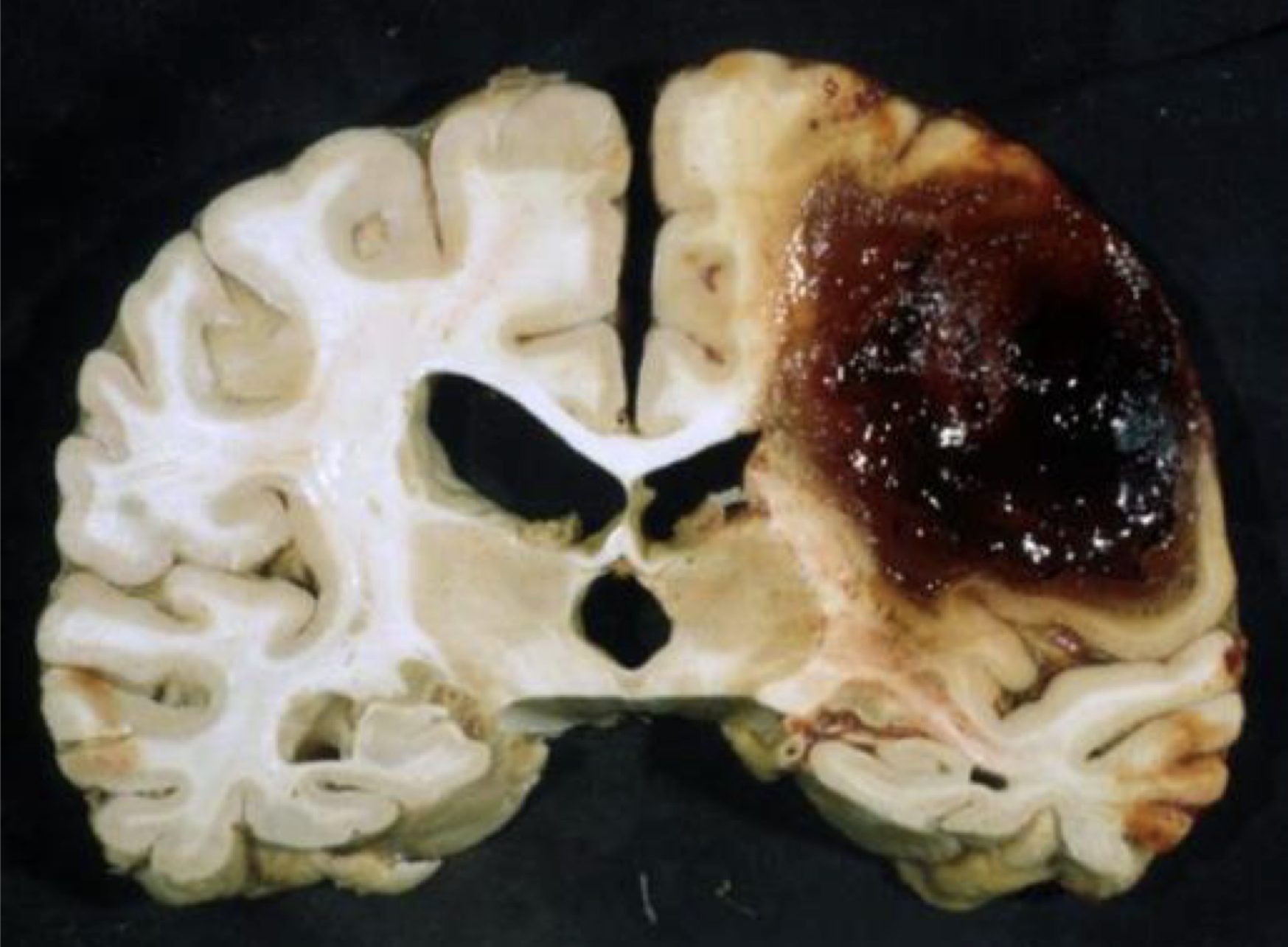
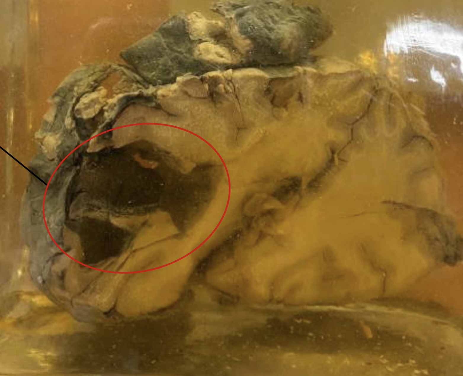
intracerebral hemorrhage
Gross findings: brown discoloration of older hematomas due to presence of hemoglobin derived pigments: hemosiderin and hematoidin
hemosiderin
hematoidin
In intracerebral hemprrhage, there are brown discoloration of older hematomas due to presence of hemoglobin derived pigments such as (2)
intracerebral hemorrhage, liquefactive necrosis
Identify and type of necrosis
intracerebral hemorrhage, liquefactive necrosis
Identify and type of necrosis
petechiae
Small, pinpoint sized, red spots that appear on skin
purpura
Patchy areas of purple or red discoloration on skin
ecchymoses
Bruises, larger areas of purple, blue, or black discoloration
disseminated intravascular coagulation
Widespread thrombosis within microcirculation. Not a specific disease but rather a complication of large number of conditions associated with systemic activation of thrombin