oral medicine/oral pathology
 Call Kai
 Learn
Learn Practice Test
 Spaced Repetition
 Match
Match Flashcards
 Knowt Play
1/98

Earn XP
Description and Tags
lecture given 8/19/2026
Name | Mastery | Learn | Test | Matching | Spaced | Call with Kai | Chat |
|---|
No analytics yet
Send a link to your students to track their progress
99 Terms
pathology
the study of diseases
histopathology
the stidy of stained slides
pathosis
individual disease entity
pathoses
multiple disease entities
pediatric dentist / general dentist
recognizes normal vs abnormal, refers when necessary, sometimes performs biopsies
oral and maxillofacial radiologists
the expert in diagnosis via imaging
oral and maxillofacial pathologist
the expert in diagnosis via histopathology, sometimes performs biopsies
oral medicine
the expert in diagnosis via lab tests, treating with medicine, performs biopsies
oral and maxillofacial surgery
the expert in treating with surgery, performs biopsies
periodontist
performs biopsies
orofacial pain
the expert in pain and sensory disturbances
epstein pearls
benign keratin filled cysts from epithelium entrapped during fusion of the palatal shelves
along the midpalatal raphe (midline of hard palate)
1-3mm firm white-yellow nodules, single or in clusters
asymptomatic
present in 60-85% of newborns
self-resolve in weeks
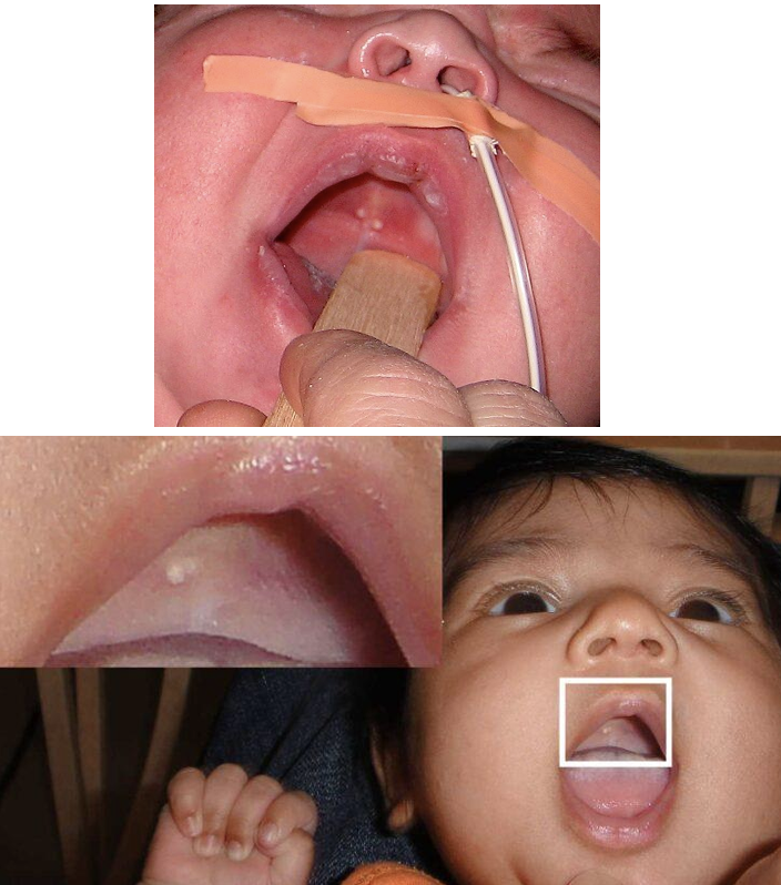
epstein pearls
bohn nodules
keratin filled cysts, remnants of odontogenic epithelium/mucous gland origin
buccal/linugal gingival (alveolar) surfaces and palate away from the raphe
small symptomatic white nodules
rupture spontaneously within weeks to months
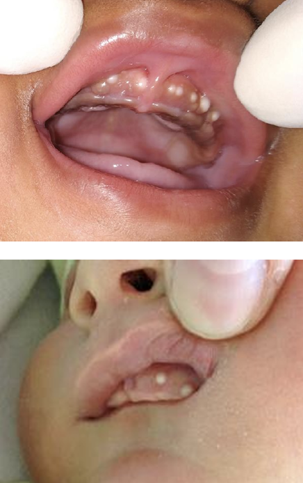
bohn nodules
gingival cysts of the newborn / dental lamina cysts
inclusion cysts from epithelial remnants of the dental lamina during tooth development
crest of the alveolar ridge, usually anterior maxilla
small (1-3mm) whitish/translucent cystic nodules, often mutliple
asymptomatic
self resolve
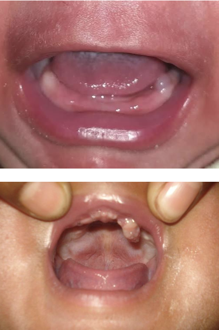
gingival cysts of the newborn / dental lamina cysts
eruption cyst
soft tissue dentigerous type cyst, dental follicle separates from the crown of an erupting tooth
overlying the erupting primary tooth (mandibular alveolar ridge common)
dome shaped bluish/translucent swelling
may become an eruption hematoma
can impair feeding/latching
usually self resolves as tooth erupts, surgical marsupialization/excison if feeding interference or pain
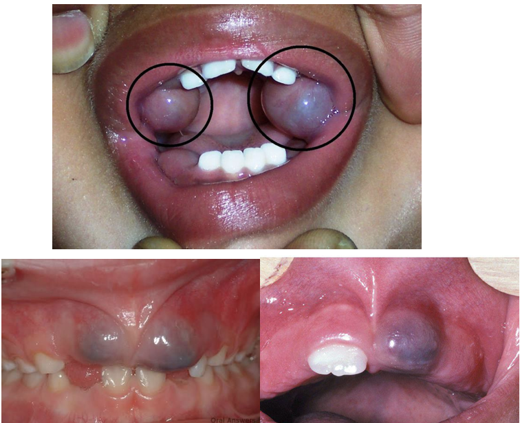
eruption cyst
congenital epulis (neumann tumor/congenital granular cell tumor)
rare benign tumor, likely mesenchymal origin
granular cell histology, distinict from adult granular cell tumor (S100-negative)
anterior maxillary alveolar ridge > mandible
pedunculated, smooth, mucosal colored mass
female predominance (~8:1)
large lesions impair feeding/rarely breathing
surgical excision (curative, no recurrence, no dentition change)
small asymptomatic lesions may be observed, some spontaneous resolution noted
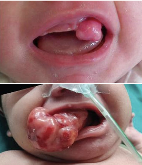
congenital epulis (neumann tumor/congenital granular cell tumor)
melanotic neuroectodermal tumor of infancy (MNTI)
rare neural crest-derived, benign but locally aggressive pigmented tumor
anterior maxilla most common (~60%), then skull, mandible
infants <1 yr
rapid enlarging expansile pigmented mass
osteolytic with tooth displacement on imaging, can mimic an eruption cyst
MRI/CT + complete surgical excision/enucleation
can disrupt tooth development
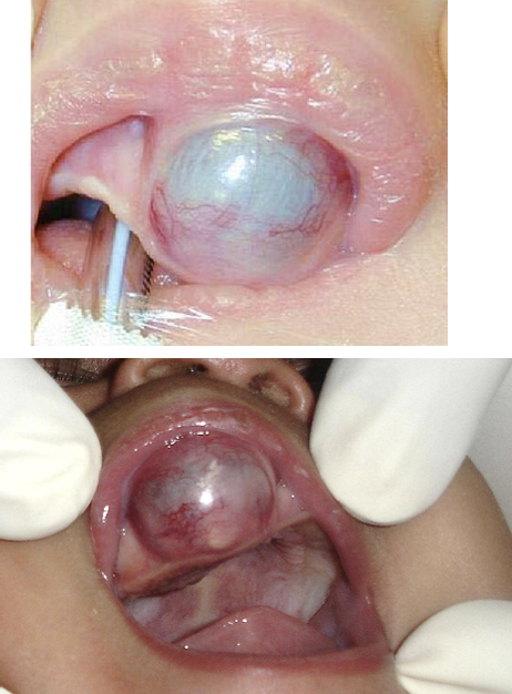
melanotic neuroectodermal tumor of infancy (MNTI)
natal and neonatal teeth
~all are mandibular central incisors (<10% supernumerary)
often mobile with hypoplastic enamel/poor root formation
retain if stable, extract if excessively mobile (aspiration risk) or supernumerary
radiograph to distiguish primary vs supernumerary
may cause breast trauma during feeding- can smooth, cover with composite, use breast shield, switch to formula, or extract
natal teeth
present at birth
neonatal teeth
erupt within first 30 days
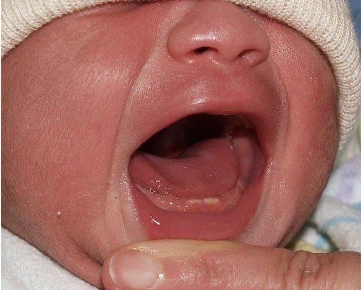
natal / neonatal teeth
riga fede disease
insensitivity to pain
benign traumatic ulceration of oral mucosa from repetitive rubbing against primary teeth in infants
typical presentation- healthy infant (6-15 mo), ulceration of ventral tongue (can become firm scar tissue), coincides with tooth erutpion, responds to conservative treatment (smoothing tooth edges, protective barriers)
red flag for extensive RFD- involves both lip and tongue, fails to respond to treatment, suspect underlying pain insensitivity syndrome
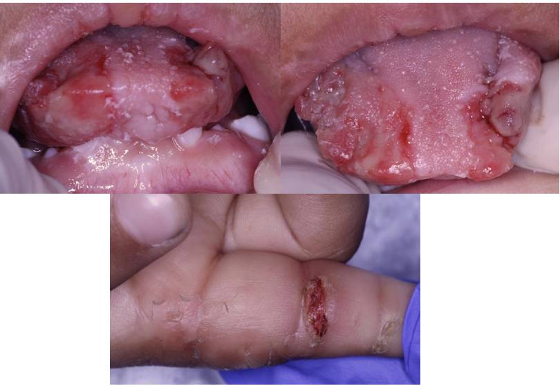
riga fede disease
autism spectrum disorder- self injurious behavior
affects 26-50% of individuals with ASD
pain paradox- many individuals show pain hypersensitvity not indifference, pain perception is highly variable, variability relates to perceptual noise rather than consistently elevated thresholds
oral manifestations- biting lips, tongue, buccal mucosa, hands, self extraction of teeth, mouthing objects causing mucosal trauma, higher rates of dental trauma and bruxism
SIB in ASD is often a response to pain or distress elsewhere
what can cause oral ulcerations?
recurrent aphthous stomatitis (RAS)
periodic fever, aphthous stomatitis, pharyngitis, and cervical adenitis (PFAPA) syndrome
inflammatory bowel disease (IBD)
celiac disease
hand, foot, mouth disease
herpangina
erythema multiforme (EM)
reactive infectious mucocutaneous eruption (RIME)
cyclic neutropenia
herpes simplex virus (HSV)
minor recurrent aphthous stomatitis
80% of cases
<1cm
located on nonkeratinized mucosa
lasts 7-10 days, no scar
major recurrent aphthous stomatitis
10-15% of cases
>1cm
located on lips, oropharynx, soft palate
lasts >4 weeks, often scars
herpetiform recurrent aphthous stomatitis
5-10% of cases
mutliple 0.1-0.3cm
located on nonkeratinized mucosa
lasts 7-14 days, no scar
what is the clinical appearance of all recurrent aphthous stomatitis lesions?
painful round/oval ulcer with gray-white fibrin base and erythematous halo
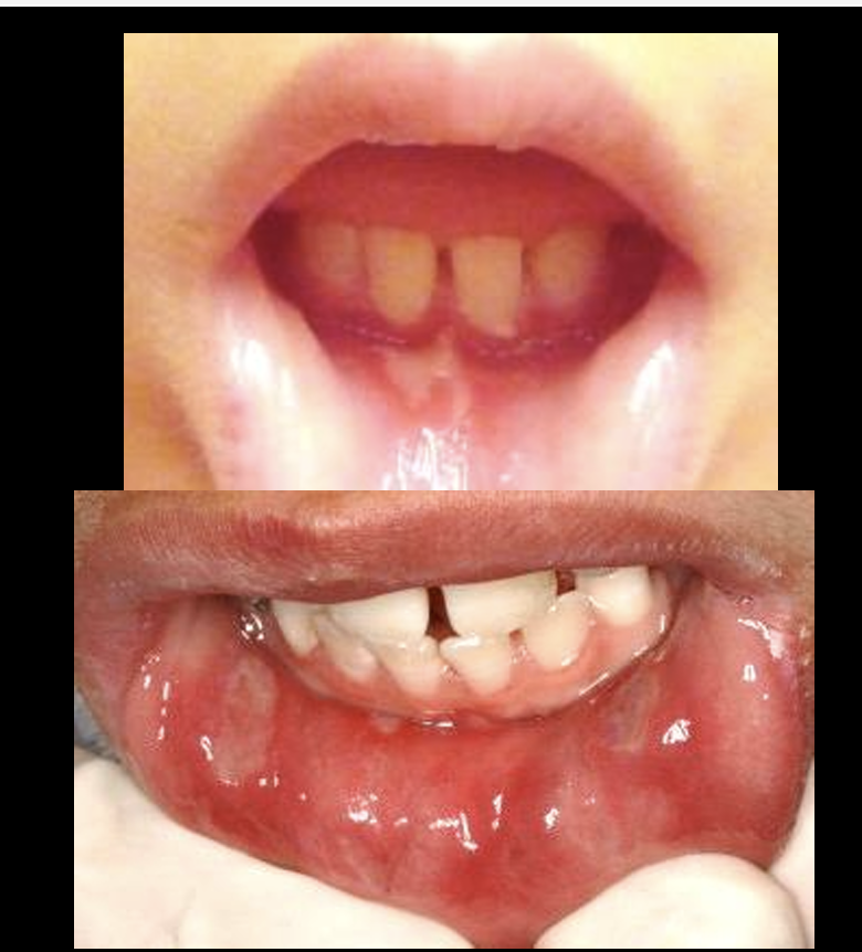
recurrent aphthous stomatitis
what is the pathophysiology of recurrent aphthous stomatitis?
mostly idiopathic but can be related to underlying systemic conditions
mutlifactorial T cell mediated immune dysregulated disease
children with affected parents have 90% chance of developing
underlying systemic conditions: celiac disease, behcet disease, IBD, iron deficiency anemia, cyclic neutropenia, PFAPA syndrome
periodic fever, aphthous stomatitis, pharyngitis, and cervical adenitis (PFAPA) syndrome
most common periodic fever syndrome in children, particularly in non-mediterranean populations
usually manifest before age 5
clusters in families → autosomal dominant inheritance
hallmark: remarkably regular periodicity- typically ever 3-8 weeks (mean 28 days/4 weeks), parents can often anticipate the next episode
each episode is characterized by- high fever (39-41 C) lasting 3-7 days, pharyngitis (most frequent symptom, ~89% of pts), cervical adenitis, apthous stomatitis
key clinical recognition points: between episodes completely well with normal growth and development, episodes abort dramatically with a single dose of corticosteroids, negative infectious workup during episodes, elevated CRP and mild leukocytosis during attacks, normal between episodes
pathophysiology- innate immune dysregulation, RAS-PFAPA-Behcet spectrum all have shared susceptibility loci, 10-30% of adult behcet’s disease patients may have had PFAPA-like sypmtoms in childhood
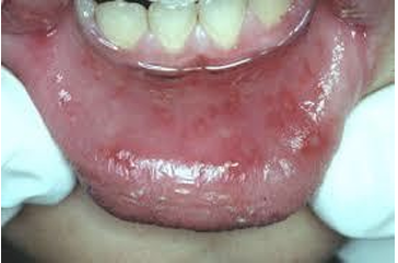
periodic fever, aphthous stomatitis, pharyngitis, and cervical adenitis (PFAPA) syndrome
inflammatory bowel disease (IBD)
crohn disease (patchy in small and large bowel) and ulcerative colitis (uniform colon) - increasingly common in children and adolescents
25% diagnosed before the age of 20
primarily affects the intestinal tract
up to 50% of ped pts with IBD present with oral manifestations
oral manifestations can appear before GI symptoms by months or years
what are the oral manifestations that are highly specific for IBD (particularly crohn disease)?
indurated mucosal tags
cobblestoning
deep linear ulcerations with vertical fissures
lip swelling with vertical fissures
mucogingivitis
what are the oral manifestations that are highly suspicious for IBD?
pyostomatitis vegetans- snail track pustules, more specific for UC, considered virtually pathognomonic for IBD when present
what are the oral manifestations that are non specific for IBD (occur in both CD and UC)?
aphthous stomatitis- most common oral manifestations
angular chelitis- more common in UC
atrophic glossitis
periodontitis
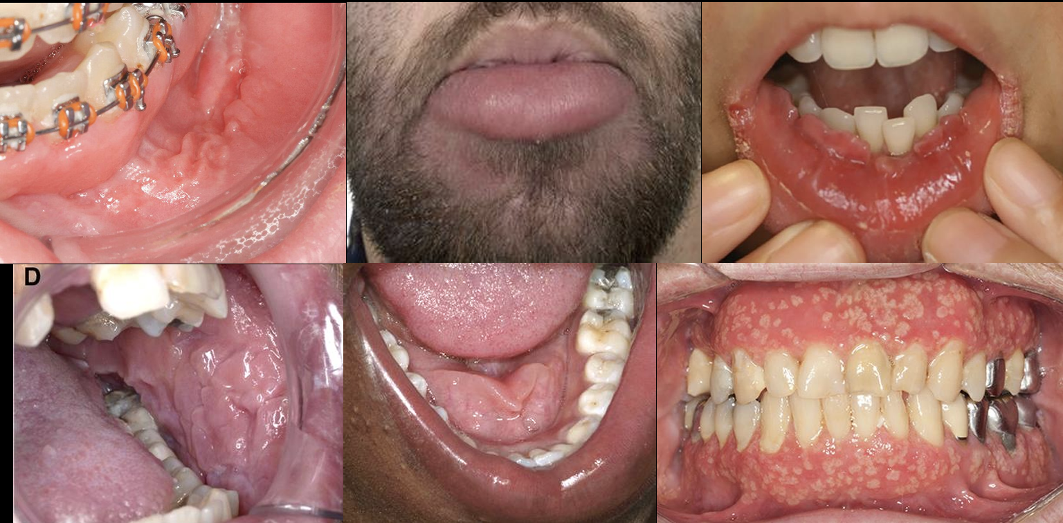
oral manifestations of IBD
celiac disease
chronic immune-mediated systemic disorder caused by ingestion of gluten found in wheat, rye, and barley
affects approx 1% of children- up to 90% of pts are undiagnosed
GI: diarrhea, bloating, weight loss, abdominal pain
oral symptoms may be the only presenting symptoms in some pts
symmetric enamel defects + ulcers are highly suspcious, esp with any growth concerns or family history
what are the key oral manifestations of celiac disease?
oral ulcerations, dental enamel defects, delayed dental eruption, hyposalivation, atrophic glossitis and geographic tongue, oral dysesthesia/glossodynia, angular chelitis- related to iron and vitamin B deficiencies
why is it important for celiac disease to be diagnosed early?
initiation of gluten free diet may prevent progression of oral manifestions
established enamel damage is irreversible
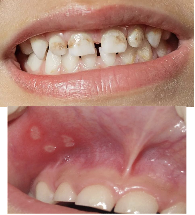
oral manifestions of celiac disease
hand, foot, and mouth disease
common, highly contagious viral illness- predominantly affects children <5 yr
causative agents- coxsackievirus A16, A6, A10, enterovirus 71
transmission- fecal oral, oral oral, respiratory droplets
seasonality- spring to fall outbreaks
incubation is 3-6 days, self limited illness resolving in 7-10 days
clinical presentations of hand, foot, and mouth disease
prodrome- low grade fever, malaise
oral enanthem- painful vesicles → shallow ulcers on buccal mucosa, tongue, hard palate
cutaneous exanthem- paulovesicular rash on palms, soles, may involve buttocks, knees, elbows
delayed sequelae (weeks later)- onychomdesis (nail shedding), beau’s lines, palmoplantar desquamation

hand, foot, and mouth disease
how can you distinguish hand, foot, and mouth disease from herpangina? (characteristics of HFMD)
anterior oral ulcerations- buccal mucosa, tongue, lips
skin rash on hands, feet, and buttocks
causative agents- same enteroviruses (CVA16, CVA6, EV71)
how can you distinguish hand, foot, and mouth disease from herpangina? (characteristics of herpangina)
posterior oral ulcerations- soft palate, uvula, tonsils, pharyngeal folds
no skin rash
causative agents- same enteroviruses (CVA16, CVA6, EV71)
CVA6 associated hand, foot, and mouth disease can present with…
more extensive and atypical features
more extensive vesicobullous eruptions
widespread distribution beyond hands/feets
eczema coxsackium (localizes to atopic dermatitis areas)
high fevers, more severe pain
more common in adults

hand, foot, and mouth disease
erythema multiforme (EM)
acute, immune mediated mucocutaneous reaction
mean age in children is 5.6 yr
EM minor- skin only
EM major- skin and mucosal involvement
self limiting, long term sequelae rare
triggers- infecitons are the primary triggers (unlike SJS which is drug induced), HSV 17.9%, mycoplasma pneumoniae 15.7%, drugs 24.1%, vaccines 3.2% overall (most common trigger in infants)
recurrent EM: 61% HSV- associated
how can you distinguish EM vs RIME (reactive infectious mucocutaneous eruption)? (EM symptoms)
raised target lesions (3 rings)
prominent, acral distribution on skin
variable on mucosa (minor none, major > 2 sites)
HSV ~70% common trigger
young adults
how can you distinguish EM vs RIME (reactive infectious mucocutaneous eruption)? (RIME symptoms)
prominent mucositis
minimal or absent on skin
always prominent on mucosa (oral, ocular, genital)
mycoplasma, other respiratory pathogens are common trigger
children/adolescents
oral and cutaneous lesions of erythema multiforme?
hallmark target lesions with 3 concentric zones
symmetric acral distribution (hands, feet, extensor surfaces)
fixed lesions > 7 days
spread centripetally
oral cavity = most commonly affected mucosal site
vesicles → painful, irregular erosions
non-keratinized mucosa- lips, buccal mucosa, ventral tongue
hemorrhagic crusting of vermilion border
gingiva typically spared (distinguishes from primary HSV)
resolves in 2-3 weeks without scarring
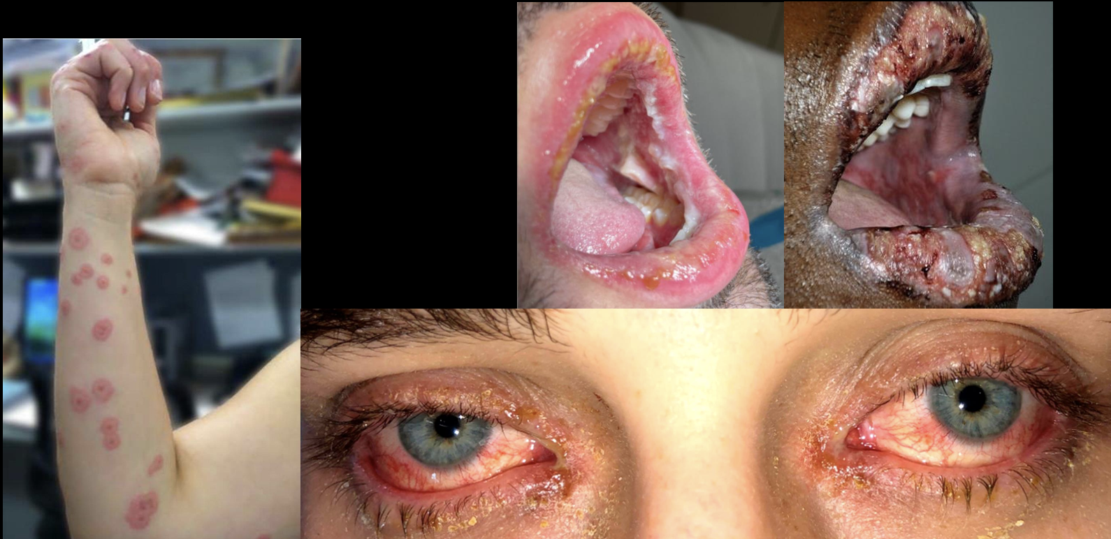
erythema multiforme
cyclic neutropenia
rare autosomal dominant hematologic disorder
usually diagnosed within first year of life
characterized by ~21 day cycles of severe neutropenia (ANC <200/uL) lasting 3-6 days
cycle length can range from 14-36 days in ~30% of pts
pts are completely well between episodes
during neutropenic nadir (lowest time)- recurrent fever, aphthous ulcers, gingivitis, pharyngitis, skin infections
more severe in children than adults
what are the oral manifestations of cyclic neutropenia?
>60% experience oral ulcerations, gingivitis, tooth abscesses >5 times/year
ulcers appear at predictable ~21-day interval
severe, progressive periodontal disease disproportionate to age/hygiene
permanent tooth loss from chronic gingivitis, recurrent abscesses, and alveolar bone loss in adolescence/young adulthood is common

cyclic neutropenia
primary herpes simplex virus (acute herpetic gingivostomatitis)
HSV1 » HSV2
6 mo to 5 yrs
cervical LAD, chills, fever of 101-103F, nausea, anorexia, irritability
oral manifestations- severe pain, pinhead vesicles, collapse and coalesce into an ulcer, keratinized tissue
lasts 5 days to 2 weeks
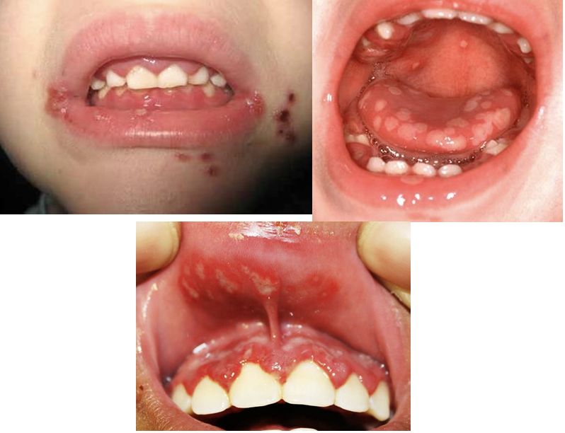
primary herpes simplex virus (acute herpetic gingivostomatitis)
secondary (recurrent herpes simplex virus; herpes labialis)
site of primary inoculation or adjacent
herpes labialis = cold sore = fever blister (15-45% of US)
prodrome: itching, tingling, warmth, erythema (6-24 hrs)
vesicles rupture and crust within 2 days- highly transmissible when vesicles rupture- avoid manipulation until scabbed
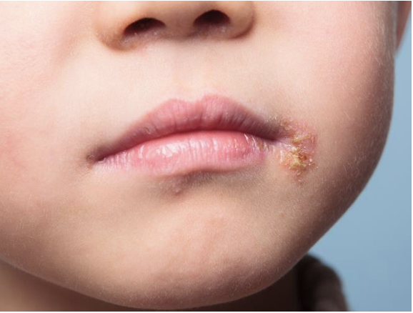
secondary (recurrent herpes simplex virus; herpes labialis)
soft tissue masses
mucocele
ranula
fibroma
squamous papilloma
verruca vulgaris
multifocal epithelial hyperplasia
pyogenic granuloma
peripheral ossifying fibroma
peripheral giant cell granuloma
juvenile spongiotic papillary gingival hyperplasia
leukemia
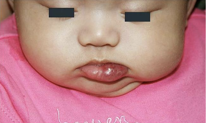
mucocele
extravasation pseudocyst from trauma to a minor salivary gland duct, no epithelial lining
lower lip most common
soft, dome shaped, bluish-to-translucent swelling, fluctuates in size
surgical excision to muscle layer (remove associated gland)
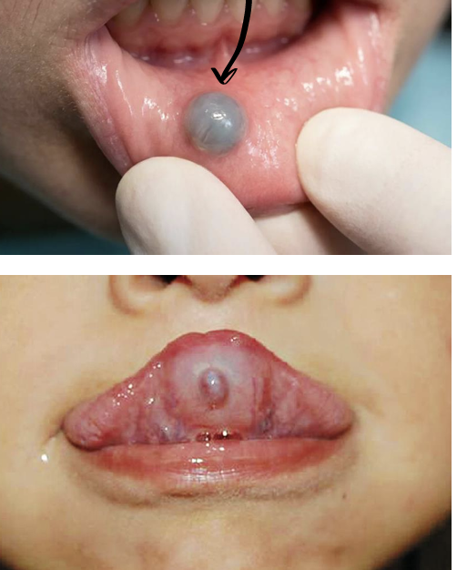
what is this, and why is the lesion blueish?
mucocele
tyndall effect
ranula
mucocele of the sublingual gland
floor of the mouth- lateral to midline
plunging- submandibular/cervical swelling (herniates through mylohyoid into neck)
soft bluish translucent floor of mouth swelling; can enlarge and impair feeding/speech
treatment must address the sublingual gland- marsupialization, sublingual gland excision, untreated lesions may become plunging ranulas
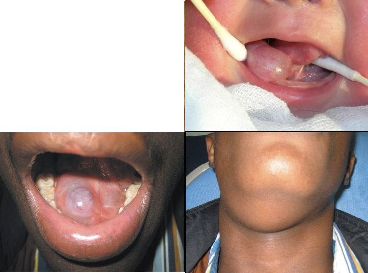
ranula
fibroma (irritation/traumatic fibroma & giant cell fibroma)
reactive fibrous hyperplasia from chronic mucosal trauma (traumatic/irritation fibroma)
buccal mucosa at line of occlusion, lateral tongue, lips
giant cell fibroma is a distinct variant not clearly linked to irritation
giant cell fibroma favors tongue, gingiva, alveolar mucoas (retrocuspid papilla)
firm, smooth, sessile or pedunculated, pink asymptomatic nodule
giant cell fibroma may be papillary/bosselated
conservative surgical excision; remove the source of trauma
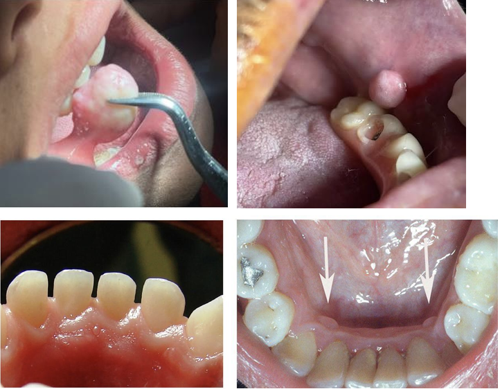
fibroma (irritation/traumatic fibroma & giant cell fibroma)
squamous papilloma
benign HPV associated epithelial neoplasm (HPV6/11)
in infants consider caregiver- fetal/neonatal transmission, in older children autoinoculation
soft palate/uvula, tongue, lips, gingiva
solitary, soft, exophytic, pink to white cauliflower/finger like front surface
usually 1cm, painless
simple surgical excision (curvative), recurrence uncommon

squamous papilloma
verruca vulgaris (oral wart)
HPV driven (commonly HPV2 and 57) benign hyperkeratotic lesion
often autoinoculated from skin warts (finger sucking, nail biting)
keratinized surfaces- labial mucosa, anterior tongue, gingiva, hard palate
painless, white, papillary/verrucous hyperkeratotic papule, may be multiple, more heavily keratinized than squamous papilloma
excision or destructive therapy (cryotherapy, cautery, laser), look for and treat concurrent skin warts
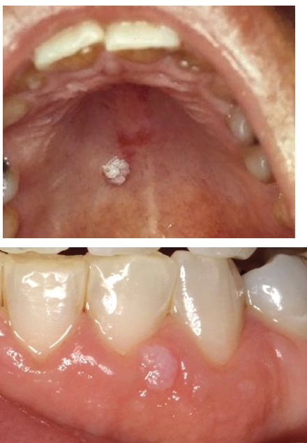
verruca vulgaris (oral wart)
multifocal epithelial hyperplasia (heck disease)
benign HPV-driven epithelial proliferation (HPV13 and 32), transmitted via salivary contact, strong genetic/familial predisposition
2-13 yrs
female predominance (~4:1- 5:1)
endemic in indigenous populations (native americans, central/south americans, inuit/yupik)
immunosuppression/HIV and low SES
labial mucosa, buccal mucosa, and tongue
multiple soft, painless, sessile papules/nodules 1-10mm, pink to mucosa-colored (occasionally whitish), papulonodular or papillomatous, may coalesce into plaques, usually asymptomatic
often non- spontaneous regression (mean 18 mo)
treat for cosmetic/functional/traumatic reasons via excision, cryotherapy, CO2 laser, or topical agents
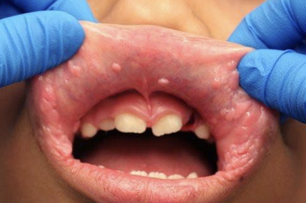
multifocal epithelial hyperplasia (heck disease)
pyogenic granuloma (lobular capillary hemangioma)
reactive vascular proliferation (not pyogenic, not granulomatous)
triggered by trauma/irritation/hormones
gingiva » lips, tongue, buccal mucosa
rapidly growing, friable red/purple papule or nodule that bleeds easily, may ulcerate
excision with removal of irritant- laser, sclerotherapy, cryotherapy, and topical timolol in children, recurrence ~0-15%
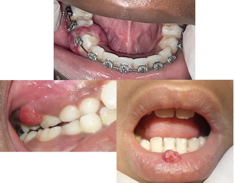
pyogenic granuloma (lobular capillary hemangioma)
peripheral ossifying fibroma (POF)
reactive gingival lesion with fibroblastic proliferation and mineralization (bone/cementum) product
thought to arise from periodontal ligament
may represent maturation of a PG
gingiva exclusively, typically interdental papilla, anterior maxilla common
peaks in adolescents/young adults
firm, sessile or pedunculated gingival nodule, pink to red, may ulcerate
excision down to periosteum/PDL with scaling of adjacent teeth; recurrence higher than the other Ps if base is not fully removed

peripheral ossifying fibroma (POF)
peripheral giant cell granuloma (PGCG)
reactive lesion of multinucleated giant cells arising from periosteum/PDL, often after local trauma
gingiva/alveolar ridge only (anterior to molars), can occur on edentulous ridge
red to bluish purple, sometimes ilcerated gingival nodule
may cause superficial cupping bone resorption
must exclude a brown tumor of hyperparathyroidism if multiple/recurrent
excision to the periosteum with removal of local irritants
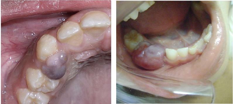
peripheral giant cell granuloma (PGCG)
parulis
nodule of inflamed granulation tissue at the mucosal opening of a draining sinus tract from an underlying odontogenic infection
attached/alveolar gingiva or vestibular mucosa overlying ot apical to the offending tooth
small yellow-red papule/nodule that may intermittently discharge pus and recur
often relatively painless once draining
pulpectomy/pulpotomy or extraction of the offending tooth

parulis
localized juvenile spongiotic gingival hyperplasia
distinct benign reactive inflammatory gingival lesion- not plaque induced
anterior maxillary (labial) gingiva, usually unifocal
mean age ~13-16 yr
slight female predominance
bright red, well demarcated, finely granular/papillary enlargement, bleeds easily
persists despite excellent oral hygiene and debridement
surgical excision is most common (cryotherapy/laser alternatives), recurrence can occur
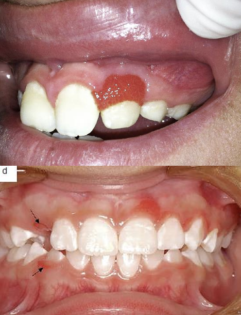
localized juvenile spongiotic gingival hyperplasia
leukemia
most common pediatric malignancy
oral changes result from direct leukemic infiltration and/or marrow failure (thrombocytopenia, neutropenia)
may be the first presenting sign
gingiva » palate, tongue, buccal mucosa
diffused boggy gingival enlargement (begins at interdental papillae), spontaneous gingival bleeding, petechiae, mucosal pallor, ulceration
systemic signs: fever, fatigue, lymphadenopathy, hepatosplenomegaly
non-plaque gingival enlargement with bleeding in a child warrants urgent CBC and hematology referral
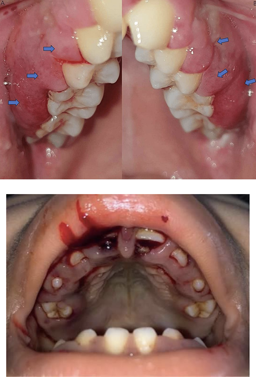
leukemia
vascular and lymphatic pathoses
hemangioma
lymphatic malformation
hemangioma
positive on diascopy
require doppler US prior to excision- high flow v low flow
congenital hemangioma- RICH, PICH, NICH
infantile hemangioma- rapidly grows, plateaus, then involutes by school age
hemangioma
infantile hemangioma
most common benign vascular tumor of infancy, from endothelial cell proliferation
usually not present at birth (a pale macule/telangiectatic patch may herald it)
rapid proliferation over the first 1-3 mos, then slow involution over years
lips, buccal mucosa, and tongue
associated with female sex, prematurity, low birth weight
bright red superficial (strawberry) or bluish deep component
most need only observation/reassurance- medical management with oral propranolol, topical timolol or laser/surgery for residual changes
infantile hemangioma
lymphatic malformation (microcystic lymphangioma / macrocystic cystic hygroma)
low flow congenital vascular malformation of maldeveloped lymphatic channels
not a tumor and does not involute
childre <2yr, ~50% present at birth
~75% in head/neck, oral cavity, oropharynx, and tongue commonly involved
large lesions cause macroglossia, airway obstruction, and dysphagia
mucosal microcystic lesions = clusters of clear or dark purple frog egg vesicles that leak chyle or blood
classified macrocystic (>1-2cm), microcystic (<1-2cm), or mixed
complications- intralesional bleeding, infection, sudden enlargement
doppler ultrasound, MRI
multidisciplinary and individualized- observation (spontaneous regression is possible, mainly macrocystic), sclerotherapy, surgical resection/debulking, CO2 laser, systemic sirolimus (mTOR inhibitor) alpelisib (PI3K inhibitor)

lymphatic malformation (microcystic lymphangioma / macrocystic cystic hygroma)
both are macro