Patho Lab - Cell Injury
 Call Kai
 Learn
Learn Practice Test
 Spaced Repetition
 Match
Match Flashcards
 Knowt Play
1/62

There's no tags or description
Looks like no tags are added yet.
Name | Mastery | Learn | Test | Matching | Spaced | Call with Kai | Chat |
|---|
No analytics yet
Send a link to your students to track their progress
63 Terms
1.3 to 1.5 cm
Normal left ventricular artery thickness
2 to 2.8 cm
Left ventricular hypertrophy thickness
Increase in the TRANSVERSE diameter of myocytes
Microscopic findings in left ventricular hypertrophy
Increase sytemic pressure due to hypertension
Cause of left ventricular hypertrophy in the case where the patient who’s hypertensive, diabetic, and has a history of cardiomegaly died due to hypovolemic shock secondary to a vehicular accident
Increase in workload
Most common stimulus for cardiac muscle hypertrophy
hypertrophy, hyperplasia, atrophy, metaplasia
Four adaptive changes in cell injury
Physiologic hypertrophy
Physiologic/Pathologic & Hypertrophy/Hyperplasia:
Hormonal: uterus - pregnancy; breast - lactation
Physiologic hypertophy
Physiologic/Pathologic & Hypertrophy/Hyperplasia:
Functional: skeletal muscles - exercise
Pathologic hypertrophy
Physiologic/Pathologic & Hypertrophy/Hyperplasia:
Heart - hypertension and faulty valves
Physiologic hyperplasia
Physiologic/Pathologic & Hypertrophy/Hyperplasia:
Hormonal: increased functional capacity, breast during puberty/pregnancy, uterus during pregnancy
Pathologic hyperplasia
Physiologic/Pathologic & Hypertrophy/Hyperplasia:
Excess hormones or growth factors
e.g. endometrial, benign prostatic, viral infections
F: do not divide
T/F: heart muscle divides and undergoes hypertrophy as a compensatory mechanism when injured
Fibrosis, fibrous
Repair in ventricular hypertrophy takes the form of _______; patient with old MI has ______ scar formation
T
T/F: increase resistance in the outflow track, where lipid deposition in the wall occludes blood flow, can also cause hypertrophy
Biventricular hypertrophy
Both right and left ventricles hypertrophied
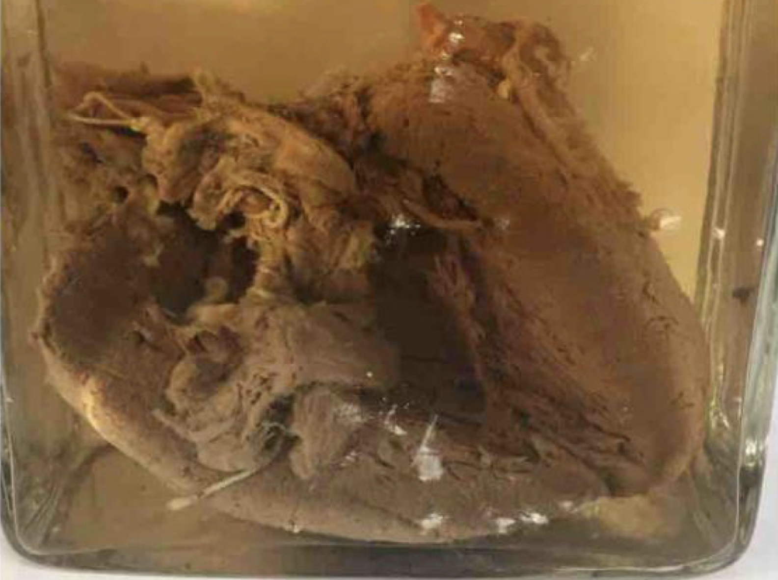
left ventricular hypertrophy; hypertrophy (increase in cell size)
Pathologic condition and cellular adaptive change associated with this specimen
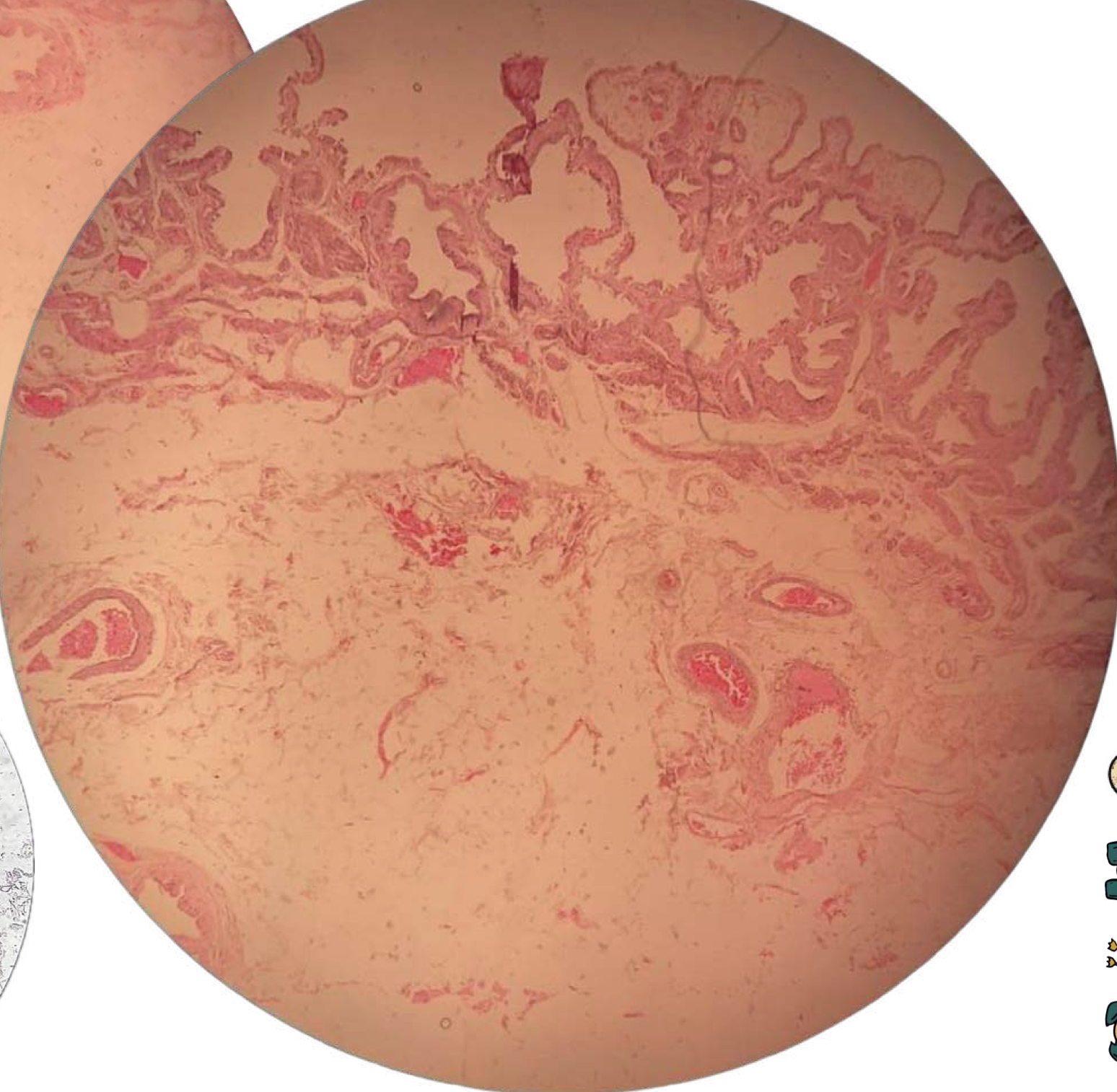
Chronic cervicitis with squamous metaplasia
Gross findings of this condition are:
Mucopurulent cervical discharge with a reddened and edematous cervix
Chronic cervicitis with squamous cell metaplasia; reversible
Diagnose and identify metaplasmic change
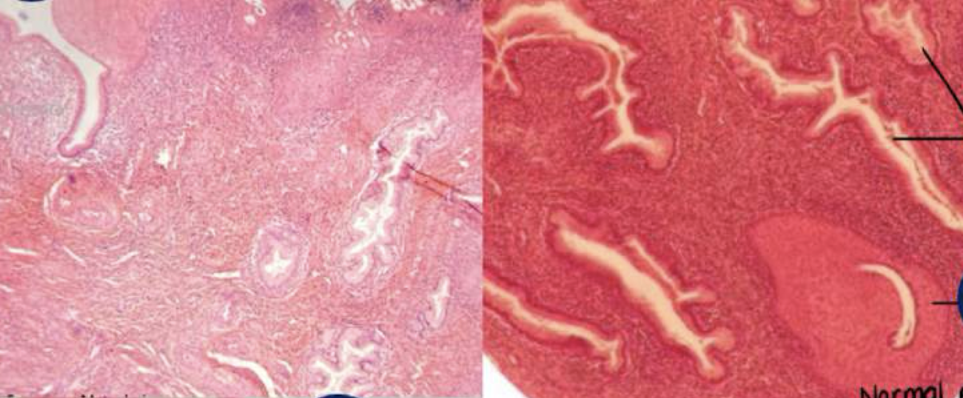
Chronic cervicitis with squamous cell metaplasia; reversible
Diagnose and identify metaplasmic change
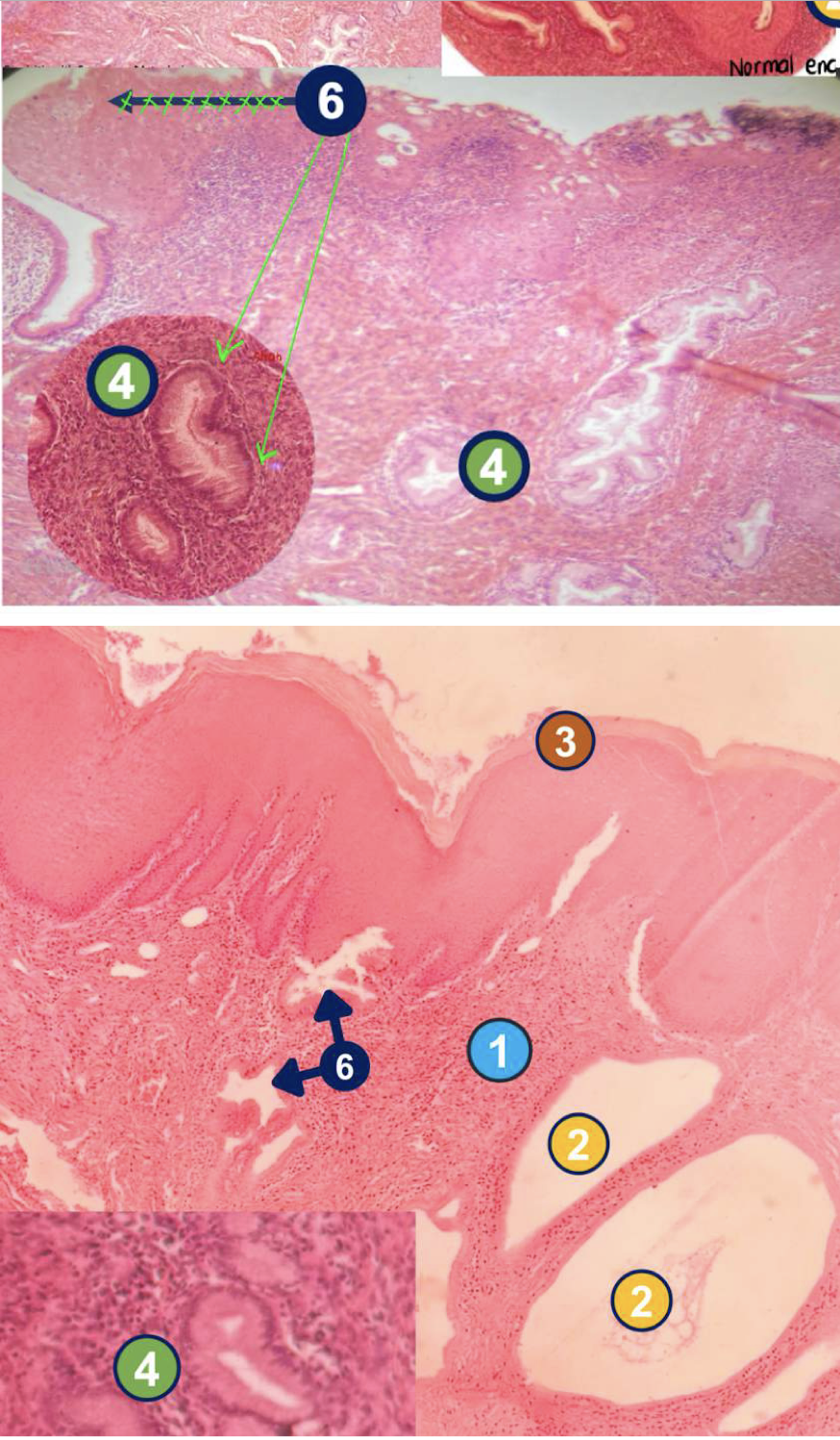
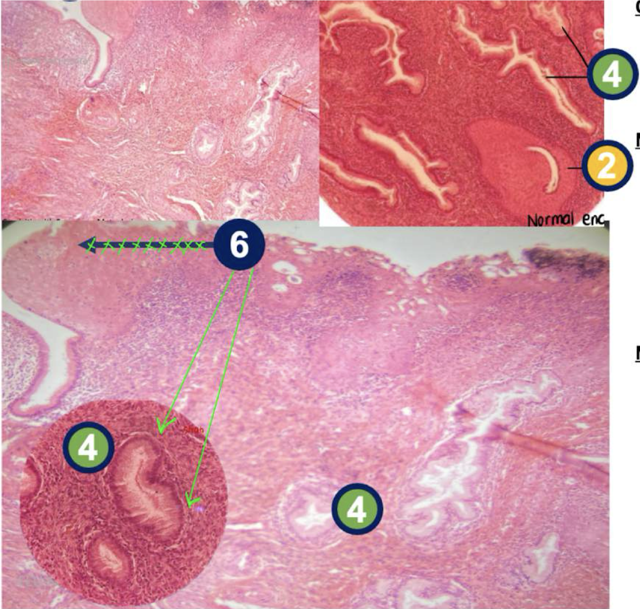
Chronic cervicitis with squamous cell metaplasia
2: Nabothian cyst
4: Endocervical glands
6: Squamous metaplasia of glands
Diagnose and identify labeled microscopic features
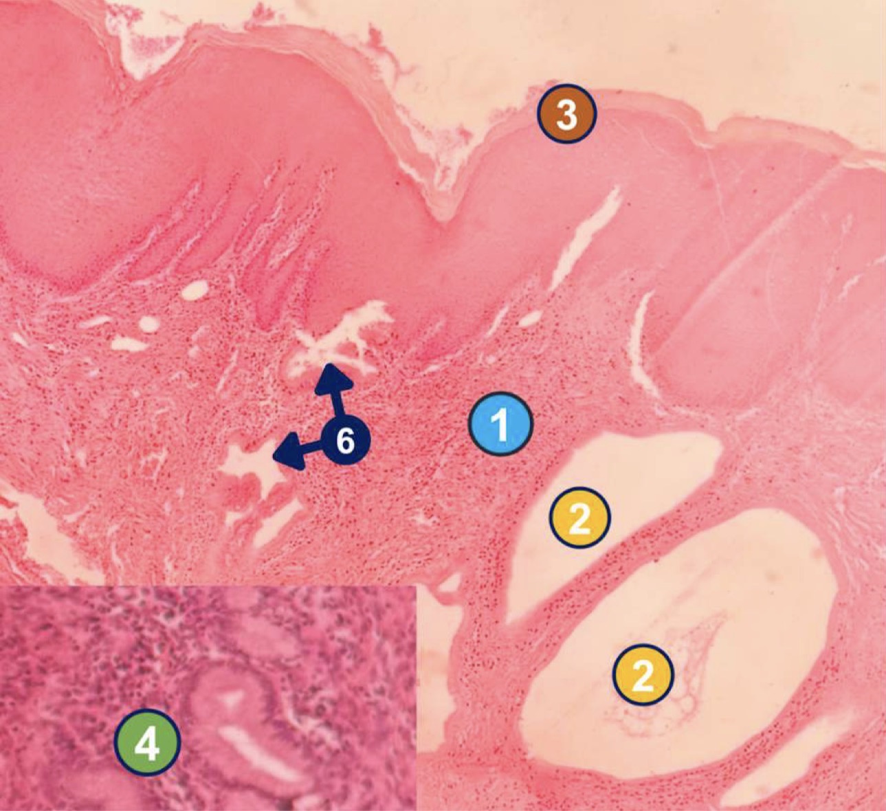
Chronic cervicitis with squamous cell metaplasia
1: lymphocytes & plasma cells
2: Nabothian cyst
3: squamous lining epithelium
4: endocervical glands
6: squamous metaplasia of glands
Diagnose and identify labeled microscopic features
Metaplasia: squamous to columnar
Morphological change in BARRETT ESOPHAGUS
Myositis ossificans
Connective tissue metaplasia that is a repair mechanism and not a true adaptation, associated with extraosseous bone formation
Metaplasia: columnar to squamous
Morphological change in bronchi of heavy smokers
Metaplasia
Reprogramming of stem cells; change of differentiated cell type
Columnar to squamous
Most common type of epithelial metaplasia
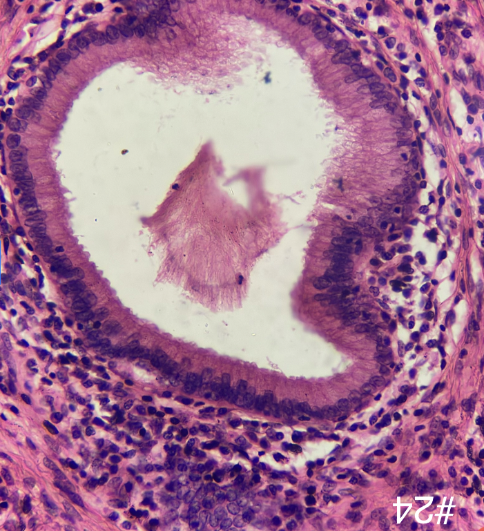
Chronic cervicitis with squamous metaplasia
Diagnose
Cellular swelling
Earliest manifestation of almost all forms of injury to cells
hydrophobic change or vacuolar degeneration
other term for cellular swelling
cellular injury
This condition causes pallor, increased turgor, increased weight of affected organ
cellular swelling of the kidneys
gross findings of this condition includes swollen tubules, granular cytoplasm, and displaced nuclei of tubules
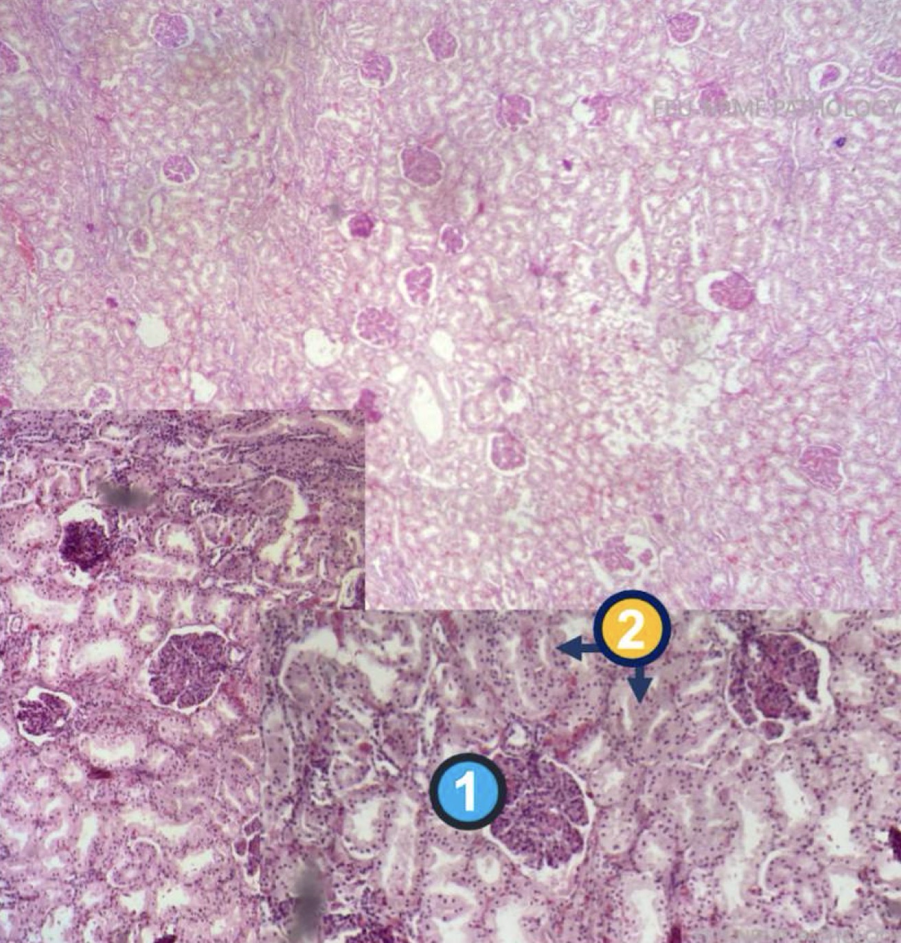
Cellular swelling of the kidneys
1: glomerulus
2: swollen tubules
Reversible
Diagnose, identify the pointed structures, and identify if reversible or irreversible
cellular swelling of kidneys
diagnose
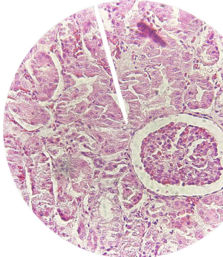
cellular swelling of kidneys
diagnose
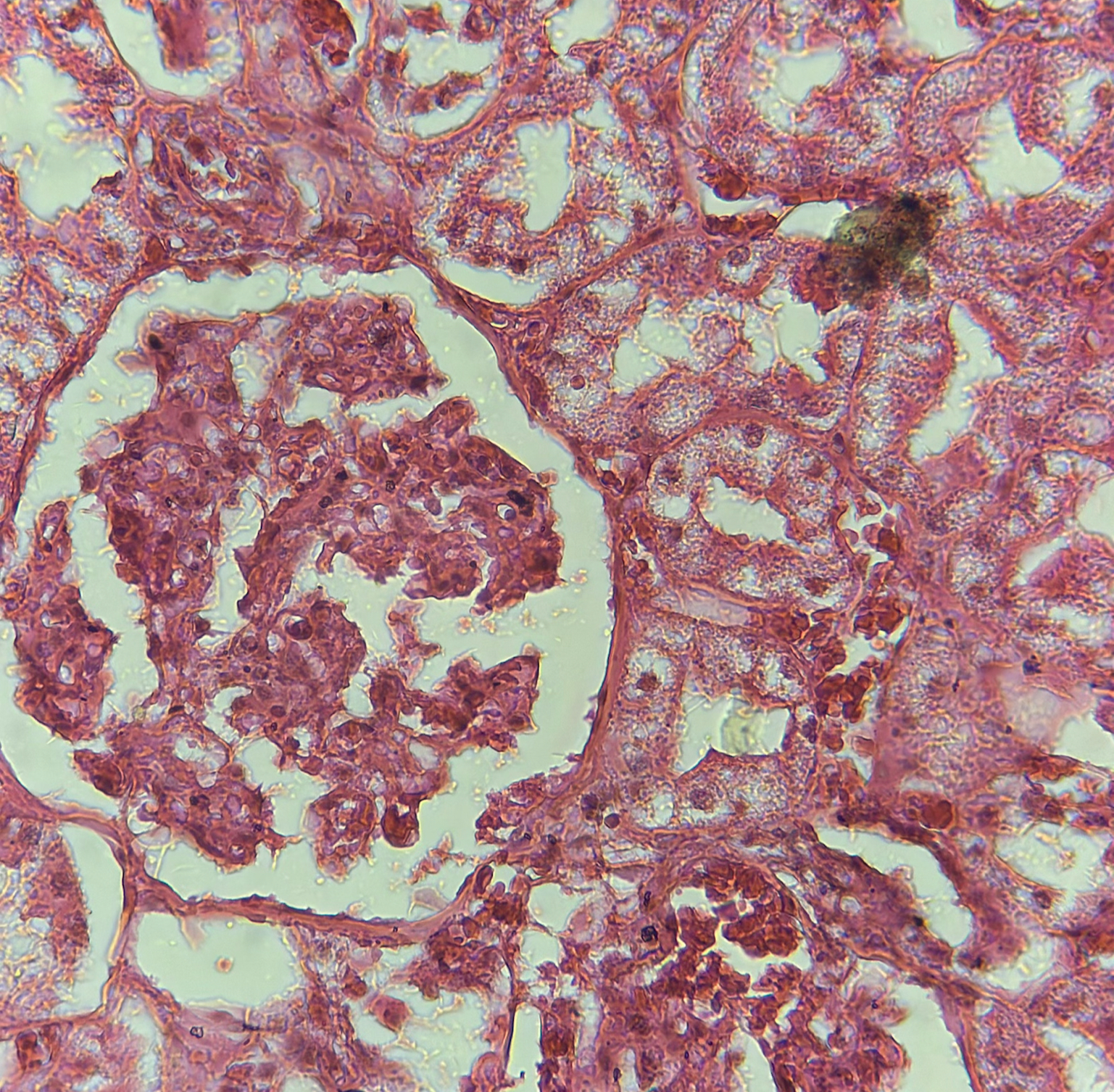
plasma membrane alterations
mitochondrial changes
accumulation of myelin figures
dilation of endoplasmic reticulum
nuclear alterations
ultrastructural changes of reversible cell injury
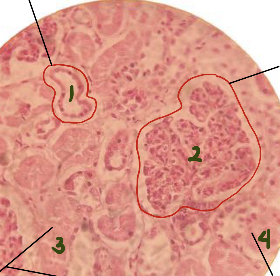
cellular swelling of kidneys
1: displaced nucleus
2: glomerulus
3: swollen tubules
4: granular cytoplasm
diagnose and identify the pointed structure
coagulation necrosis in renal infarct
(image shows wedge-shaped kidney infarct)
diagnose
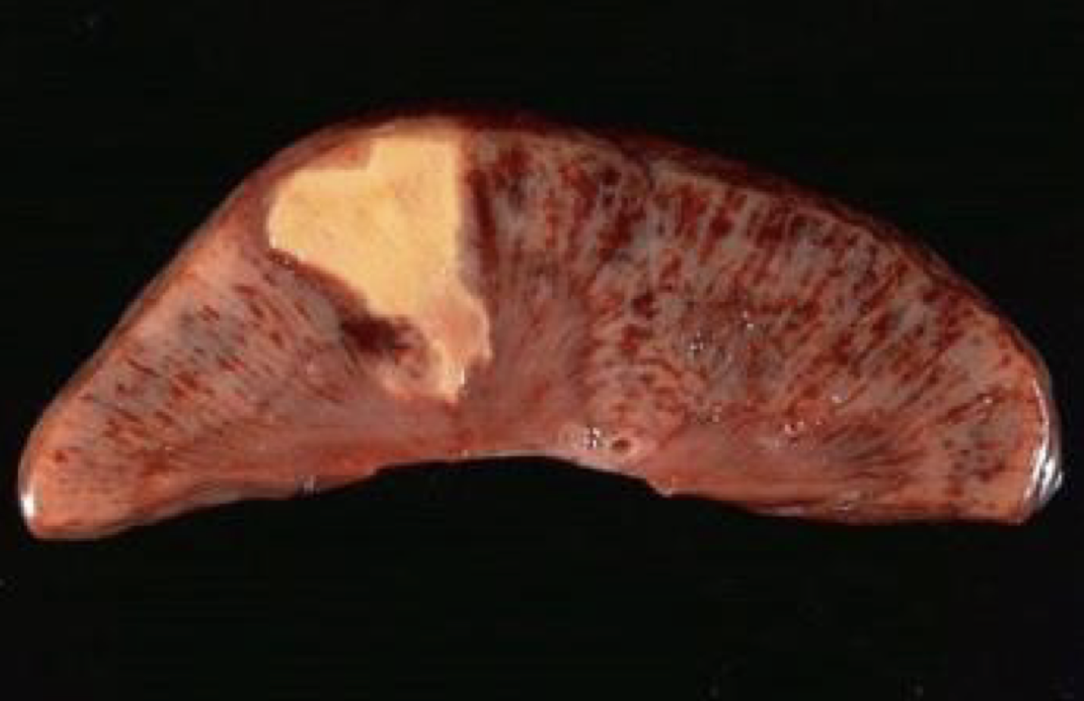
coagulation necrosis in renal infarct, coagulative necrosis, irreversible
diagnose, type of cell injury, reversible/irreversible
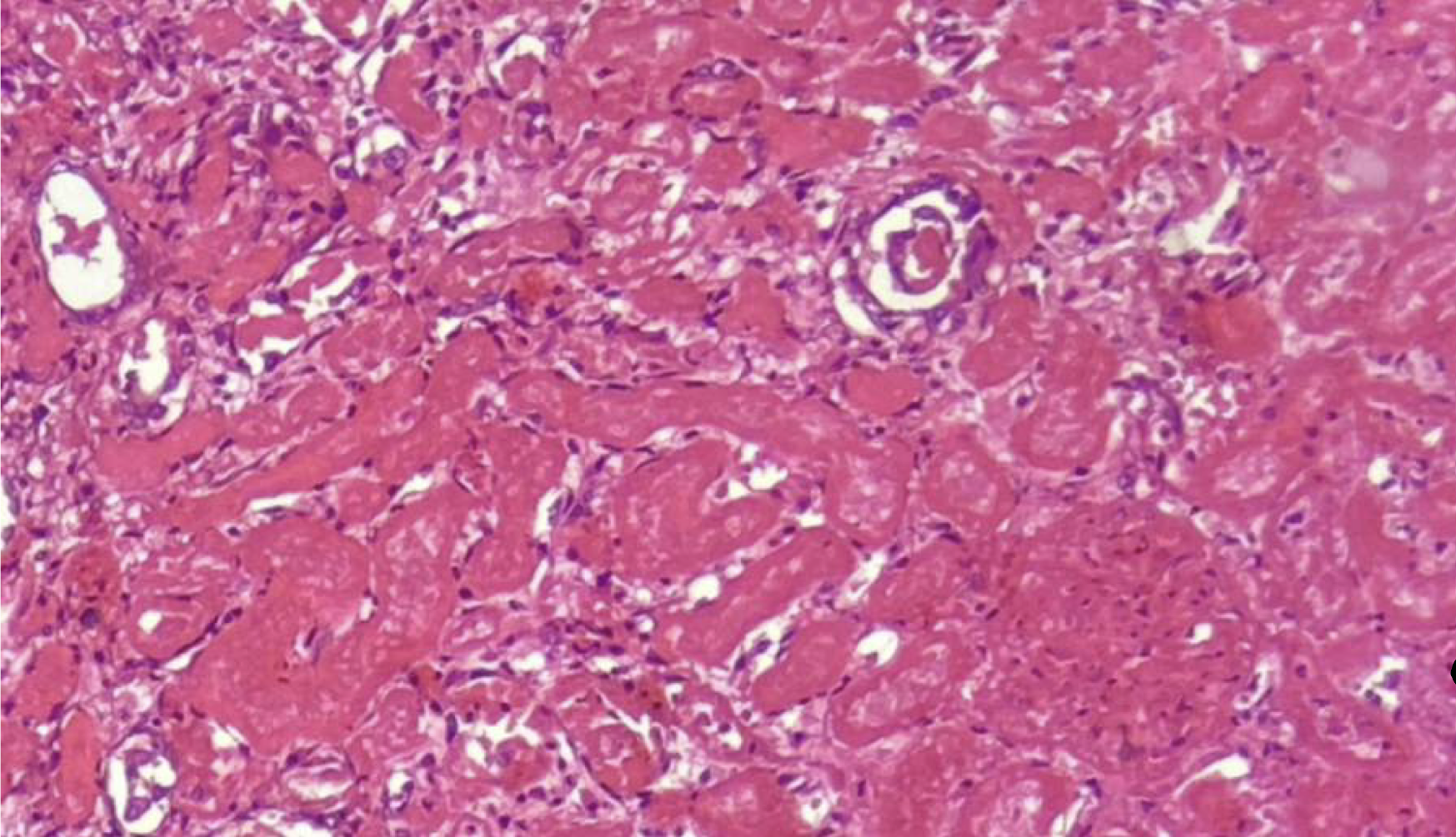
coagulation necrosis in renal infarct, coagulative necrosis, irreversible
diagnose, type of cell injury, reversible/irreversible
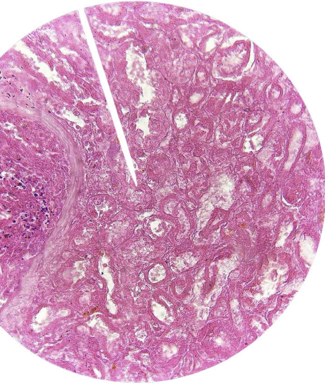
coagulation necrosis in renal infarct, coagulative necrosis, irreversible
diagnose, type of cell injury, reversible/irreversible
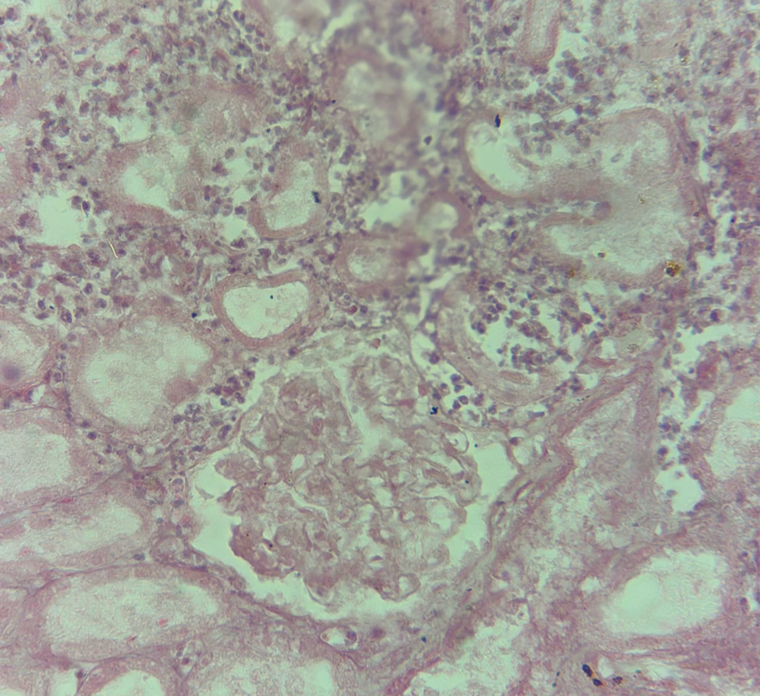
coagulative
caseous
liquefactive
fibrinoid
gangrenous
enzymatic fat
six types of necrosis
caseous necrosis
necrosis often seen in tuberculosis infection
enzymatic fat necrosis
necrosis often seen in pancreatitis
caseous necrosis
A friable yellow-white necrotic area and cellular outlines cannot be discerned
coagulative necrosis
Necrosis that has a firm structure and affected tissue and has maintained cellular outline
cellular swelling of kidney, coagulative necrosis in renal infarct
Displaced nuclei is associated with ______, while absence of nuclei is ______ in kidney injuries
enzymatic fat necrosis of omentum
cell injury with gross findings of chalky white appearance
saponification
this mechanism requires both fatty acids and calcium
pancreatic lipase
release of these enzymes leads to fat necrosis in the momentum
dystrophic calcification
calcification expected to develop in the omentum
dystrophic
deposition of calcium at sites of cell injury and necrosis, serum calcium is normal
metastatic
deposition of calcium in normal tissue caused by hypercalcemia
hyperparathyroidism, vitamin d deficiency, renal failure
conditions that leads to metastatic calcification
enzymatic fat necrosis of omentum
1: necrotic fat cells
2: normal adipocytes
diagnose and identify pointed structure
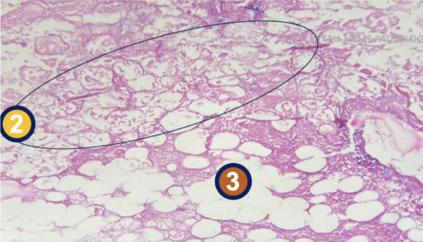
enzymatic fat necrosis of omentum
1: necrotic fat cells
2: normal adipocytes
diagnose and identify pointed structures
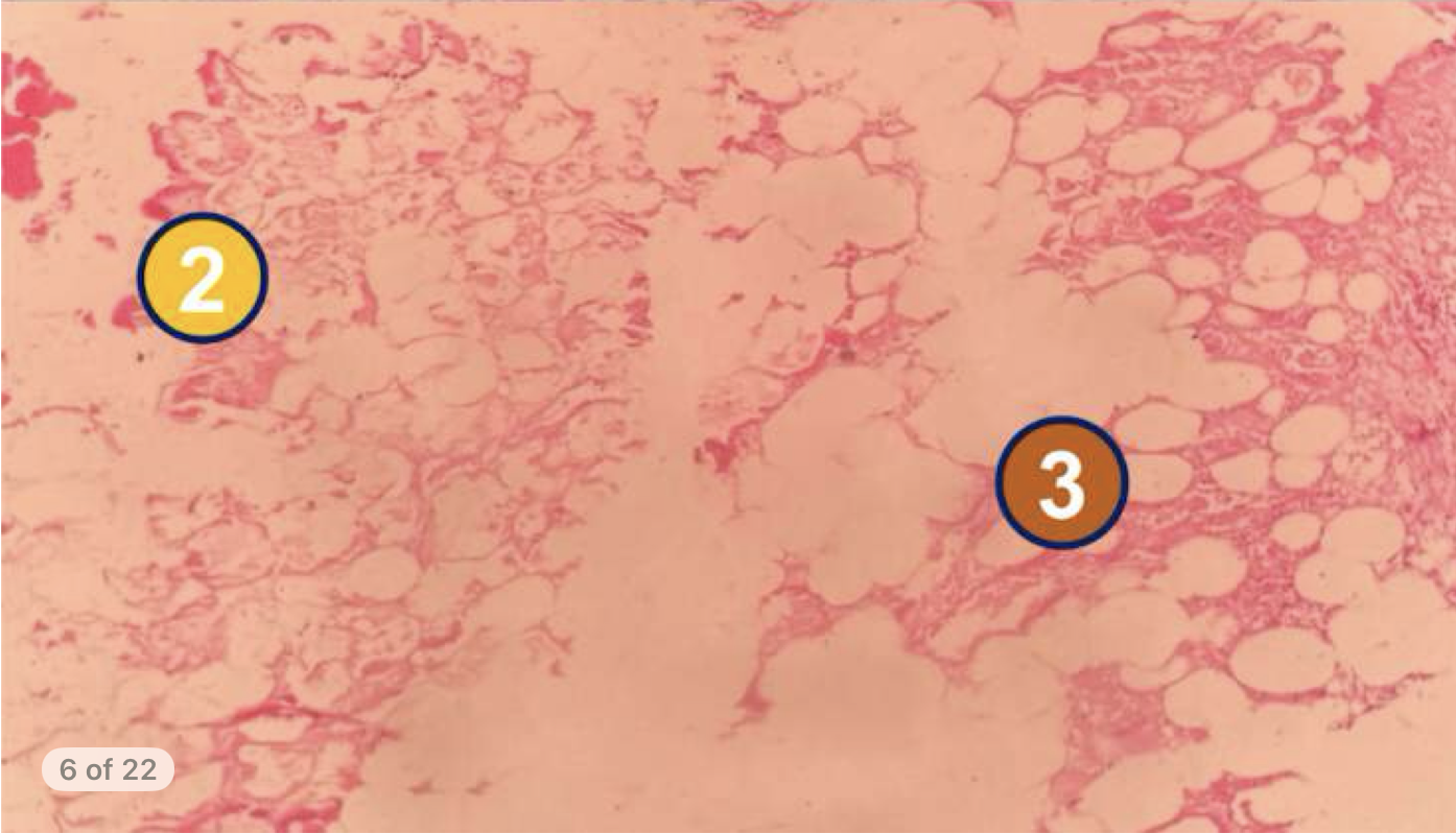
enzymatic fat necrosis of omentum
1: necrotic fat cells
2: normal adipocytes
Diagnose and identify pointed structure
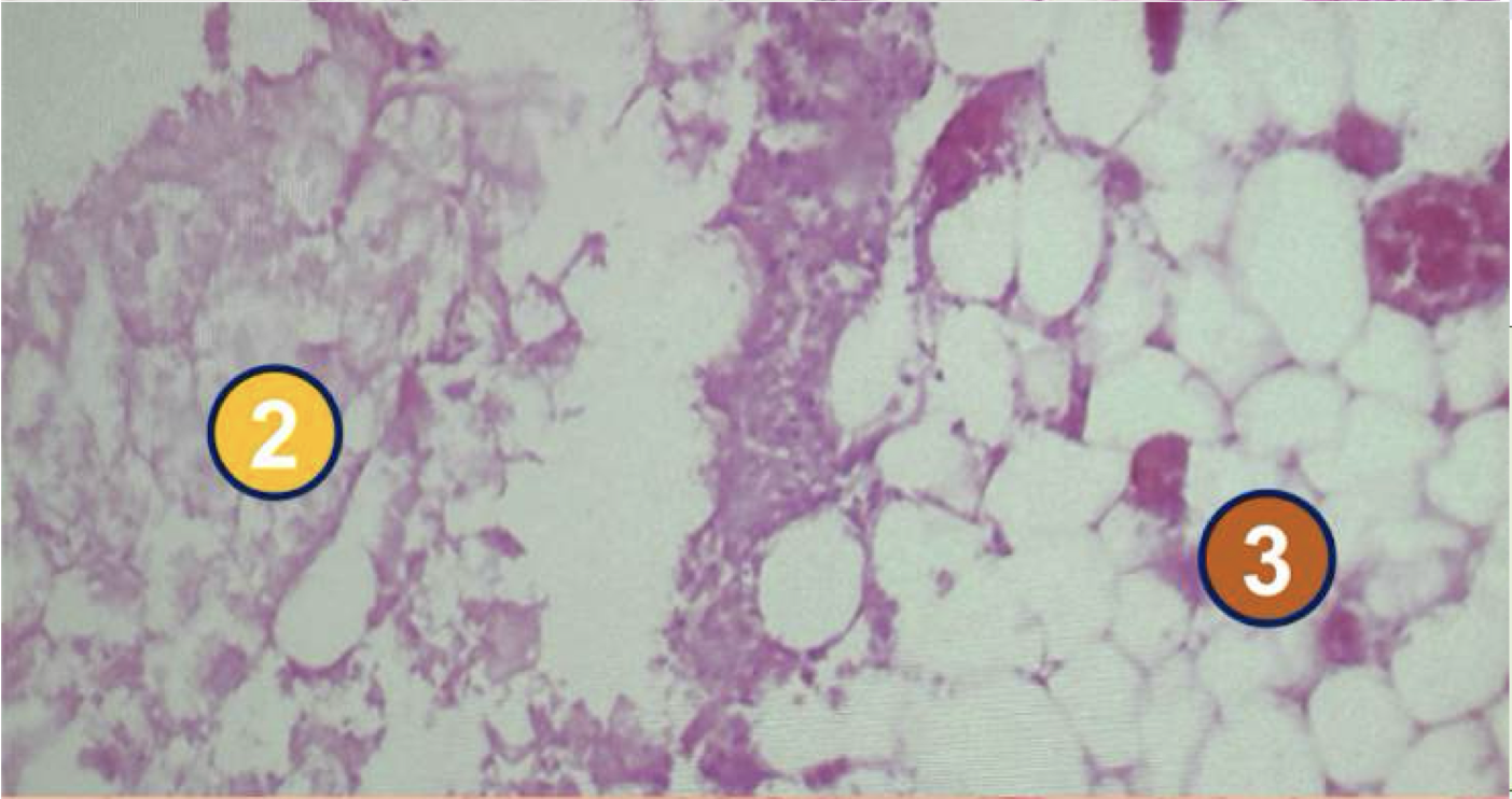
enzymatic fat necrosis of omentum
identify pointed structure

chronic cholecystitis with cholesterolosis
condition with gross findings of subserosal fibrosis, thickened walls, and dense fibrous adhesions
endogenous lipids
abnormal metabolism of _______ are the nature of intracellular accumulation in chronic cholecystitis with cholesterolosis
chronic cholecystitis with cholesterolosis
in a tissue section, accumulation of lymphocytes, rokitansky-aschoff sinuses, cholesterol laden macrophages, and fibrosis is indicative of
chronic cholecystitis with cholesterolosis
3: rokitansky-aschoff sinuses
4: lymphocytes, eosinophils, plasma cells
6: chronic inflammation
diagnose and identify pointed structures

chronic cholecystitis with cholesterolosis
2: endogenous lipid/foamy microphage/cholesterolosis
diagnose and identify pointed structure

chronic cholecystitis with cholesterolosis
diagnose