BIEN 1100
0.0(0)
Studied by 5 peopleCard Sorting
1/98
Earn XP
Last updated 2:38 AM on 12/13/22
Name | Mastery | Learn | Test | Matching | Spaced | Call with Kai | Chat |
|---|
No analytics yet
Send a link to your students to track their progress
99 Terms
1
New cards
Definition of Biophotonics/Biomedical Optics
Interaction between light and biological systems, such as human cells and tissues
2
New cards
How does light modify tissue?
-Laser Surgery
-Laser Thermal Ablation
-Low Level Laser Therapy (LLLT)
-Photodynamic Therapy (PDT)
-Laser Thermal Ablation
-Low Level Laser Therapy (LLLT)
-Photodynamic Therapy (PDT)
3
New cards
How does tissue modify light?
-Optical spectroscopy
-Imaging
-Biosensing
-Imaging
-Biosensing
4
New cards
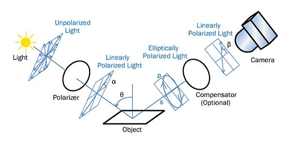
Polarization Imaging Definition
A light beam that is scattered by an object will contain a variety of information about the object's surface structure
5
New cards
What is Diffuse Optical Tomography (DOT)?
Non-invasive technique/proceudure that uses light (near the infrared spectral region) to measure optical properties of tissue and create a 3-D picture of the tissue.
6
New cards
What is diffuse optical tomography typically used to create an image of?
it works best on soft tissues such as breast and brain tissue
7
New cards
pixel definition
Pixels are a grid of dots or picture elements, each pixel contains a number (binary format=zeros and ones) representing the color (tone) of the pixel.
8
New cards
pixel size
When you zoom in you can't see any details smaller than the pixel size, the closer you zoom in the more likely you will start to see pixels
9
New cards
Bit depth
The greater the bit depth, the more tones in the image
10
New cards
File-Size Equation
Number of Pixels * bitdepth
11
New cards
Spatial Resolution defintion
The ability to depict small details; number of pixels used to comprise an image
12
New cards
Contrast definition
The difference in gray level value between the anatomy of interest and the background
13
New cards
Noise definition
The variance in gray level value; presence of artifacts in an image
14
New cards
Artifacts definition
Distortions and/or streaks; column of pixels darker than other
15
New cards
Higher spatial resolution
=may be more noise since there are a greater number of pixels
16
New cards
Lower spatial resolution
= less noise
17
New cards
Less noise
=higher contrast
18
New cards
More noise
=lower contrast
19
New cards
How can we measure image quality?
Phantoms
20
New cards
Anthropomorphic Phantoms
Model realistic anatomy
21
New cards
What do we need to image the body?
- Waves/material that can penetrate and exit the body
- Waves/material that interacts differently with different tissues to create contrast
- Safe
- Relatively fast
- Waves/material that interacts differently with different tissues to create contrast
- Safe
- Relatively fast
22
New cards
What imaging uses X-Rays from the electromagnetic spectrum?
X-rays
23
New cards
What imaging uses gamma rays from the electromagnetic spectrum?
Nuclear Medicine, PET (positron emission tomography)
24
New cards
What imaging uses infrared radiation (IR) from the electromagnetic spectrum?
Thermal imaging
25
New cards
What imaging uses radio waves from the electromagnetic spectrum?
MRI
26
New cards
Ionizing Radiation Modalities
-X-ray
-CT
-Nuclear Medicine/ Positron Emission Tomography (PET)
-CT
-Nuclear Medicine/ Positron Emission Tomography (PET)
27
New cards
Nonionizing radiation modalities
-Ultrasound
-MRI
-MRI
28
New cards
X-ray Radiography
* A beam of x-rays travels through the body
\-The amount of attenuation depends on tissue depth, atomic number, atomic weight, and density
\-Resulting image is a projection or shadow of the body
\
* Application - used to diagnose fractures, scan bones, pneumonia, and cancers
\-The amount of attenuation depends on tissue depth, atomic number, atomic weight, and density
\-Resulting image is a projection or shadow of the body
\
* Application - used to diagnose fractures, scan bones, pneumonia, and cancers
29
New cards
Pros of X-ray radiography
- high spatial resolution (=0.1 mm)
- cheap
- fast (real time)
- cheap
- fast (real time)
30
New cards
Cons of X-ray Radiography
-ionizing radiation
- 2D projection (shadow imaging)
- 2D projection (shadow imaging)
31
New cards
The problem with X-ray
- projection (shadow) image
- 3D anatomy projected onto 2D image
- 3D anatomy projected onto 2D image
32
New cards
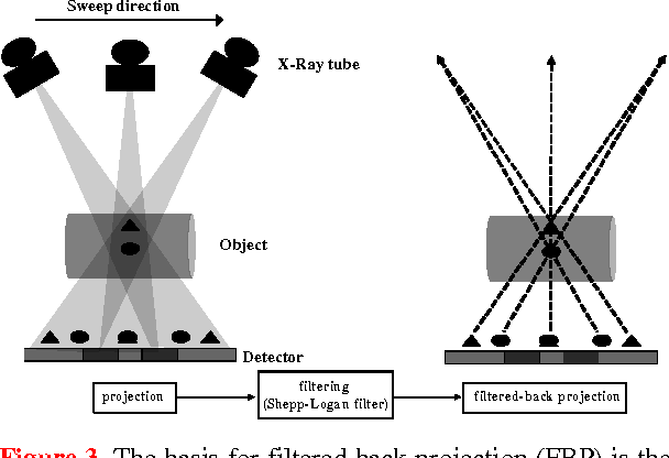
X-ray Tomosynthesis
A digital X-ray advanced application that involves the acquisition of a series of low-dose projection images during a single sweep of the x-ray tube over a limited angle
33
New cards
Reconstructed image of x-ray tomosynthesis
the individual images are reconstructed into thin slices for viewing as a single image or in a cine mode
34
New cards
What is X-ray tomosynthesis?
Tomosynthesis acquires multiple projections, covers a range of projection angles, and provides multiple image plane reconstructions or "slices."
35
New cards
Tomosynthesis Pros
- Relatively fast
- Cheaper than CT
- Partial 3D info
- High spatial resolution (=0.5mm)
- Cheaper than CT
- Partial 3D info
- High spatial resolution (=0.5mm)
36
New cards
Tomosynthesis Cons
- More ionizing radiation than x-ray
- More expensive than x-ray
- Partial 3D info
- More expensive than x-ray
- Partial 3D info
37
New cards
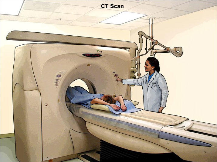
Computed Tomography (CT)
X-ray projections acquired at multiple view angles. Cross-sectional images reconstructed.
Image contrast depends on DENSITY of tissue
Image contrast depends on DENSITY of tissue
38
New cards
Clinical applications of a CT scan
Primarily used to scan for diseases in soft tissue and organs
* Trauma
* Brain perfusions
* Oncology
* Abdominal
* Cardiac
* Lung
* Trauma
* Brain perfusions
* Oncology
* Abdominal
* Cardiac
* Lung
39
New cards
CT history
- Invented by Sir Godfrey Hounsfield and Alan Cormack (invented in 1972)
- Nobel Prize in Medicine 1979
- Nobel Prize in Medicine 1979
40
New cards
CT pros
- Fast
- 3D
- High spatial resolution (=0.4mm)
- 3D
- High spatial resolution (=0.4mm)
41
New cards
CT cons
- Lots of ionizing radiation
- Only measures X-ray attenuation (density)
- Only measures X-ray attenuation (density)
42
New cards
Nuclear Medicine/ PET
- Radioactive tracers injected into the body
- Tracers targeted to specific tissues
- Tracers emit radiation (gamma rays) that are imaged using a detector
- Functional imaging
- Can merge w/ CT to provide function and anatomical
- Tracers targeted to specific tissues
- Tracers emit radiation (gamma rays) that are imaged using a detector
- Functional imaging
- Can merge w/ CT to provide function and anatomical
43
New cards
Clinical applications of nuclear medicine/ PET
- Oncology
- Brain perfusion
- Cardiac
- Brain perfusion
- Cardiac
44
New cards
Nuclear Medicine/PET pros
- Functional imaging
- Limitless applications
- Limitless applications
45
New cards
Nuclear Medicine/PET cons
- Radiation that stays with patient (1/2 life < 4h)
- Noisy
-Low spatial resolution
- Noisy
-Low spatial resolution
46
New cards
Radiation Equivalence of 2 X-ray Exams
- 2 round-trip flight from NY to LA
- Moving from East coast to Denver for 2.5 months
- Moving from East coast to Denver for 2.5 months
47
New cards
Chest CT equivalence
100x chest x-ray
48
New cards
Radiation, risk of death (1 in 1 million) equivalent to:
- 10 mi on a bike
- 6 mins in a canoe
- Smoking 1.4 cigarettes
- Eating 100 charcoal broiled steaks
- 6 mins in a canoe
- Smoking 1.4 cigarettes
- Eating 100 charcoal broiled steaks
49
New cards
Radiation in chest x-ray
4 mRem
50
New cards
Radiation in X-ray coronary angiogram
460-1600 mRem
51
New cards
Radiation in head CT
200 mRem
52
New cards
Radiation in chest CT
800 mRem
53
New cards
Radiation in various PET FDG studies
1400 mRem
54
New cards
Radiation in Bone Nuclear Medicine
420 mRem
55
New cards
Radiation in heart stress test (nuclear medicine)
585 mRem
56
New cards
Ionizing Radiation modalities
- X-ray
- Computed Tomography (CT)
- Nuclear Medicine
- Positron Emission Tomography (PET)
- Computed Tomography (CT)
- Nuclear Medicine
- Positron Emission Tomography (PET)
57
New cards
Nonionizing Radiation Modalities
- Ultrasound
-MRI
-MRI
58
New cards
Ultrasound Definition
High frequency sound waves are transmitted through the body. Echoes are recorded and converted to image
59
New cards
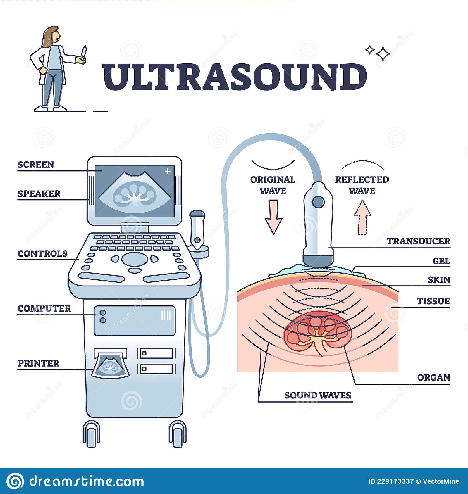
Ultrasound physics
A pulse (sound wave) is sent, the echoes from the sound waves reflecting occurs. The longer the echo the farther away the tissue.
60
New cards
3D Ultrasound
Created by moving transducer, many 2D images acquired. A computer reconstructs the 3D images
61
New cards
Ultrasound pros
* Safe
* 3D
* Inexpensive
* Real time
* Nonionizing
* 3D
* Inexpensive
* Real time
* Nonionizing
62
New cards
Ultrasound cons
- limited spatial resolution
- need acoustic window
- need acoustic window
63
New cards
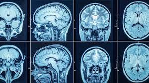
Magnetic Resonance Imaging (MRI)
Uses magnetic field and radio waves to take images of your body's interior, and can diagnose/investigate conditions that affect soft tissue, such as tumors or brain disorders.
64
New cards
MRI physics (what our body contains)
Since our bodies are made mostly of water, and water contains hydrogen photons, those photons act as small magnets.
65
New cards
MRI physics (magnetic fields)
- Normally, magnetic fields point in all directions= no net magnetization
- Placed in a magnetic field, more protons align with field= net magnetization
- Placed in a magnetic field, more protons align with field= net magnetization
66
New cards
MRI physics (Larmour Frequency)
- Net magnetization
- Protons precess about main magnetic field at Larmour frequency
-Larmour frequency depends on material and magnetic field strength
- Protons precess about main magnetic field at Larmour frequency
-Larmour frequency depends on material and magnetic field strength
67
New cards
MRI physics (MRI image created)
* Radio frequency (RF) pulse applied to magnetized tissue, tuned to Larmour frequency
* Larmour frequency- specific range where the radio frequency does not disrupt living tissue (much lower than X-ray)
* Radio frequency pulse causes protons to RESONATE
* When RF pulse is turned off, protons return to equilibrium
\-Protons emit an RF pulse
* Image created by manipulating magnetic field and RF pulses
* Many different tissue contrasts possible
68
New cards
MRI Physics (2D image)
-Created by a proton spin frequency depends on the magnetic field
- Apply a varying magnetic field across the patient
- Frequency encodes location
- Fourier Transform used to decompose signal into frequencies
- Apply a varying magnetic field across the patient
- Frequency encodes location
- Fourier Transform used to decompose signal into frequencies
69
New cards
MRI applications
- Multiple Sclerosis
- Knee Imaging ACL
- MRI of the Spine
- Knee Imaging ACL
- MRI of the Spine
70
New cards
MRI pros
- Great soft tissue contrast
- Nonionizing radiation (safe)
- Limitless contrast possibilities
- Nonionizing radiation (safe)
- Limitless contrast possibilities
71
New cards
MRI cons
- Slow
- Can't image people with magnetic materials
- Claustrophobic
- Loud
- Can't image people with magnetic materials
- Claustrophobic
- Loud
72
New cards
mm to pixel
0.127 mm in one pixel
73
New cards
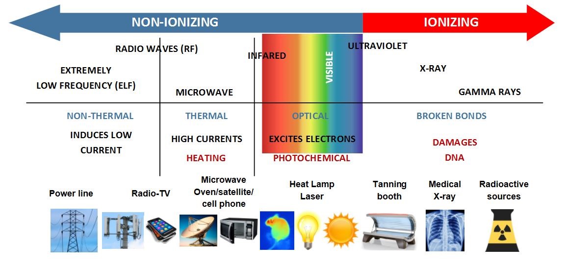
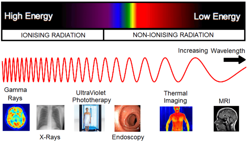
Ionizing radiation on electromagnetic spectrum
Uses more energy (more harmful)
74
New cards
Non-ionizing radiation on electromagnetic spectrum
uses less energy (less harmful)

75
New cards
Phantom definition
model used in the place of human anatomy
76
New cards
How are phantoms used?
used rather than testing a protoype on a human body
77
New cards
What are the 3 kinds of phantoms?
spatial resolution, contrast detail, anthropomorphic
78
New cards
Where are X-rays on electromagnetic spectrum
Towards the left end, high energy, decreasing wavelength
79
New cards
Where are Gammas (PET) on electromagnetic spectrum?
All the way to the left

80
New cards
Where is the MRI on the electromagnetic spectrum?
towards the right side (radio waves)

81
New cards
Compare and Contrast X-ray and CT
X-ray (ONLY)- 2d, inexpensive, used primarily to see bones and scan for pneumonia and cancers.
\
CT (ONLY)- full 3d projection, more radiation, used primarily to diagnose diseases in soft tissues and organs
\
BOTH- ionizing radiation \[X-rays\], high spatial resolution, fast scans
\
CT (ONLY)- full 3d projection, more radiation, used primarily to diagnose diseases in soft tissues and organs
\
BOTH- ionizing radiation \[X-rays\], high spatial resolution, fast scans
82
New cards
Why is it important for MRI systems that our bodies are mostly made of water?
Each water molecule contains two hydrogen protons that act as magnets. MRI generates a magnetic field that aligns the protons in water. The protons resonate when radiofrequency pulse is applied. The MRI manipulates magnetic field and RD pulse to create images.
83
New cards
During which phase of the design process do you identify the scope of the problem?
Problem identification
84
New cards
What are other stages of the design process?
- identification of customer needs
- establishment of target specifications
-generation of concepts
-construction of protoype
-design validation and verification
- establishment of target specifications
-generation of concepts
-construction of protoype
-design validation and verification
85
New cards
What is the term for a method for adding contrast to images by only allowing grayscale values within a certain range in?
__Windowing__
* grayscale pixel values outside the window are displayed as black or white
* narrower windows= starker contrasts
* grayscale pixel values outside the window are displayed as black or white
* narrower windows= starker contrasts
86
New cards
2D Fourier transform: image to frequency
- If the dots are closer together, then the lines are in bigger chunks (there are less lines)
- If the dots are farther apart, then the lines are in smaller chunks and there are more lines.
-The lines are perpendicular to the dots
- If the dots are farther apart, then the lines are in smaller chunks and there are more lines.
-The lines are perpendicular to the dots
87
New cards
Filtering images- salt and pepper noise
Equation: sum of pixels/number of pixels
88
New cards
What are the four required sections of a student resume?
1)personal/contact information
2)education
3)experience
4)activities/interests
2)education
3)experience
4)activities/interests
89
New cards
Median Filtering
Removes outlier values - artifacts
90
New cards
How to filter using the median
- Sort the pixels in the neighborhood from lowest to highest
- The value at the pixel of interest is REPLACED by the MEDIAN value in the neighborhood
-This removes the artifacts
- The value at the pixel of interest is REPLACED by the MEDIAN value in the neighborhood
-This removes the artifacts
91
New cards
Filtering
\- Way to remove unwanted info
\- Preserves integrity of underlying image
\- Works differently for different types of noise
\- Has many forms
\- Preserves integrity of underlying image
\- Works differently for different types of noise
\- Has many forms
92
New cards
Difference in filter design. Can one remove all types of noise?
In image processing lab the filter only worked on salt and pepper noise and did not work for right-left streak noise.
93
New cards
Pseudocode
Artificial and informal language that helps programmers develop algorithms (fake code, text-based)
94
New cards
Customer needs
1. phantom models knee joint anatomy (femur and tibia) and soft tissue
2. Materials used resemble bone and tissue
3. Max JSW will model a healthy person, min JSW will model a person with cartilage degradation
4. JSW must be adjustable
5. Represents bone and tissue size of average to large middle ages male
6. Comprised of affordable and available products
7. Must stand unassisted
\
95
New cards
Specifications for Phantom
Target specifications - converts customer needs into quantitative and measurable specification
1. minimum joint space width - 0mm
2. maximum joint space width - 1cm.
3. diameter of femur and tibia bone at joint - 5-10cm
4. material density is equivalent to bone - 2 g/cm^3
5. material density is equivalent to soft tissue - 1.2 g/cm^3
6. total phantom height- 15-30cm
7. Width or diameter of soft tissue - 12-18cm
8. Free standing
9. Common/non-toxic materials
10. Max budget - $25
96
New cards
window width/level settings for phantom
* increase/decrease brightness
* Create contrast for bone and soft tissue
* Create contrast for bone and soft tissue
97
New cards
Concept generation of the phantom
* Each person generated their own ideas (no comparison)
* Share with group and come up with new ideas
* Choose three ideas
* compare and contrast the candidates to determine the final design concept (technical feasibility, economic feasibility, usability constraints)
\
* Share with group and come up with new ideas
* Choose three ideas
* compare and contrast the candidates to determine the final design concept (technical feasibility, economic feasibility, usability constraints)
\
98
New cards
X-ray images reconstruction
The x-ray will not pass through dense materials (bone) = shows up white
\
The x-ray will pass through less dense material (tissue)= shows up darker
\
The x-ray will pass through less dense material (tissue)= shows up darker
99
New cards
2D space patterns
* frequency space is symmetric
* spatial frequency increases the further from the center of 2D Fourier space
* vertical axis results in streaks that appear in the “up-down” direction
* horizontal axis results in “left-right” streaks
* changing the angle in frequency space changes the orientation
* spatial frequency increases the further from the center of 2D Fourier space
* vertical axis results in streaks that appear in the “up-down” direction
* horizontal axis results in “left-right” streaks
* changing the angle in frequency space changes the orientation