Procedures 1: Chapter 1 PPT Diagram | Quizlet
 Call Kai
 Learn
Learn Practice Test
 Spaced Repetition
 Match
Match Flashcards
 Knowt Play
1/148

There's no tags or description
Looks like no tags are added yet.
Name | Mastery | Learn | Test | Matching | Spaced | Call with Kai | Chat |
|---|
No analytics yet
Send a link to your students to track their progress
149 Terms
Osteology
the study of bones
Anthrology
the study of joints
206
the number of separate bones in the adult skeletal system
Sesamoid Bones
small, round/oval-shaped bones embedded in certain tendons, mostly found near joints. NOT counted as part of the normal axial or appendicular skeleton, except for the patellae (the largest of this kind of bone)

Bone shape classifications
long, short, flat, irregular
Long Bones
consist of a body (shaft) and two ends or extremities; supports weight and movement; femur, tibia, humerus, radius

Short Bones
found only in wrists and ankles; roughly cuboidal (cubic in shape); nearly equal in length, width, and thickness; ex: 8 carpal bones in a wrist & 7 tarsal bones in a foot

Flat Bones
thin, flat, and often curved bones; provide protection for interior contents and broad surfaces for muscle attachment; ex- skull bones, sternum, ribs, & scapulae

Irregular Bones
any bone with a peculiar shape; ex- vertebrae, facial bones, bones of the base of the cranium, pelvis bones; protects internal organs, give support, muscle attachment

Classification of Joints
synarthrosis, amphiarthrosis, diarthrosis
Synarthrosis
immovable joint; stationary (skull)

Amphiarthrosis
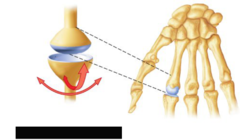
joint with limited movement; ex: pubic sythesis

Diarthrosis
freely moveable joint (think Dancing)
(eg, elbow, knee, fingers, etc)
Body Habitus
the build, physique, and general shape of the human body
Classifications of Body Habitus
sthenic, hyposthenic, hypersthenic, asthenic
Sthenic
body shape considered average in shape and internal organ location; approximately 50% of the population

Hyposthenic
thin body style; approximately 35% of the population

Asthenic
very thin or slender patient with long and narrow body build; approxiamately 10% of the population

Hypersthenic
massive body style with a large and broad frame; approximately 5% of the population: "the hulk" appearance

Radiography
the process and procedures of producing a radiograph to help doctors diagnose and treat a variation of medical conditions
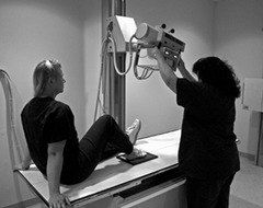
Radiograph
an image of a patient's anatomic part(s) produced by the action of x-rays on an image receptor (bones absorb more x-rays appearing white)
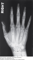
Image Receptor (IR)
the device that captures the radiographic image that exits the patient

Central Ray (CR)
refers to the centermost portion of the x-ray beam emitted from the x-ray tube; "bull's eye" of the x-ray beam

Anatomic position
an upright position with arms abducted slightly (down), hands by side with palms forward, and head and feet together and directed straight ahead

4 Common Planes in Medical Imaging
sagittal, coronal, horizontal (axial), oblique
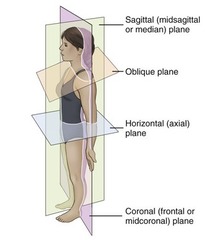
Plane
a straight line surface connecting 2 points
Sagittal Plane
any longitudinal plane that divides the body into right and left parts; parallel to the midsagittal/median plane
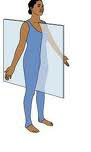
Midsagittal/Median Plane
a midline sagittal plane that divides the body into equal left and right parts
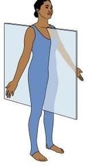
Coronal Plane
any longitudinal plane that divides the body into anterior and posterior parts' passes through the coronal suture of the skull; any plane parallel to the midcoronal or frontal plane

Midcoronal Plane
Divides the body into equal anterior and posterior parts
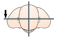
Horizontal (axial) Plane
any transverse plane that passes through the body at right angles to a longitudinal plane; divides the body into superior and inferior portions

Oblique Plane
a longitudinal or transverse plane that is at an angle or a slant; NOT parallel to the sagittal, coronal, or horizontal plane

Posterior/Dorsal
refers to the back half of the patient, or the part of the body seen when the person is viewed from the back (includes the bottoms of feet & the backs of hands when in the anatomic position)

Anterior/Ventral
refers to the front half of the patient, or the part seen when viewed from the front (includes the tops of the feet and the palms of the hands when in the anatomic position)

Plantar
refers to the sole or posterior surface of the foot
Palmer
refers to the palm of the hand; in the anatomic position, the same as the anterior or ventral surface of the hand
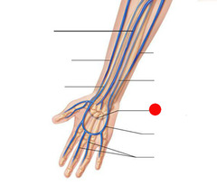
Dorsal (foot)
foot: refers to the top or anterior surface (dorsum pedis); hand: refers to the back or posterior aspect of the hand (dorsum manus)
Radiographic Projections
the direction or path of the CR of the x-ray beam as it passes through the patient, projecting and image onto the image receptor (IR)
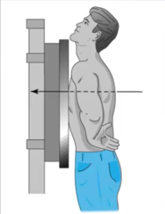
Posteroanterior (PA) Projection
refers to a projection of the CR from posterior to anterior; the CR enters at the posterior surface and exits the anterior surface
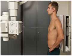
Anteroposterior (AP) Projection
projection of CR from anterior to posterior; the CR enters at an anterior surface and exits at a posterior surface
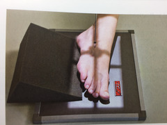
AP Oblique Projection
A projection of a body part that is rotated, shot anterior to posterior (should also contain a qualifying "medial" or "lateral" term); CR enters the anterior surface and exits the posterior surface of the body part
PA Oblique Projection
A projection of a body part that is rotated, shot posterior to anterior (should also contain a qualifying "medial" or "lateral" term); CR enters the posterior surface and exits the anterior surface of the body part

Mediolateral Projection
CR enters the medial aspect and exits the lateral aspect

Lateromedial Projection
CR enters the lateral aspect and exits the medial aspect

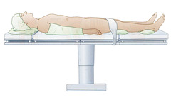
Supine
patient lying on back and facing upward
Prone
patient lying on abdomen and facing downward; *head may be turned to one side

Erect
patient in an upright position; to stand or sit erect

Recumbent
lying down in any position (prone, supine, or on side)
Dorsal Recumbent
lying on back (supine)

Ventral Recumbent
lying face down (prone)

Lateral Recumbent
lying on side (right or left lateral)
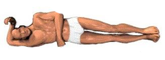
Trendelenburg
a recumbent position with the body tilted with the head lower than the feet; *position is sometimes beneficial if your patient is experiencing dizziness

Fowler
recumbent position with the body tilted with the head higher than the feet
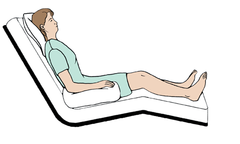
Sims
recumbent oblique position with the patient lying on the left anterior side, with the right knee and thigh flexed and the left arm extended down behind the back (semiprone position); typical position for barium enema

Lithotomy
a recumbent (supine) position with knees and hip flexed and thighs abducted and rotated externally, supported by ankle supports; *this position is seen frequently in the surgical suite for certain urinary studies
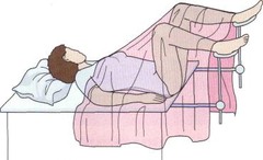
Lateral Position
the body part closest to the IR or the body part from which the CR exits; always 90 degrees or perpendicular to a true AP or PA projection

Decubitus (Decub) Position
patient laying down; always performed with the CR horizontal; body position is designated according to the surface on which the patient is lying; patient lying on one of the following body surfaces- back (dorsal), front (ventral), or side (right or left lateral)
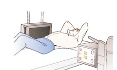
Oblique Position
refers to an angled position; positions of the thorax, abdomen, or pelvis are described by the body part closest to the IR or the body part from which the CR exits; NOT 90 degrees or perpendicular
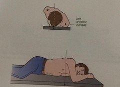
Left & Right Posterior Oblique Positions (LPO & RPO)
the specific oblique positions in which the left or right posterior aspect of the body is the closest to the IR
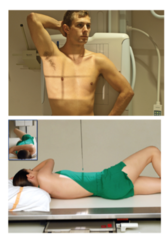
Left & Right Anterior Oblique Positions (LAO & RAO)
positions in which the left or right anterior aspect of the body is closest to the IR and can be erect or recumbent general body positions

Axial Projection
any angle of the CR of 10 degrees or more along the long axis of the body or body part
Inferosuperior Axial Projection
Central ray enters inferiorly and exits superiorly

Superoinferior Axial Projection
CR enters above or superiorly and exits below or inferiorly

Tangential Projection
touching a curve or surface at only one point; the CR skims a body part to project the anatomy into profile free of superimposition of surrounding body structures
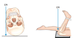
Medial
Toward the midline of the body
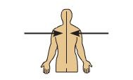
Lateral
Away from the midline of the body

Proximal
Closer to the point of attachment

Distal
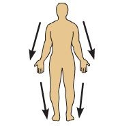
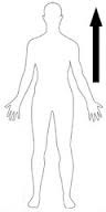
Cephalad
toward the head (superior)
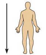
Caudad
away from the head (inferior)
Cephalad Angle
any angle toward the head
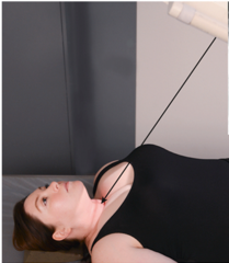
Caudad Angle
any angle away from the head

Flexion
Decreases the angle of a joint
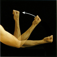
Extension
increases the angle of a joint

hyperextension
extension of a joint beyond anatomical position

Dorsiflexion of foot
To decrease the angle (flex) between the dorsum (top of foot) and the lower leg, moving foot and toes upward

Plantar Flexion of Foot
Extending the ankle joint, moving foot and toes downward from the normal position; flexing or decreasing the angle toward the posterior surface

Eversion
an outward stress movement of the foot at the ankle joint

Inversion
an inward stress movement of the foot as applied to the foot without rotation of the leg

Valgus Stress
describes an abnormal position in which a part or limb is forced outward from the midline of the body

Varus Stress
describes an abnormal position in which a part or limb is forced inward toward the midline of the body

Medial Rotation
rotation or turning of a body part with movement of the anterior aspect of the part toward the inside/median plane

Lateral Rotation
a rotation of an anterior body part toward the outside, or away from the median plane

Abduction
the lateral movement of the arm or leg away from the body

Adduction
a movement of arm or leg toward the body, draw toward a center or medial line

Supination
a rotational movement of the hand into the anatomic position (palm up in supine position or forward in erect position); "SUPINE"

Pronation
a rotation of the hand into the opposite of the anatomic position (palm down or back); "PRONE"

Rotate
to turn or rotate a body part on its axis
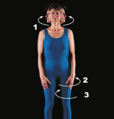
Tilt
a slanting or tilting movement with respect to the long axis
Position
used to indicate the patient's general physical position, such as supine, prone, recumbent, or erect; also described by the body part closest to the IR
Projection
a correct positioning term that describes or refers to the path or direction of the CR, projecting an image onto an IR
Types of markers on every radiographic image
1. patient identification & date
2. Anatomic side markers
Anatomic Side Marker
must be placed prior to exposure to indicate the patient's left or right side or which limb is being radiographed; need to be placed on the IR inside the lateral portion of the collimated border of the part being x-rayed; marking after image processing is considered unacceptable practice

1st General Rule for Determining Positioning Routines
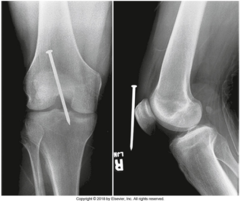
Minimum of 2 projections taken as near to 90 degrees from each other
Exceptions due to only one projection providing adequate information: AP portable chest, single AP abdomen (KUB), and AP pelvis
Reasons for minimum of two projections (3)
1. superimposition of anatomic structures
2. localization of lesions or foreign bodies
3. determination of alignment of fractures
2nd General Rule for Determining Positioning Routines
All radiographic procedures of the skeletal system involving joints require a minimum of 3 projections (AP/PA, Lateral, & Oblique)
Reasoning: there are multiple surfaces and angles of the bones making up the joint, and a small oblique chip fracture or other abnormality within the joint space may not be visual on the AP/PA & lateral views, but may be seen on the oblique view
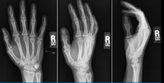
exams that generally requires three projections as routine
fingers
toes
hand
wrist
elbow
ankle
foot
knee
exams that generally require two projections as routine
forearm
humerus
femur
hips
tibia-fibula
chest
Topographic (Bony) Landmarks
found on a patient through palpatation; important for the technologist to be able to position the body parts correctly

Palpation
process of applying light pressure with the fingertips directly on the patient to locate positioning landmarks; always inform the patient of the purpose of palpitations and gain permission
