Lab 8: The Respiratory System
 Call Kai
 Learn
Learn Practice Test
 Spaced Repetition
 Match
Match Flashcards
 Knowt Play
1/76

There's no tags or description
Looks like no tags are added yet.
Name | Mastery | Learn | Test | Matching | Spaced | Call with Kai | Chat |
|---|
No analytics yet
Send a link to your students to track their progress
77 Terms
sequence of inhalation
inspiratory muscles contract (diaphragm descends; rib cage rises)
thoracic cavity volume increases
lungs are stretched; intrapulmonary volume increases
intrapulmonary pressure drops
air flows into lungs down its pressure gradients until intrapulmonary pressure is 0 (= to atmospheric pressure)
during inhalation (ribs + sternum)
ribs are elevated and sternum flares as external intercostals contract
during inspiration (diaphragm)
diaphragm moves inferiorly during contraction
during inspiration (external intercostals)
external intercostals contract
pressure and volume relationship
pressure is inversely proportional to volume
bucket handle effect
ribs and sternum elevate due to contraction of external intercostal muscles
increases both width and depth of thoracic cavity
effect is to increase volume of thoracic cavity
inhalation is…
active
exhalation is…
passive, but can become active during exercise
sequence of events in exhalation
inspiratory muscles relax (diaphragm rises; rib cage descends due to recoil of costal cartilages)
thoracic cavity volume decreases
elastic lungs recoil passively; intrapulmonary volume decreases
intrapulmonary pressure rises
air flows out of lungs down its pressure gradient until intrapulmonary pressure is 0
during exhalation (ribs + sternum)
ribs and sternum are depressed as external intercostals relax
during exhalation (diaphragm)
diaphragm moves superiorly as it relaxes
during exhalation (external intercostals)
external intercostals relax
exhalation during exercise
becomes active; abdominal muscles and internal intercostals contract and force air out of lungs
bainbridge effect
during inhalation, thoracic pressure decreases (because volume increases)
venous blood return to heart increases as abdominal pressure is greater than thoracic pressure
pressure in right atria increases due to more blood
atrial stretch receptors signal this to SA node and medulla oblongata
increased efferent sympathetic signals to SA node increase HR during inhalation
tidal volume (TV)
amount of air inspired during normal released breathing
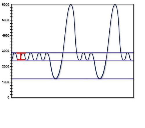
Inspiratory Reserve Volume (IRV)
the additional air that can be forcibly inhaled after the inspiration of a normal tidal volume

Expiratory Reserve Volume (ERV)
the additional air that can be forcibly exhaled after the expiration of a normal tidal volume
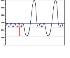
Residual Volume (RV)
the volume of air still remaining in the lungs after the expiratory reserve volume is exhaled
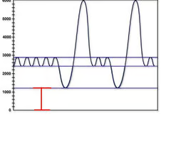
Total Lung Capacity (TLC)
the maximum amount of air that can fill the lungs
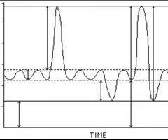
TLC equation
TV+IRV+ERV+RV
Vital Capacity (VC)
the total amount of air that can be expired after fully inhaling
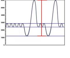
VC equation
TV+IRV+ERV
basic respiratory waveforms =
volumes
respiratory capacities =
sum of 2 or more respiratory volumes
functional residual capacity (FRC)
the amount of air remaining in the lungs after a normal expiration

FRC equation
RV + ERV
inspiratory capacity (IC)
the maximum amount of air that can be inspired
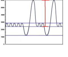
IC equation
TV + IRV
minute ventilation
total volume of gas that flows into our out of respiratory tract in 1 minute to give you a flow rate
normal MVV at rest
6 L/min eupnea
eupnea
normal breathing
normal MVV with exercise
up to 200 L/min
cellular respiration
produces CO2
bicarbonate buffering system characteristics
occurs in RBCs
maintains blood pH level
enzyme carbonic anhydrase
bicarbonate buffering system—pH too high
HCO3- is created in systemic capillaries
HCO3- diffuses from RBCs into plasma
HCO3- combines with H+ to form H2CO3
bicarbonate buffering system—pH too low
H2CO3 dissociates to release H+
hyperventilation
increased depth and rate of breathing that exceeds body’s need to remove CO2
effects of hyperventilation
hypocapnia + alkalosis
cerebral vasoconstriction + ischemia
hypocapnia
decreased blood CO2 levels
alkalosis
increased blood plasma pH
treatment for hyperventilation
breathing into paper bag increases CO2 levels
why does hyperventilating not increase O2 levels?
O2 levels do not change much as RBCs are saturated
composition of air inhaled
21% O2
0.04% CO2
78-79% N2
Other gases (argon, etc) less than 1%
composition of air exhaled
16% O2
4-5% CO2
78-79% N2
Other gases (argon, etc) less than 1%
hypoventilation
decreased depth and rate of breathing that does not meet the body’s need to remove CO2
effects of hypoventilation
hypercapnia
acidosis
hypoxia
stimulate respiratory control centers
hypercapnia
increased blood CO2
acidosis
decrease in blood plasma pH
hypoxia
drop in O2 levels
increase in H+, CO2, and/or big decrease in O2…
stimulate central and peripheral chemoreceptors that send afferent signals to DRG to increase minute ventilation
decrease in H+, CO2, and/or increase in O2…
silences chemoreceptors. DRG influence on VRG is lessened and minute ventilation decreases
pontine respiratory centers
interact with the medulla respiratory centers to smooth the respiratory pattern
ventral respiratory group (VRG)
contains rhythm generators whose output drives respiration
dorsal respiratory group (DRG)
integrates peripheral sensory input and modifies the rhythms generated by the VRGs
phrenic nerves (C3, C4, C5)
innervates the diaphragm
spirometry/forced expiratory volume tests distinguish between…
obstructive pulmonary diseases
restrictive lung diseases
obstructive pulmonary diseases
increased airway resistance
traps air
asthma, bronchitis
FRC and RV increase due to hyperinflation of lungs
normal overall volume
reduced flow rate
restrictive lung diseases
reduced total lung capacity
due to disease (tuberculosis), posture (kyphosis), or damage from lung toxins (fibrosis)
thoracic cavity small or lungs unable to expand
VC, TLC, FRC, RV decline
reduced volume
normal or increased flow rate
diameter of airways affects…
flow rates, not the volume of air you can fit in your lungs
pulmonary ventilation
air is inhaled through the nasal and oral cavities
air moves through pharynx, larynx, and trachea into lungs
then it is exhaled, flowing back through the same pathway
internal respiration
bloodstream delivers O2 to cells and removes CO2
respiratory system anatomy

nasal cavity
composed of the chambers of the internal nose that are apart of upper respiratory system

laryngopharynx
most posterior part of pharynx
shared by respiratory + digestive systems
front merges with larynx

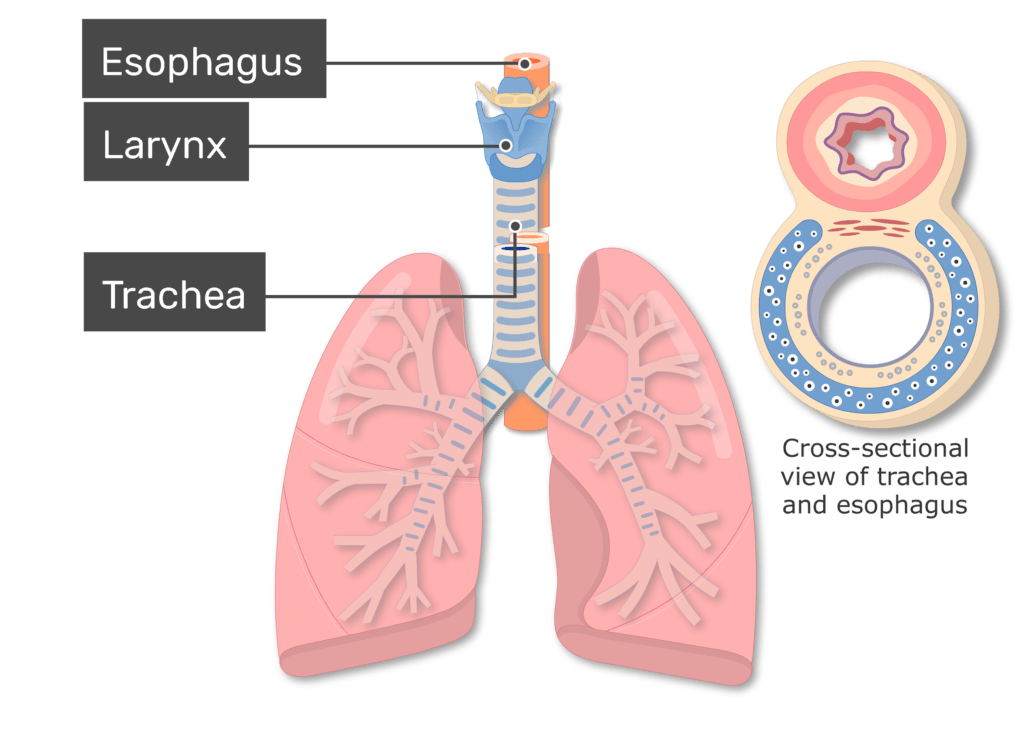
trachea
conveys air between upper and lower respiratory structures
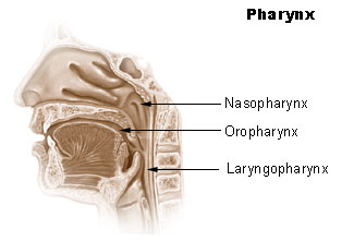
nasopharynx
airway of upper respiratory system
always open
lungs
two organs responsible for gas exchange
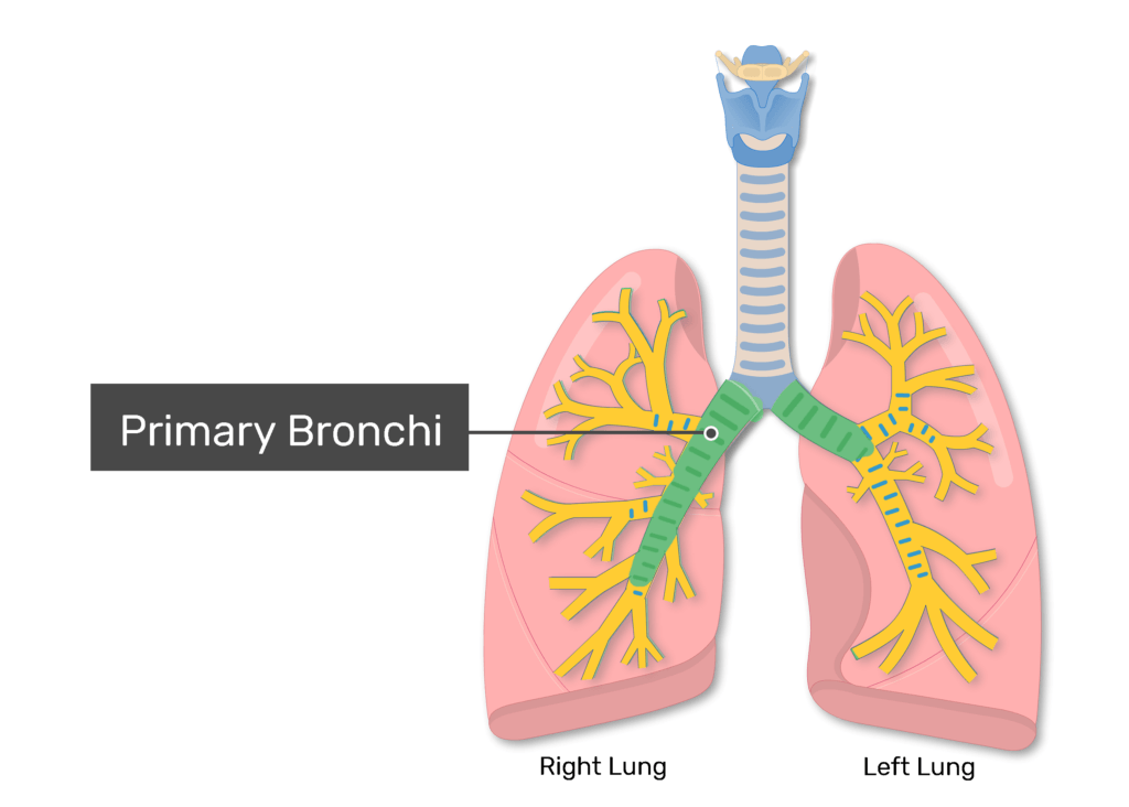
primary bronchi
major airways of the lower respiratory system

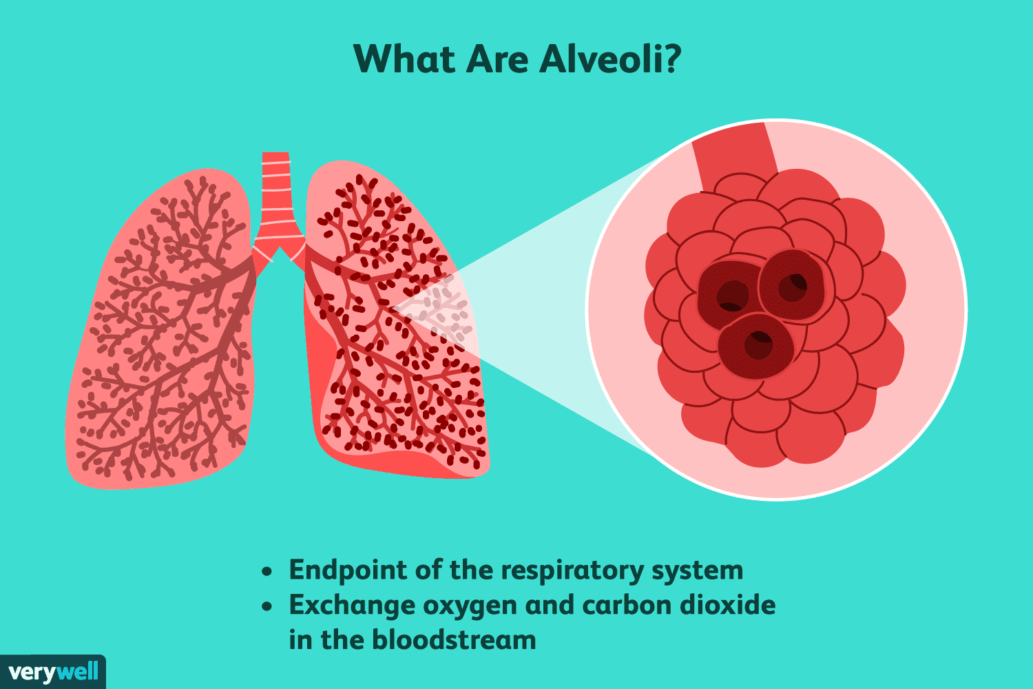
alveoli
major sites of gas exchange, where O2 is brought into the bloodstream and CO2 is removed
oropharynx
shared by respiratory + digestive
airway in upper respiratory

bronchi
major airways in lower respiratory system
link trachea with R and L lungs
wrapped in rings of hyaline cartilage
interiors lined with mucous membrane
minute ventilation equation
tidal volume x respiratory rate
forced expiratory volume
measures the max amount of air you can forcefully exhale in the 1st second of a full breath
tidal volume and exercise
increases significantly
reserve volume (IRV and ERV) and exercise
decrease
vital capacity and exercise
stays the same
which lung volume remains in the lungs after someone has exhaled all the air they can exhale?
residual volume