Mycology Introduction
1/31
There's no tags or description
Looks like no tags are added yet.
Name | Mastery | Learn | Test | Matching | Spaced |
|---|
No study sessions yet.
32 Terms
Fungi exist as …
Molds, Yeasts, or Dimorphic fungi
Mold vs Yeast
Mold =
Multicellular
Sexual or Asexual reproduction (spores / conidia)
Hyphae → Fuzzy / Wooly colonies
Various colors
Yeast =
Single-celled
Asexual reproduction = Budding (blastoconidia)
Pseudohyphae (formed by continous budding) or No hyphae
White, smooth, “bacteria-like” colonies
Dimorphic fungi =
Fungi that has a yeast + mold phase
Grows as Yeast at Body temp. (37°C in vitro + in vivo)
Grows as Mold at Room temp. (22-30°C in vitro)
most common yeast isolated from blood cultures =
Candida spp.
if testing is delayed, how do you store CSF?
keep at Room Temp.
how to process nail vs skin specimen
Nails : dissolve w/ 20% KOH + may need to use heat
Skin : break down w/ 10% KOH
Why is it important to know how a urine specimen was collected for culture?
Clean Catch : some normal flora contamination is normal ; >100,000 CFU/mL is considered a UTI
Catheter : NO normal flora contamination should occur ; any growth is a concern
purpose of using KOH
KOH dissolves human cells lacking keratin cell walls (hair, nails, skin)
leaves any fungus to be viewed
Calcofluor White stain
Fluorescent blue stain → binds to Fungi cell walls
causes fungal elements to fluoresce under fluorescent scope
(used w/ KOH)
India ink is used on what specimen to identify what fungus ?
India ink stains Cryptococcus neoformans in CSF
C. neoformans has visible “Halo” = thick capsule lacks india ink surrounding fungi
what stain is used for Fungal tease or cellophane mounts?
Lactophenol aniline blue
Most common media used to culutre fungi =
Sabouraud dextrose agar (SDA)
Sabouraud Dextrose Agar (SDA / SAB) media
Non-selective media for Fungi cultures
acidic pH inhibits bacteria growth
incubation temperature for Fungal cultures
30°C
Rugose vs Verrucose colonies
Rugose = wrinkles, corrugated
Verrucose = rough elevations
Umbonate colony appearance =
“Round knob” above media
Mycelium =
network of hyphae (fungal threads)
Aerial vs Vegetative Hyphae
Aerial Hyphae =
Reproductive (produce conidia)
Above media surface
Vegetative Hyphae =
Inside media → Absorbs nutrients inside agar

What is the name for this hyphae structure?
Racquet hyphae = enlarged, club-shaped
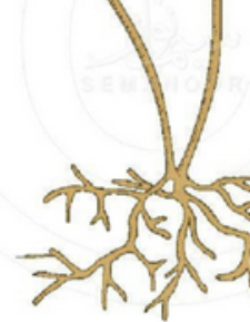
What is the name for this hyphae structure at the bottom?
Rhizoid hyphae = “Root-like” hyphae that grow on stolons
Hyaline vs Dematiaceous hyphae
Hyaline = BLUE stained (Nonpigmented hyphae)
Dematiaceous = BROWN stained (dark pigmented due to melanin in cell wall)
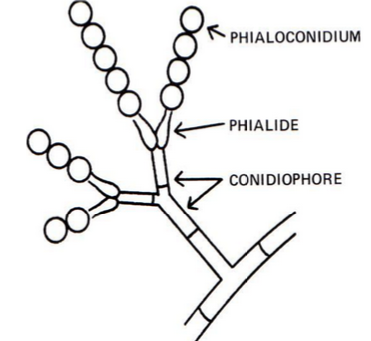
Conidiogenous cells =
Conidiogenous cells are attached to Conidiophore (extension of hyphae)
Conidiogenous cells produce Conidia (spores)
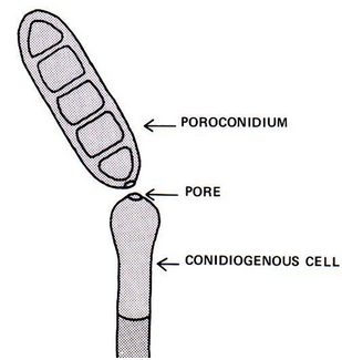
Phialides =
“Vase-like” structures → attached to conidiophores ; produce phialoconidia
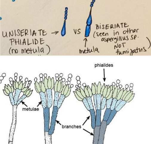
Uniseriate vs Biseriate phialides
Uniseriate = One row of phialides
Biseriate = Two rows of phialides
bottom row = Metulae
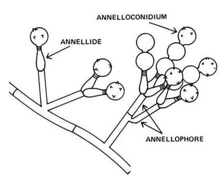
Annellides =
similar to phialides, but have rings around tip (where annelloconidia forms)
annelloconidium can become spiny (echinulate) w/ age
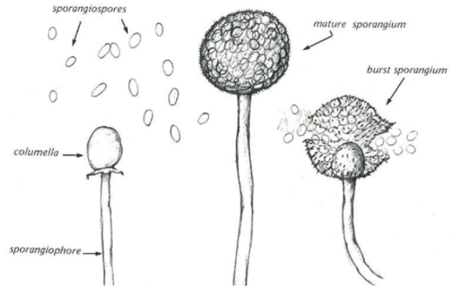
Sporangium
Sac-like cell containing spores
form on Sporangiophores + produce Sporagniospores
Columella
Structure that spores attach to inside sporangium
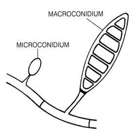
Micro- vs Macroconidia
Microconidia = small, unicellular spores
Macroconidia = larger, multi-septate spores
Arthroconidia
spores formed by fertile Hyphae Fragmentation
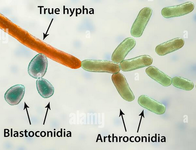
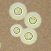
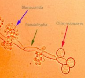
Blastoconidia vs Chlamydoconidia
both = asexual reproduction of YEAST
Blastoconidia =
spores produced by budding
develop in clusters along hyphae
Chlamydoconidia =
thick-walled survival spores forming under certain growth conditions
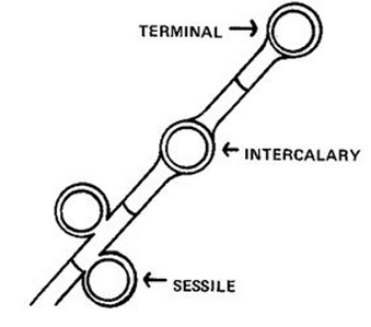
Terminal, Sessile, Intercalary chlamydospores form at tips, side, or inside hyphae
Differentiate the types of Chlamydocondia → Terminal, Sessile, Intercalary
based on location in relation to Hyphae
Terminal = tip
Intercalary = inside
Sessile = on the side
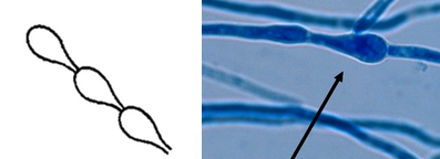
Pseuodhyphae =
structure of YEAST
yeast budding continues w/out conidia separation → forms long filament of attached blastoconidia
shows constrictions between cells (TRUE hyphae = NO constrictions w/ parallel sides)
