Frozens and Cytology EX 1
1/49
There's no tags or description
Looks like no tags are added yet.
Name | Mastery | Learn | Test | Matching | Spaced | Call with Kai |
|---|
No analytics yet
Send a link to your students to track their progress
50 Terms
Most tissue temp
-13 to -20
Fatty and softer tissue (breast, skin, pancreas) temp
-25 to -30
Dryer/harder tissue (brain, liver, spleen, lymph node, bloody tissues) or fixed tissue temp.
-5 to -15
Peltier element
-60 C
Rapid freezing
If anti-roll plate angle is below…
sections curls on edge of knife
If anti-roll plate angle is above…
the tissue hits the plate on downstroke
Freeze artifact
Caused by slow freezing
Ice crystals become holes in tissue
Common in brain, lung, kidney, bowel
Liquid Nitrogen artifact
Liquid nitrogen vapor bubbles
Best method for freezing
Isopentane chilled in liquid N
Slowest freezing method
Cryostat freezing and causes most freeze artifacts
Freeze spray
Freezes tissue but easily produces freeze artifact.
Will also aerosolize infectious tissue
Frozen storage
Short term - in cryostat or regular freezer
Extended - store in -80C freezer.
Wrap in foil
Difficulty getting a section
Alter temp
Sharpen/change blade
Rotate spec (shortest path is usually best)
Tighten/oil/clean components
Section shreds causes
Dull knife
Wrong temp
Tissue has hard components (calcifications, etc.)
Compression
Blade too warm
Blade holder or knife surface dirty
Incorrect knife angle
Dull blade
Thick/Thin section causes
Loose blade/specimen chuck
Dull Blade
Wrong temp
Incomplete section causes
Insufficient facing
Tissue depletion
Improper orientation
Varying tissue densities (different optimal temps)
Tissue chips out causes
Aggressive cutting
Hard tissue
Loose cryostat
Tissue or OCT is too cold (especially when freezing in isopentane)
Sections not laying flat causes
Anti-roll plate is not positioned correctly/dirty/marred
Frost on blade
Static
Mohs advantages
Preserves tissue, minimizes re-growth of tumor
Less scarring
Reliable and accurate
Mohs disadvantages
Time consuming
Costly
Diagnostic cytology
Assessment of morphological details and chromatin patterns
Determines if cells are normal/abnormal
Nucleus:Cytoplasm ratio
Liquid based cytology
GYN spec received in preservative or fixative
Cell block
Aggregate cells processed histologically
And/or when IHC needed
GYN Spec
Female genital spec.
PAP smears (slide smears and liquid brushings)
Non-GYN specs
Body fluids, discharges, CNS fluid, washings/brushings, urine, FNAs, Cyst aspirates
Fixation of choice in Cytology
95% ETOH
Will dehydrate and sharpen nuclear membrane.
Methanol as a fixative
100% is used as alt to 95% ETOH
80% isopropanol as a fixative
Shrinks cells more than 95% ETOH
Cytolyt as a fixative
Methanol based, less shrinkage than ETOH
Commercial spray fixatives
Alcohol - fix
Acetone - dry
Polyethylene - protective coating (remove w/alcohol before staining)
Saccomanno Fluid as a pre-fixative for transportation
50% ETOH + Carbowax
Alcoholic saline as a pre-fixative for transportation
Equal Saline + 50% ETOH
Commercial transport fluid as a pre-fixative for transportation
Alcoholic based
Pull apart method - Prep smear
Preferred, nice monolayer, cells will not dry out
Cross Hatch - Preparing smear
Use loop to smear specimen on slide
Crush - Preparing smear
Used for mucoid spec
For sparsely cellular specimens…
Centrifuge to separate cells (button) and supernatant
For bloody specimens…
Lyse RBCs w/commercial lysing agent or with Carnoy’s acetic alcohol solution
For Cell adhesion to slide…
Spray adhesives, but not needed with pull-apart method
To remove excess mucoid…
Use a mucolytic agent.
DTT (Dithiotheritol) → reduces protein disulfide bonds
Cell Block Preparation steps
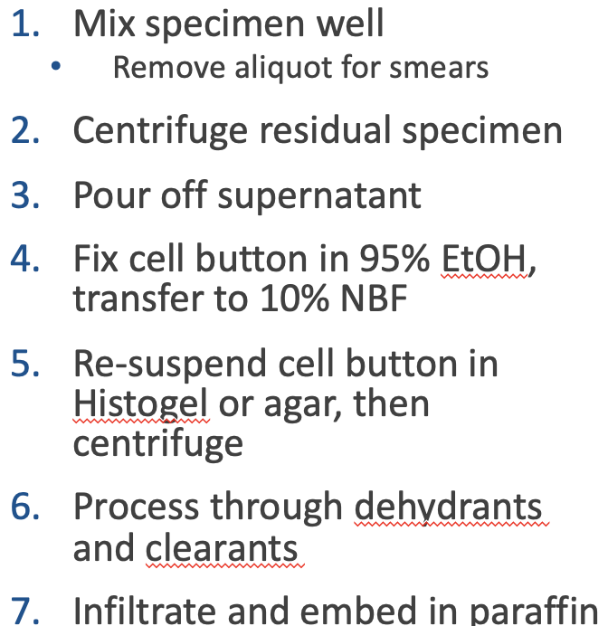
Centrifuge → Pour off supernatant → Fix button in 95% ethanol then 10% NBF → Embed in histogel/agar or infiltrate and embed w/paraffin
Why use cell blocks?
Diagnostic information - correlation w/smears
Special stains and IHC - Cell block spec can withstand harsh chemical contact better than smears
PAP Stain Reagents
Hematoxylin - nuclear
OG (Orange G) - Keratin, squam cells
EA (Light Green and Eosin) - Cytoplasmic components
Hematoxylin as a PAP Stain Reagent
Nuclear detail
OG (Orange G) as a PAP Stain Reagent
Keratin, squam cells
EA (Light Green and Eosin) as a PAP Stain Reagent
Cytoplasmic components
PAP Stain purpose
Visualize and differentiate cell components.
Provides nuclear and cytoplasmic morphology
More info about cell blocks…
A cell block is a method of preparing cytology material so that it can be processed, sectioned, stained and viewed as a histology section.
It can provide diagnostic information in addition to that obtained from cytology smears.
Also, it is easier to do special stains if needed, including IHC, on a cell block than on additional smears, because smears often require adaptations of the staining protocols and different controls.
More on cell block prep
A cell block is prepared with material remaining after the cytology smears have been made.
The quality or quantity of the smears should never be sacrificed to make a cell block.
The method chosen is dependent upon the characteristics of the specimen.
If a cytology specimen contains small fragments of tissue, the fragments should be carefully picked out and submitted as a cell block.
Clots or heavy mucus in specimens should be used to prepare cell blocks. Loose cells are difficult to contain, embed, and section and therefore make poor cell blocks.
A method should be used with all loose cellular material to “block” it together so that it can be handled “en masse”.