ECG
1/17
There's no tags or description
Looks like no tags are added yet.
Name | Mastery | Learn | Test | Matching | Spaced |
|---|
No study sessions yet.
18 Terms
V1
4th intercostal space, right sternal border —> ventricular septum
V2
4th intercostal space, left sternal border —> ventricular septum
V3
Between V2 and V4 —> anterior wall of left ventricle
V4
5th intercostal space, midclavicular line —> anterior of left ventricle
V5
Lateral to V4 at the anterior axillary line —> lateral wall of left ventricle
V6
Lateral to V5 at the midaxillary line —> lateral wall of left ventricle
Artifact
Tracing on an ECG that is the result of interference, such as patient movement, rather than the heart’s electrical activity
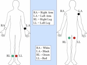
12-lead ECG electrode placement
RA electrode - right arm on the wrist
LA electrode - left arm on the wrist
LL electrode - left leg on the thigh/ankle (or lower left side of the abdomen)
RL electrode - right leg on the thigh/ankle (or right abdomen)
12-lead ECG tracing electrode placement
V1 - right side of the sternum, between 4th-5th ribs
V2 - left side of the sternum across from V1
V4 - between 5th-6th ribs in a straight line down from the middle of the clavicle
V3 - halfway between V2 and V4 in between the 5th-6th ribs
V6 - horizontally even with V4 in a straight line down from the middle of the armpit
V5 - halfway between V4 and V6
QRS complex
Largest, narrow deflections on the ECG. Represents one contraction of the ventricles. Closer they are —> faster HR
The ST segment connects the end of the QRS complex and the beginning of the_______.
T wave
Horizontal distance between each wave on the ECG represents_________.
Time passing, each small (1-mm) box represents 0.04 sec and each larger (5-mm) box represents 0.20 sec. QRS complex should be less than 3 mm wide
If 2 QRS complexes are <15 mm apart (3 large boxes), the HR is__________.
>100 bpm, tachycardia
If the QRS complexes are >25 mm apart (5 large boxes), the HR is_______.
<60 bpm, bradycardia
P wave
Small rounded wave before the QRS, represents contraction of the atria
T wave
After each QRS complex, represents the ventricles recharging
Baseline
Horizontal line between all of the waves
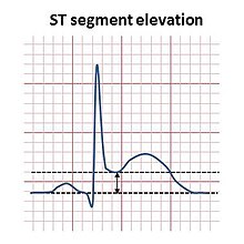
Elevation of the ST segment above the baseline in a 12-lead ECG is an indication of_________ in progress.
Myocardial infarction