chapter 13- General and special senses
 Call Kai
 Learn
Learn Practice Test
 Spaced Repetition
 Match
Match Flashcards
 Knowt Play
1/46

There's no tags or description
Looks like no tags are added yet.
Name | Mastery | Learn | Test | Matching | Spaced | Call with Kai | Chat |
|---|
No analytics yet
Send a link to your students to track their progress
47 Terms
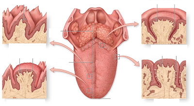
4 types of papillae
filliform, fungiform, vallate, and foliate
filliform
anterior 2/3 of tongue, no taste buds
fungiform
tip and sides of tongue, few taste buds
vallate (circumvallate)
Back of tongue, largest
Foliate
lateral tongue, taste buds during infancy
basal cells
replace the other 2 cell types, immature
gustatory cells
-chemoreceptors
replaced every 7-10 days
supporting cells
support other cells in tate bud
what do chemoreceptors do?
react to chemicals
what type of muscle is the tongue surface
stratified squamous epithelium
5 flavors detected over broad regions of the tongue
salty
sweet
sour
bitter
umani (savory)
what are the 2 nerves involved with taste
facial nerve (VII)
innervates anterior 2/3 of tongue
glossopharyngeal nerve (IX)
innervates posterior 1/3 of tongue
which type is papilae only found in infants?
folliate- lateral part of tongue
what does gustatory cells connect to
neurons
oflaction receptor neurons- what kind of receptors?
chemoreceptors, bipolar neurons
what types of cells are in olfaction
supporting cells
basal cells
loss of smell is called what?
ansomia
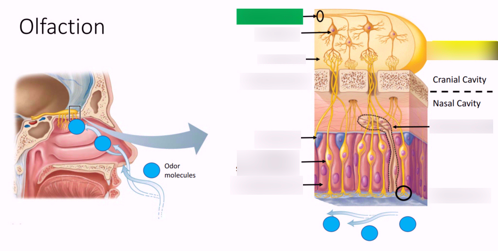
Label- axons of CN1, Olfactory bulb, cubiform plate, olfactory gland, olfactory epithelium, mucus layer, supporting cell, olfactory receptor neuron
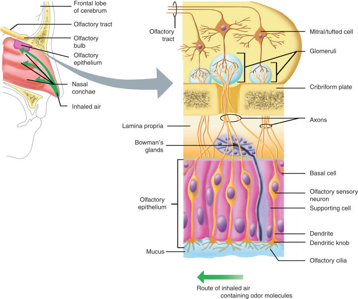
when we smell things, tiny particles (chemicals) bind to __1__ which causes __2_ to send action potential through axons into the _3__ lobe where we percieve smell
1,2-dendrites of olfactory receptor neurons, 3-temporal lobe
list the accessory structures of the eye and their functions
conjunctiva- covers eye’s anterior surface and internal eyelid surface
eyebrows, eyelashes and palpebrae(eyelids)- prevent foreign objects from contacting the eye
lacrimal system (lacrimal glands, canaliculi, lacrimal sac, and nasolacrimal duct) how tears are made and the flow of tears
tarsal glands- secretes oil to keep moisture in eye and prevents things from coming into eyes
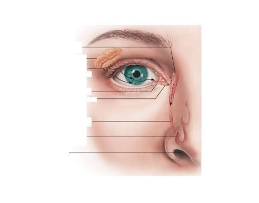
list the flow of tears in order
lacrimal glands, canalicula, lacrimal sac, and nasolacrimal duct
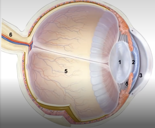
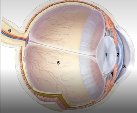
eye structures
lens
pupil
anterior chamber- filled with aqueous humor
posterior chamber- filled with aqueous humor
vitreous chamber- filled with vitreous humor
optic nerve
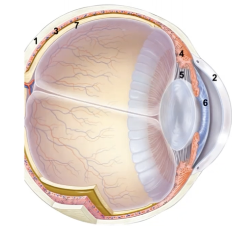
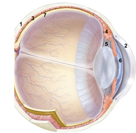
list tunics and what is included
Fibrous Tunic
sclera- white part of eye
cornea- (where contacts go)
Vascular tunic
choroid- layer of vascular tissue around eye
ciliary body- made up of muscles that connect to suspensory ligaments that hold lens
suspensory ligaments- hold lens
iris- controls amount of light that hits the lens
Neural tunic
retina
photoreceptors
when ciliary body contracts, it pulls on suspensory ligaments which helps what?
helps our lens focus on what we’re looking at
what type of muscle helps pupils constrict and dilate
smooth muscles help contract and relapse
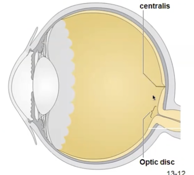
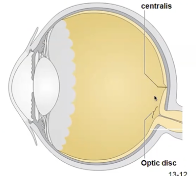
fovea centralis vs optic disc
fovea centralis- contains ONLY cones, maximal visual acuity
Optic disc- contains NO rods or cones, axons exit eye, blind spot)
Rods Vs Cones
Rods- function in dim light, don’t provide sharp vision or color vision, more numerous than cones
Cones- operate best in bright light, provide high acuity color vision
what is the part/center of the macula utea
fovea centralis
what does it mean when someone has 20/20 vision
lens magnifies and concentrates right on fovea centralis
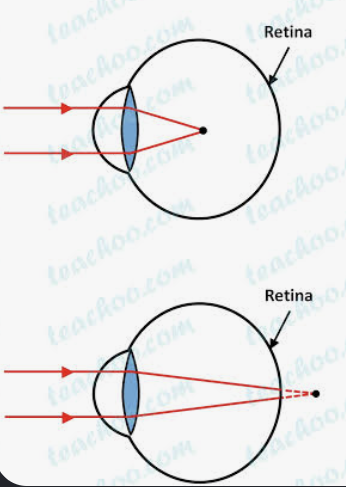
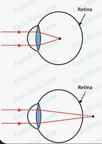
myopia vs hyperopia
mypoia- nearsightedness
hyperopia- farsightedness
name and explain structures in external ear
auricle- outer ear; groves that tunnel sound to →
external acoustic meatus
ceruminous glands
produce cerumen
earwax impedes microorganism growth
tympanic membrane
name and explain structures in middle ear
ossicles-
malleus
incus
stapes (oval window is beneath the footplate in the stapes)
stapedius muscle
tensor tympani muscle- contracts (like at a concert to keep eardrum from moving too much)
auditory tube- connects air-filled middle ear to nasopharynx
auditory tube
connects air-filled middle ear to nasopharynx
ususally closed, opens to equilibrate pressure in middle ear
otitis media- infection of the middle ear
name and explain structures of inner ear
vestibule
utricle and saccule
detects acceleration and decleration
helps sense equillibrium
semicircular canals
detect rotational movements
help sense equillibrium
cochlea
senses autdition
vestibular and cochlear nerves
when the fluid pushes all the way back in vestibule, is it acceleration or deceleration?
acceleration
semicircular canals- if you spin really fast and then stop suddenly in your chair, why do you feel dizzy
the fluid in the semicircular canals keeps spinning til it it slows down
name the journey of sound after entering external auditory canal
tympanic membrane
Malleus
Incus
Stapes pushes on oval window
cause fluid movments within cochlea
goes through scala vestibuili
waves transported and causes movement within cochlear duct
pass through cochlear duct
cause fluid to move through scala tympani
scala vestibuli and scala tympani are filled with what?
perilymph fluid
endolymph is found where?
cochlear duct
journey of sound inside cochlea
crosses scala vestibuili
vibrate vestibular membrane
cause vibration within cochlear duct
cause basilar membrane and hair cells to vibrate and hit tectorial membrane that creates perception of sound
conductive hearing loss
sound isn’t conducted efficiently through the outer or middle ear
sensorineural hearing loss
damage to the inner ear or nerve pathways
cochlear implant
the antenna, transmitter, and receive are inserted in the skin posterior and superior to the auricle. sound waves are received here and turned into electrical impulses
transmitter lead is inserted through the external ear, the middle ear, and the cochlea of the inner ear
electrical impulses from the transmitter are relayed through a lead and stimulate the cochlear nerve directly adjacent to the cochlea
cataracts
eye disorder
cloudy lense
ex: proteins in lens could start breaking down
usually old age
macular degeneration
eye disorder
area around fovea degenerates
Billy woke up one morning and realized that he had lost the sense of smell. we would diagnose that symptom of ___
anosmia