ekg class
 Call Kai
 Learn
Learn Practice Test
 Spaced Repetition
 Match
Match Flashcards
 Knowt Play
1/34

There's no tags or description
Looks like no tags are added yet.
Name | Mastery | Learn | Test | Matching | Spaced | Call with Kai | Chat |
|---|
No analytics yet
Send a link to your students to track their progress
35 Terms
1500 method
This method is the most precise, and the best method to use with fast rhythms.
The rhythm must be regular to use this method.
Count the small squares between qrs complex and take that number and 1500/# of small boxes
Sequence method
not as accurate
quick calculation
heart rate is regular
qrs complex starts on solid line, count big boxes
300/ number of big boxes
6 second method
irregular rhythm
3 black lines, 30 big boxes
qrs complex *10
maxium heart rate
220 - patients age
target heart rate
maximum heart rate * percentage to limit the rate = target
P wave
atrial depolarization
when SA node fires
positively deflected
QRS complex
ventricular depolarization
end of PR interval to the end of the J point
0.04 to 0.10 seconds
J point
where ventricular depolarization stops and ventricular repolarization starts
myocardial ischemia
J point elevate/ depress
T wave
ventricular repolarization
should be uniform
U wave
not always visible
repolarization of the Bundle of his and Purkinje fibers
PR interval
time it takes for the SA node to fire, atria to depolarize and electrivity to travel through the AV node
beginning of P wave to beginning of Q wave
0.12-0.20 seconds
RR interval
amount of time between atrial depolarization cycles
QT interval
one complete ventricular cycle
beginning of the q wave to the end of the t wave
segments
flat lines
PR segment
begins at the end of the P wave and goes to the beginning of the Q wave
ST segment
early phase of ventricular repolarization
shape is important when looking for patterns of ischemia
end of S wave beginning of t wave
time from end of ventricular depolarization to beginning of repolarization
positive deflection
wave that rises above the isoelectric line
negative deflection
wave below the isoelectric line
p wave and PR interval abnormalities
atrial dysfunction
QRS and t wave abnormalities
ventricular dysfunction
low levels of potassium and high levels
heart rate decrease
abnormal rate or rhythm
low levels of calcium and high levels
slower heart rate
longer than normal contractions
pacemakers
an artifact
Normal sinus rhythm
P wave present, upright, and rounded
amplitude is less than 2.5 mm
duration is less than 110 milliseconds
QRS complex is narrow
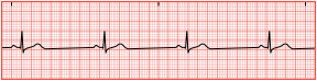
Sinus Brachycardia
less than 60 bpm
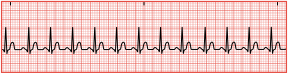
Sinus tachycardia
more than 100 bpm
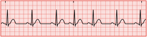
Sinus dysrhythmia
slight irregularity in rhythm
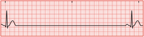
Sinus arrest
break in normal EKG
SA node failed in fire
not significant unless arrest last longer than 6 seconds
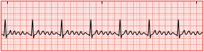
Atrial flutter
more sever than sinus arrhythmias
atria contracts faster than ventricles
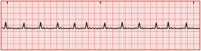
Atrial fibrillation
no organized contraction of the atria
quivering state
blood clot forms because of stagnation of blood in ventricles
Junctional arrhythmias
occur at AV node
inverted P wave configuration
SA node damaged
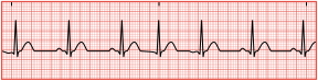
Premature junctional complex
early impulse that occurs before next beat
P wave can occur before, after, or during QRS complex
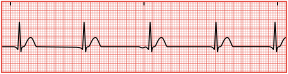
Junctional escape rhythm
absent P wave
impulse from AV node
atria and ventricles receive impulse simultaneously
heart rate does not exceed 60 bpm
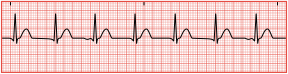
Accelerated junctional rhythm
escape rhythm but 60-100 bpm
Junctional tachycardia rhythm
escape rhythm but 100-150 bpm