BIOL201 Lecture Exam 1 CH 1-5, 7 and 15 (REMEMBER CHEAT SHEET)
1/23
Earn XP
Description and Tags
Area 1. Historical scientists and their contributions Area 2. Koch's Postulates and their use Area 3. Represent an atom using the Bohr model Area 4. Levels of protein structure Area 5. Fimbriae and their role in infection Area 6. Phospholipid bilayer as a barrier Area 7. Function of the electron transport chain Area 8. Enzymes and steps of DNA replication Area 9. Types of DNA mutations Area 10. Steps of phagocytosis Area 11. Hallmarks of inflammation Area 12. types of leukocytes and their function
Name | Mastery | Learn | Test | Matching | Spaced | Call with Kai |
|---|
No analytics yet
Send a link to your students to track their progress
24 Terms
Antoni van Leeuwenhoek
Made microscopes completely by himself in order to view a single specimen
Examined water and visualized tiny animals, fungi, algae, and single-celled protozoa; “animalcules
By end of 19th century, these organisms were known as microorganisms
His designs let us see protozoans in rainwater
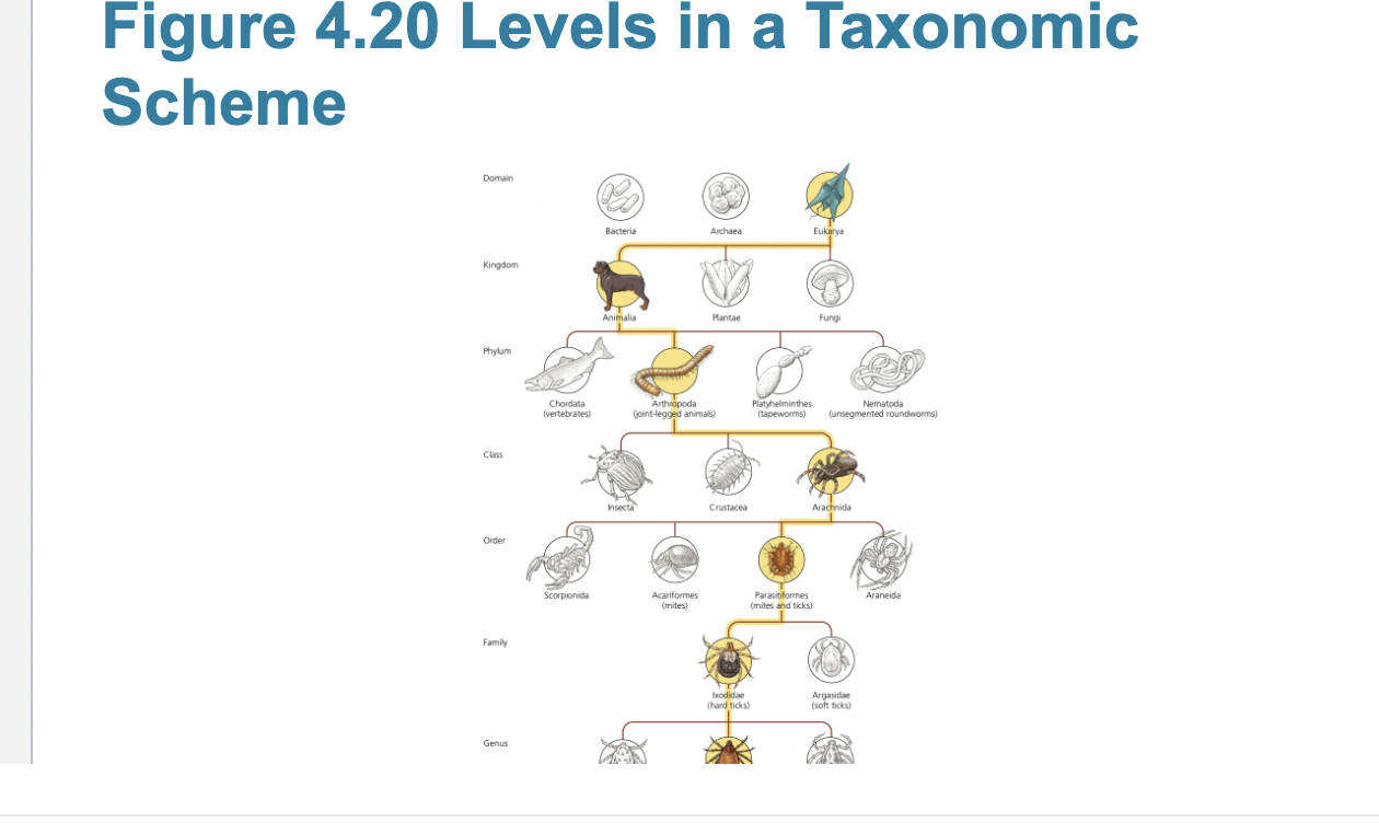
Carolus Linnaeus
How Can Microbes Be Classified?
– Carolus Linnaeus developed a taxonomic system for
naming plants and animals and grouping similar organisms
together.
– Leeuwenhoek’s microorganisms can be grouped into six
categories:
▪ Bacteria
▪ Archaea
▪ Fungi
▪ Protozoa
▪ Algae
▪ Small multicellular animals
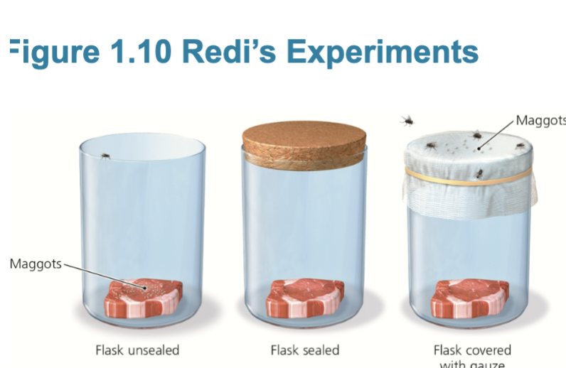
Francesco Redi, John Needham, Lazzaro Spallanzani’s
Does Microbial Life Spontaneously Generate?
– Redi’s experiments
▪ When decaying meat was kept isolated from flies,
maggots never developed.
▪ Meat exposed to flies was soon infested.
▪ As a result, scientists began to doubt Aristotle’s
view.
Does Microbial Life Spontaneously Generate?
– Needham’s experiments
▪ Scientists agreed that large animals could not arise
spontaneously, but believed microbes could.
▪ Needham’s experiments with beef gravy and
infusions of plant material reinforced this idea.
Does Microbial Life Spontaneously Generate?
– Spallanzani’s experiments
▪ His experiments contradicted Needham’s findings.
▪ Concluded that:
– Needham failed to heat vials sufficiently to kill all
microbes or had not sealed them tightly enough.
– Microorganisms exist in air and can contaminate
experiments.
– Spontaneous generation of microorganisms does not
occur; all living things arise from other living things.
▪ Critics said sealed vials did not allow enough air for
organisms to survive and that prolonged heating destroyed
the “life force.”
Louis Pasteur
Does Microbial Life Spontaneously Generate?
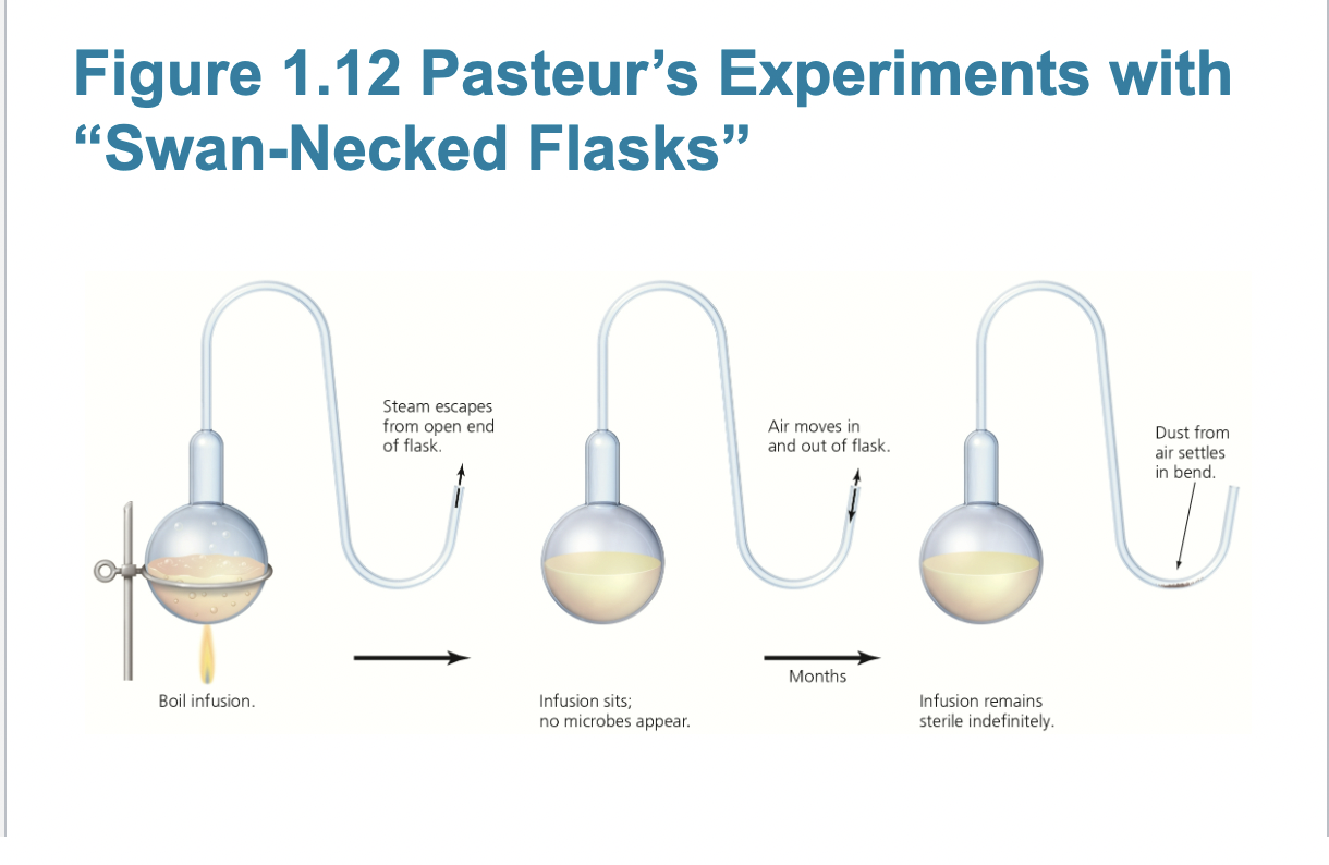
– Pasteur’s experiments
▪ Performed experiments with “swan-necked” flasks
▪ When the flasks remained upright, no microbial
growth appeared.
▪ When the flask was tilted, dust from the bend in the
neck seeped back into the flask and made the
infusion cloudy with microbes within a day.
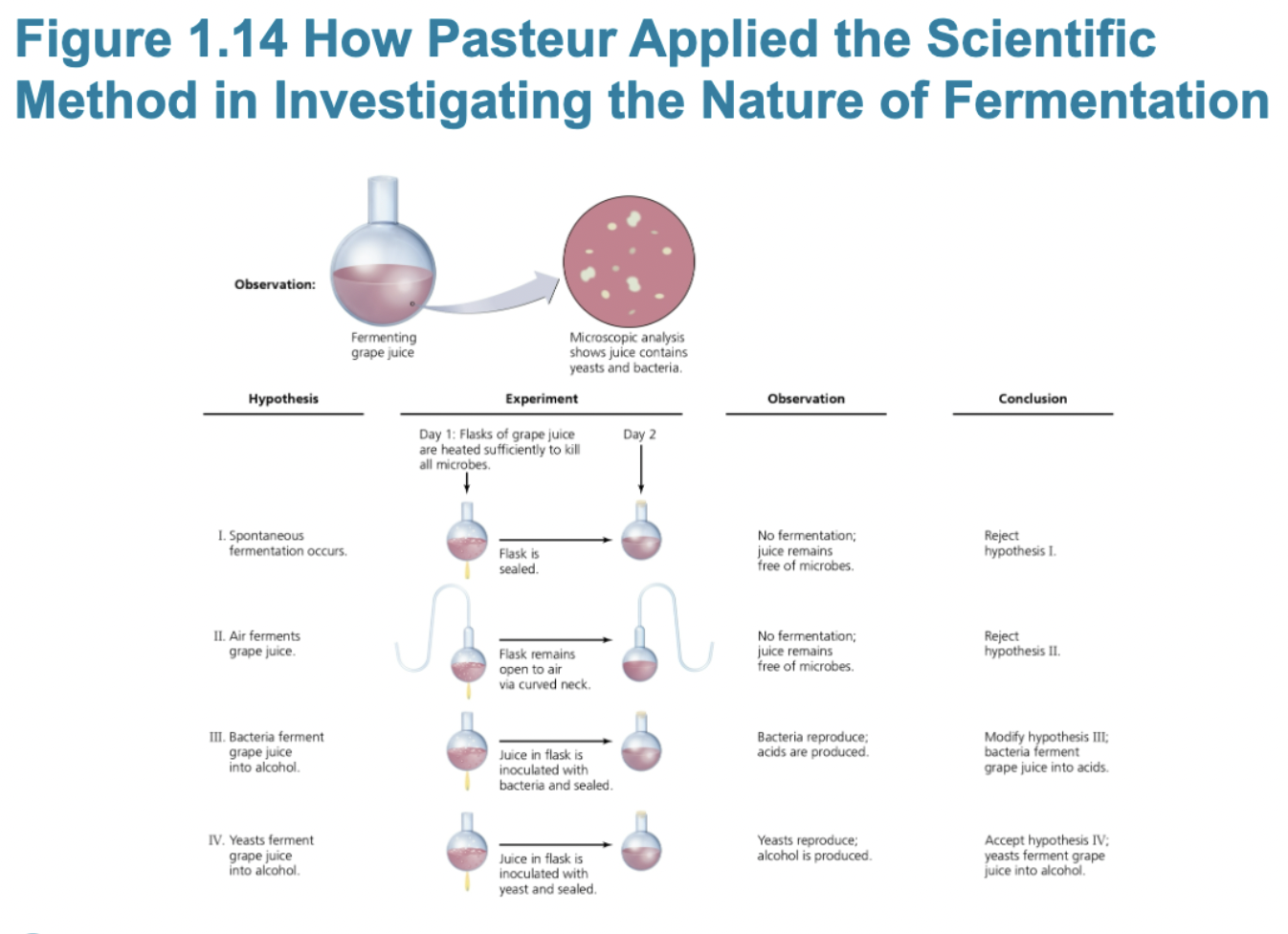
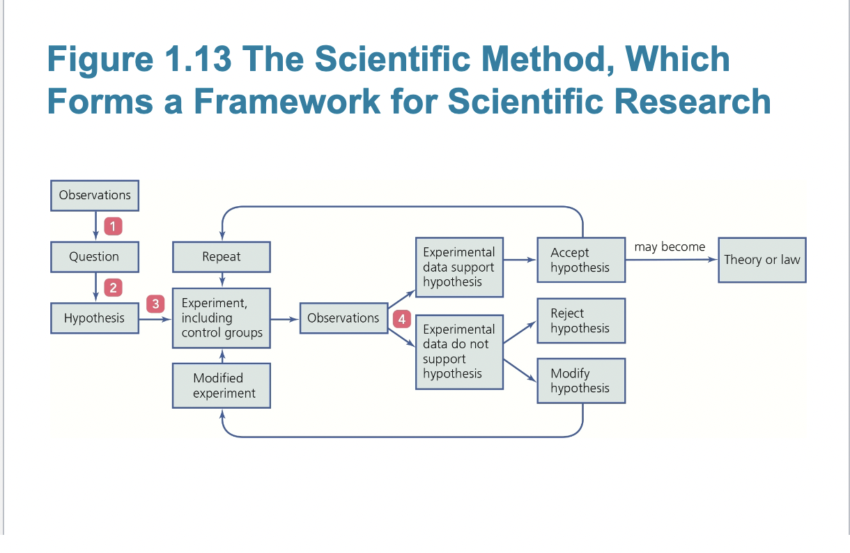
The scientific method
▪ Debate over spontaneous generation led in part to
development of scientific method.
– Observation leads to question
– Question generates hypothesis
– Hypothesis is tested through experiment(s)
– Results prove or disprove hypothesis
• Accepted hypothesis leads to theory/law
• Disproved hypothesis is rejected or modified
Some scientists believed air caused fermentation;
others insisted that living organisms caused
fermentation.
▪ Pasteur conducted a series of experiments that
addressed the cause of fermentation and led to the idea of pasteurization.
Robert Kochs Postulates
– Koch’s experiments
▪ Simple staining techniques
▪ First photomicrograph of bacteria
▪ First photograph of bacteria in diseased tissue
▪ Techniques for estimating bacterial number in a
solution
▪ Use of steam to sterilize growth media
▪ Use of Petri dishes
▪ Laboratory techniques to transfer bacteria
▪ Bacteria as distinct species
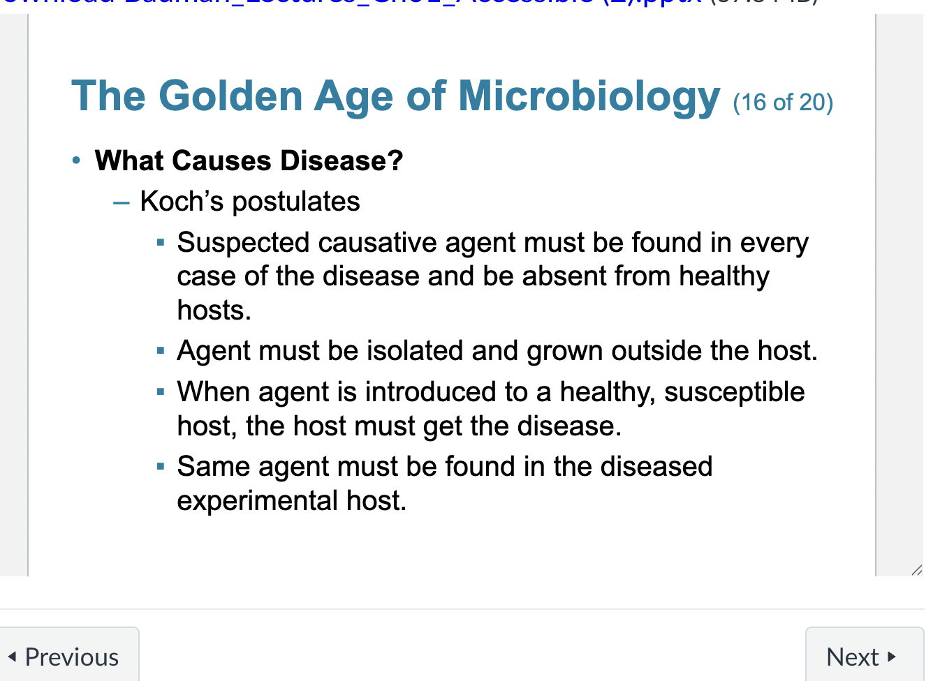
What Causes Disease?
– Koch’s postulates
▪ Suspected causative agent must be found in every
case of the disease and be absent from healthy
hosts.
▪ Agent must be isolated and grown outside the host.
▪ When agent is introduced to a healthy, susceptible
host, the host must get the disease.
▪ Same agent must be found in the diseased
experimental host.
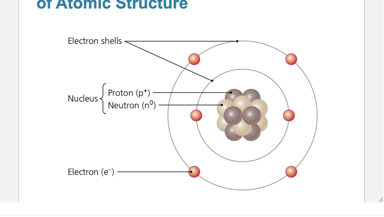
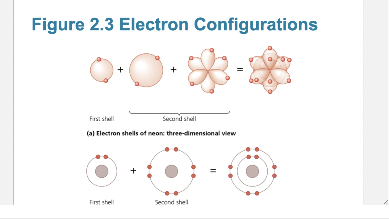
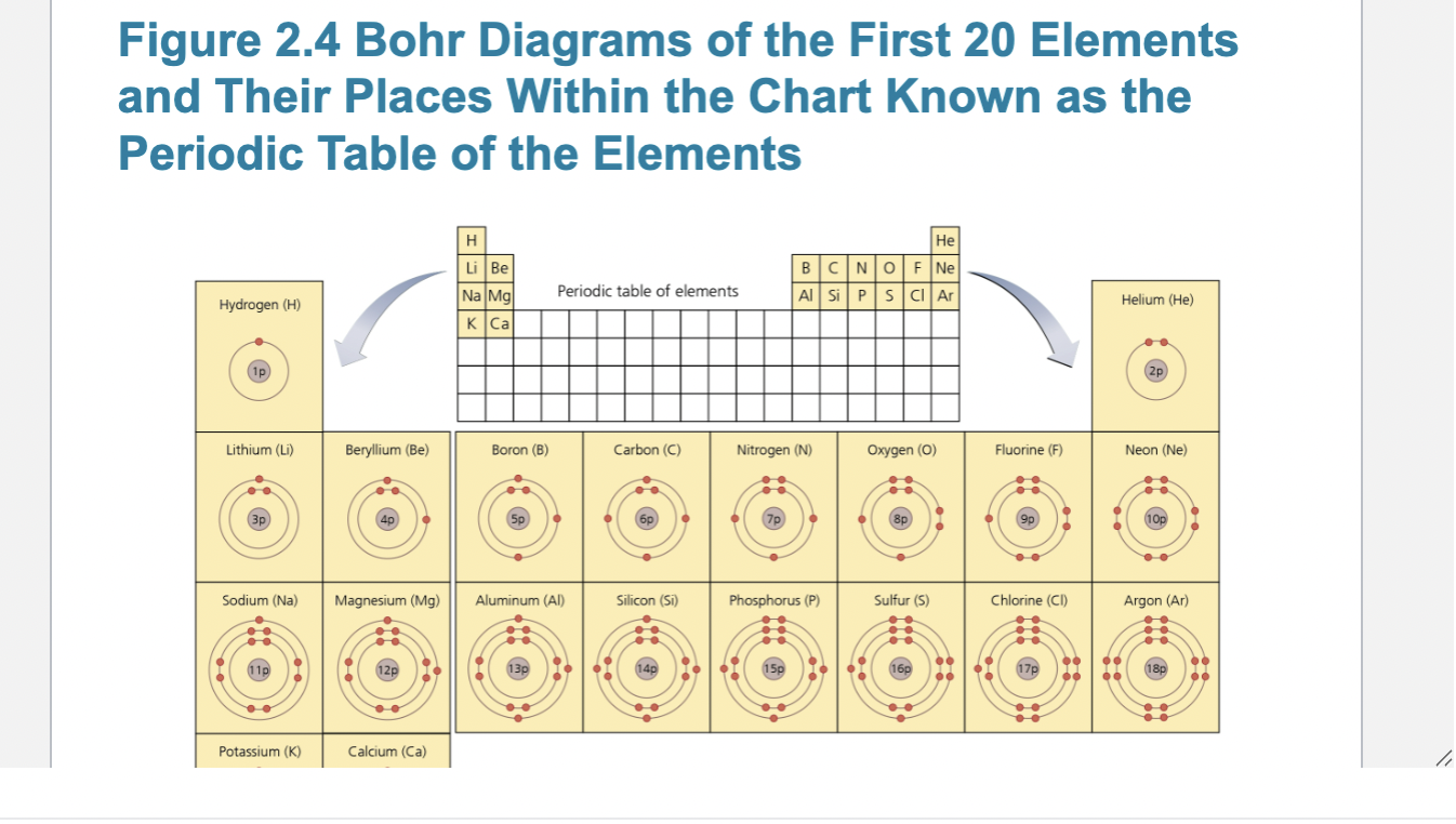
represent an atom using the bohr model
Atomic Structure
– Electrons—negatively charged subatomic particles
circling a nucleus
– Nucleus—structure containing neutrons and protons
– Neutrons—uncharged particles
– Protons—positively charged particles
Atomic Structure
– Element—composed of a single type of atom
– Atomic number—equal to the number of protons in
the nucleus
– Atomic mass (atomic weight)—sum of masses of
protons, neutrons, and electrons
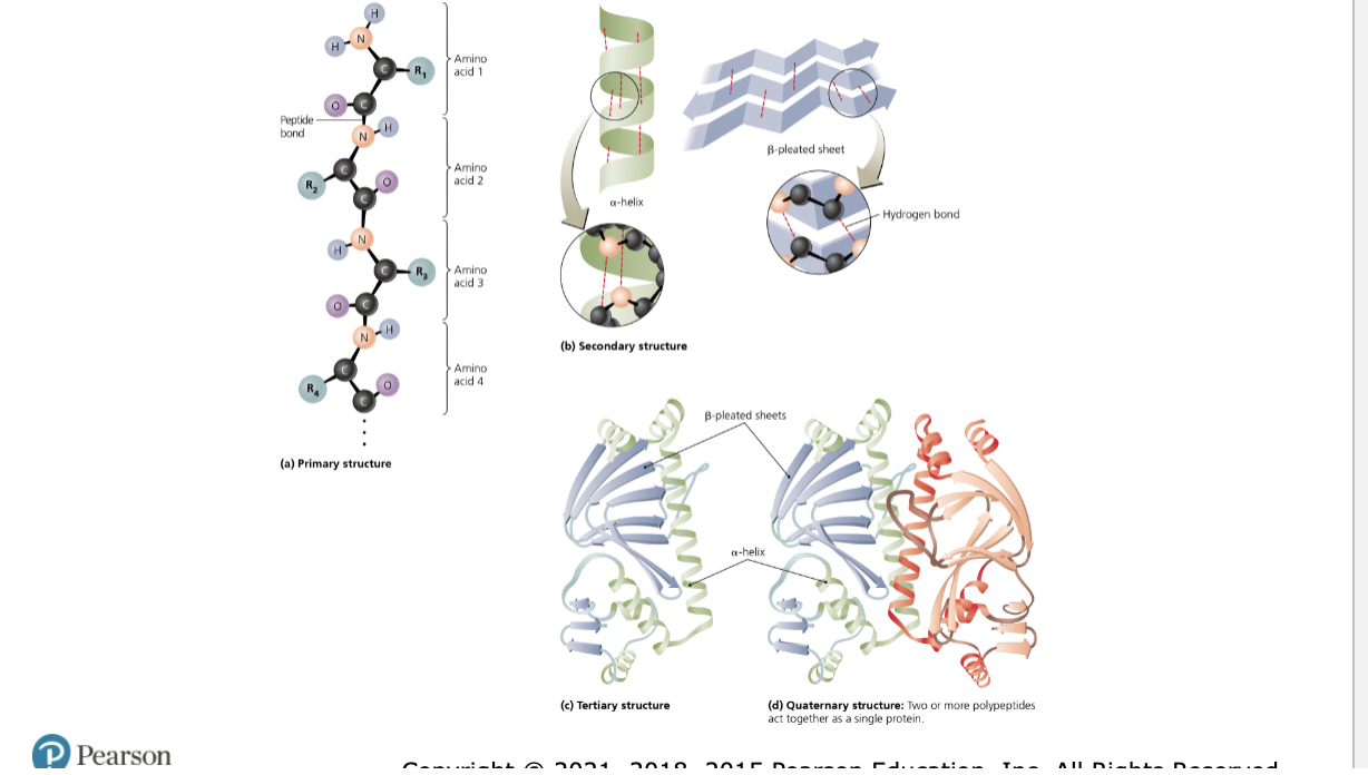
Levels of protein structure
Primary Structure: The linear sequence of amino acids linked by peptide bonds within a polypeptide chain. This level dictates all higher levels of folding.
Secondary Structure: Localized, repeating folding and coiling of the polypeptide backbone, stabilized by hydrogen bonds. The most common motifs are the α-helix and the β-pleated sheet.
Tertiary Structure: The overall three-dimensional shape of a single polypeptide chain. It is stabilized by various chemical interactions, including disulfide bridges, ionic bonds, and hydrophobic interactions between the amino acid side chains
Quaternary Structure: The three-dimensional structure formed by the assembly and aggregation of two or more separate polypeptide chains (subunits) into a single functional protein complex
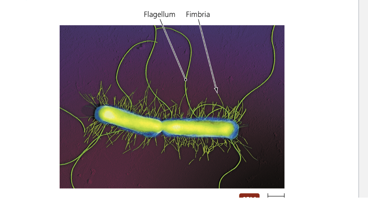
Fimbriae and their role in infection
Fimbriae and Pili
– Fimbriae
▪ Sticky, bristlelike projections
▪ Used by bacteria to adhere to one another and to
substances in environment
▪ Shorter than flagella
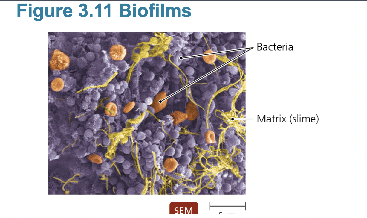
▪ Serve an important function in biofilms: Fimbriae are hair-like appendages found on the surface of bacteria used primarily for adhesion. By locking onto specific host cells and tissues, they allow pathogens to colonize, evade immune defenses, and form biofilms, making them critical virulence factors in initiating and maintaining infections.
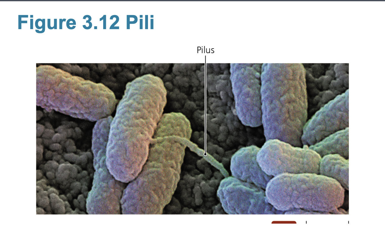
Fimbriae and Pili
– Pili
▪ Special type of fimbriae
▪ Also known as conjugation pili
▪ Longer than fimbriae but shorter than flagella
▪ Bacteria typically have only one or two per cell.
▪ Transfer DNA from one cell to another (conjugation)
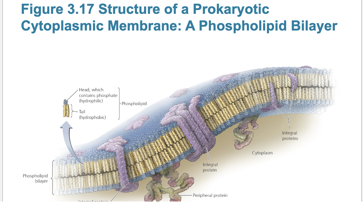
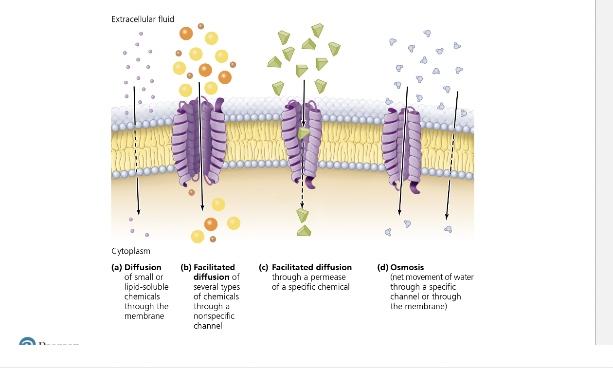
Phospholipid bilayer as a barrier
bacterial cytoplasmic membranes
Function
– Controls passage of substances into and out of the
cell
– Harvest light energy in photosynthetic bacteria
– Selectively permeable
– Naturally impermeable to most substances
– Proteins allow substances to cross membrane
– Maintain concentration and electrical gradient
-- passive processes: diffusion, facilitated diffusion, osmosis
—active processes: active transport, group translocation (substance is chemically modified during transpoirt)
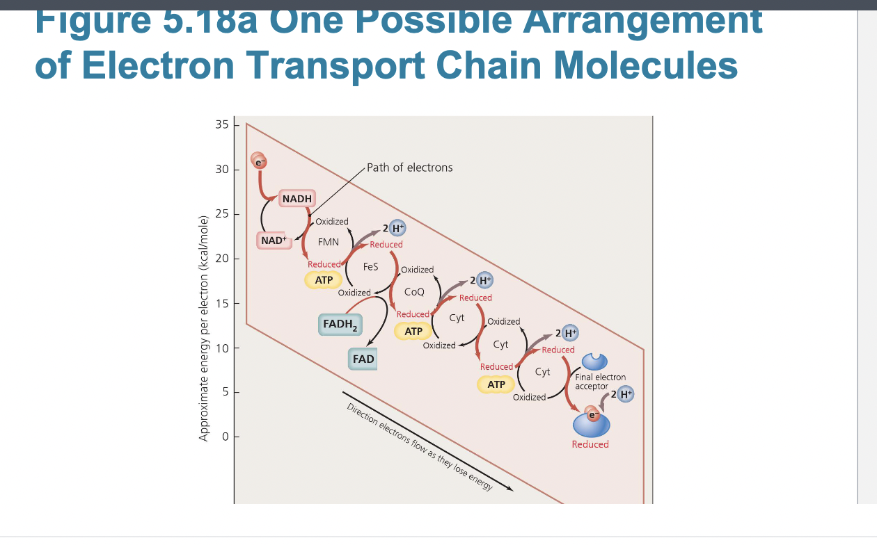
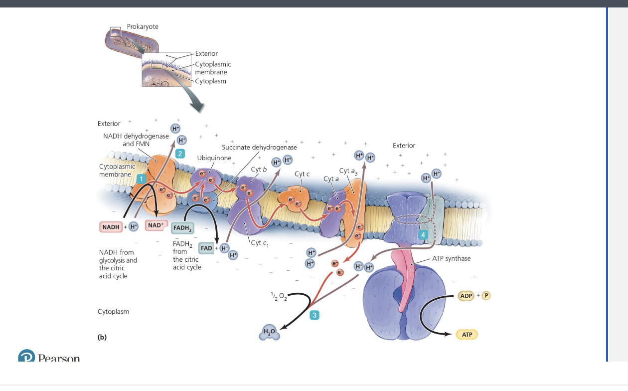
Function of the electron transport chain
Cellular Respiration
– Electron transport
▪ Most significant production of ATP occurs from series
of redox reactions known as an electron transport chain
(ETC)
▪ Series of carrier molecules that pass electrons from
one to another to final electron acceptor
▪ Energy from electrons used to pump protons (H+)
across the membrane, establishing a proton gradient
▪ Located in inner mitochondrial membrane of eukaryotes
and in cytoplasmic membrane of prokaryotes
Four categories of carrier molecules:
– Flavoproteins
– Ubiquinones
– Metal-containing proteins
– Cytochromes
▪ Aerobic respiration: oxygen serves as final electron
acceptor.
▪ Anaerobic respiration: molecule other than oxygen
serves as final electron acceptor.
factors affecting ATP yield: remove oxygen, add cyanide, add uncoupling protein
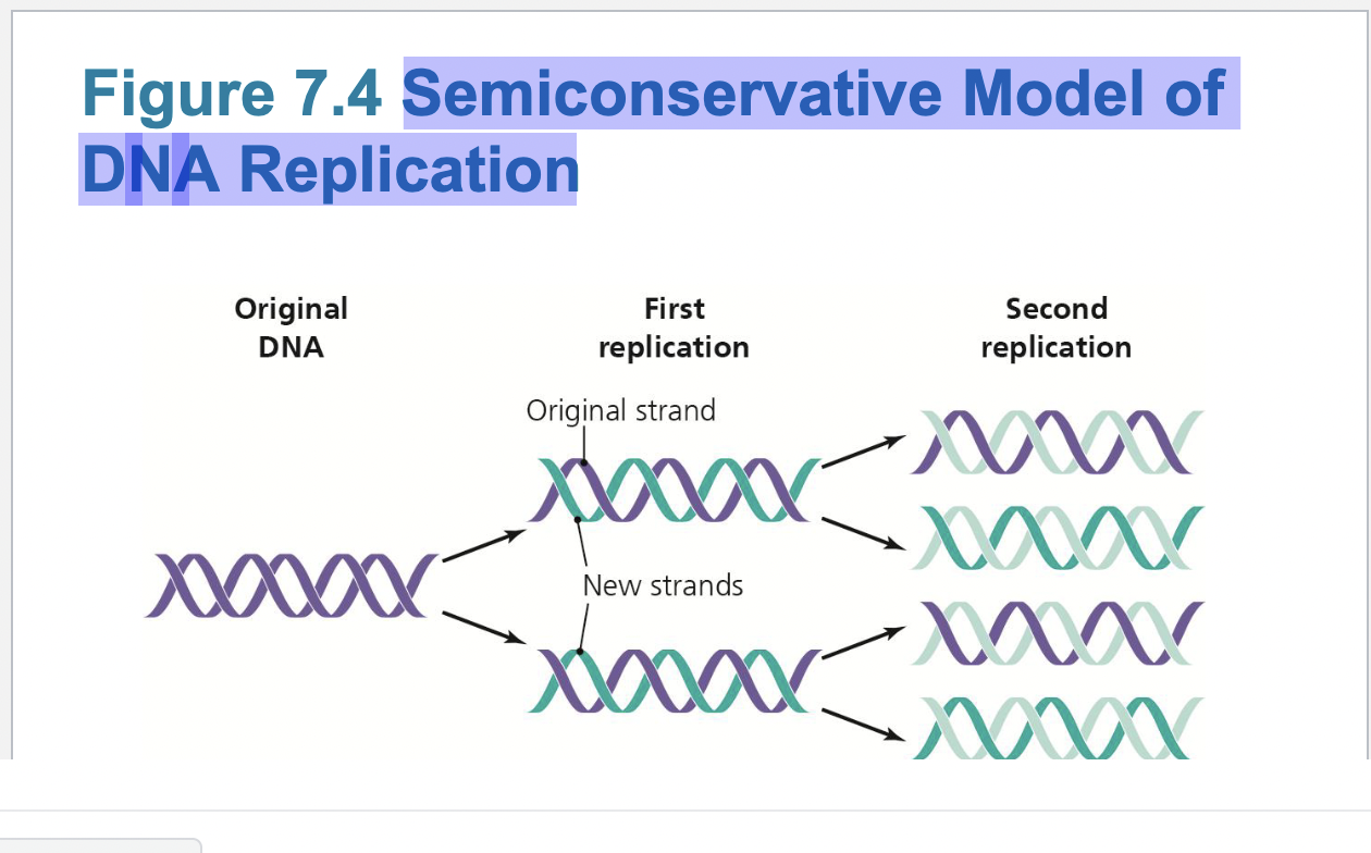
Enzymes and DNA replication
DNA Replication
– Key to replication is the complementary structure of
the two strands
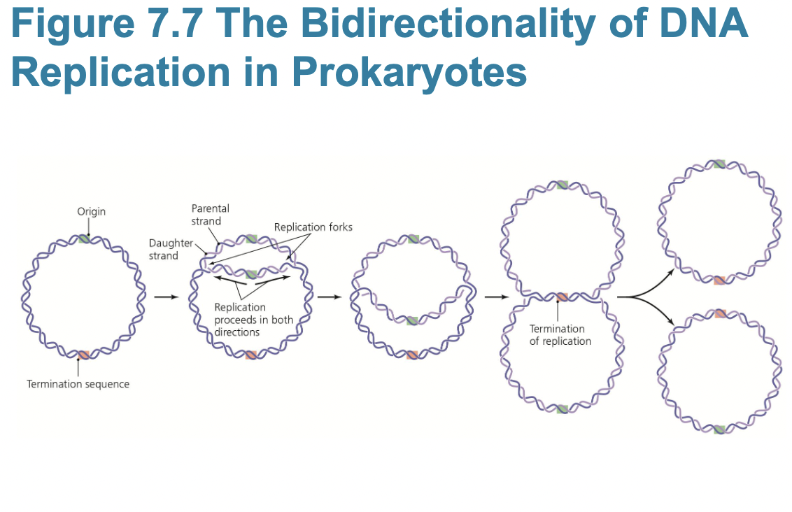
– Replication is semiconservative and bidirectional
▪ New DNA composed of one original and one
daughter strand
gyrases and topoisomerases remove supercoils in DNA
– Anabolic polymerization processes require monomers
and energy
▪ Triphosphate deoxyribonucleotides serve both
functions
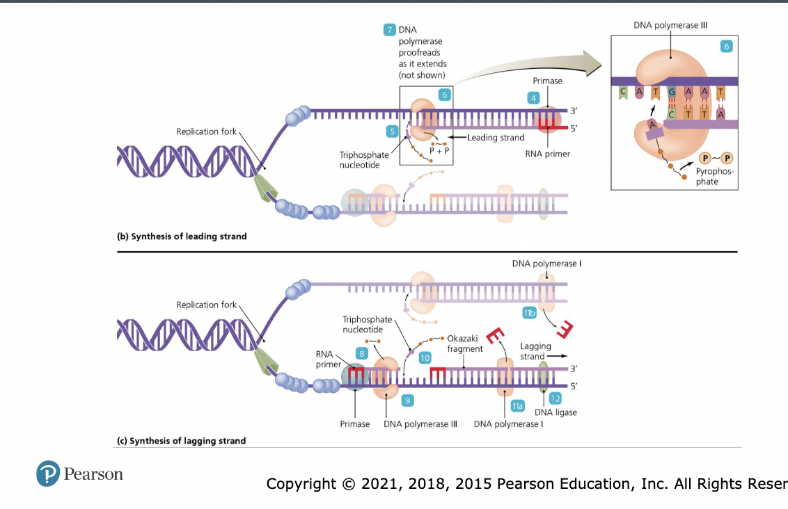
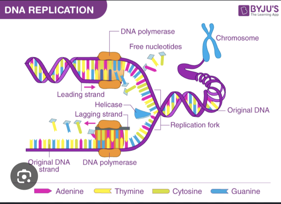
– Initial processes in bacterial DNA replication
▪ Bacterial DNA replication begins at the origin
▪ DNA polymerase replicates DNA only 5′ to 3′
▪ Because strands are antiparallel, new strands are
synthesized differently:
– Leading strand synthesized continuously
– Lagging strand synthesized discontinuously
!!Enzymes and DNA replication pt. 2
DNA Replication
– Replication of eukaryotic DNA
▪ Similar to bacterial replication
▪ Some differences:
– Uses four DNA polymerases
– Thousands of replication origins
– Shorter Okazaki fragments
– Plant and animal cells methylate only cytosine
bases
!!!what are the enzymes needed for DNA replication and what do they do?
Helicase: Binds to the DNA origin and breaks the hydrogen bonds between complementary bases. This unwinds the double helix and creates a Y-shaped structure known as the replication fork.
DNA gyrase or topoisomerase: Travels just ahead of the replication fork to relieve the physical strain and supercoiling (over-winding) that builds up in the rest of the DNA strand as it is unwound
RNA primer: Synthesizes a short RNA primer. Because the main builder (DNA polymerase) cannot start a completely new strand from scratch, it requires this primer to provide a starting 3' hydroxyl group
DNA pol 1: The primary enzyme responsible for synthesizing the new DNA strand. It adds complementary free-floating nucleotides in a \(5^{\prime }\) to \(3^{\prime }\) direction. Involved in proofreading and building the leading and lagging strand
Ligase: links Okazaki fragments, ligase seals these sugar-phosphate backbones to form a continous seamless strand of DNA
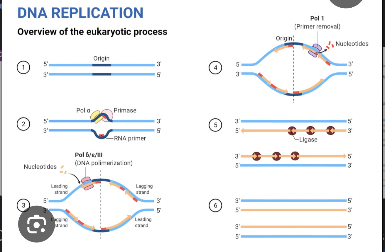
Steps of DNA replication
Initiation
DNA unwinds – The enzyme helicase breaks the hydrogen bonds between base pairs, unzipping the DNA double helix.
DNA is stabilized – Single-strand binding proteins keep the separated DNA strands from rejoining.
Topoisomerase prevents DNA from over-winding and supercoiling ahead of the replication fork
Elongation
RNA primers are added – Primase lays down short RNA primers to provide a starting point for DNA synthesis.
New DNA strands are built – DNA polymerase adds complementary nucleotides in the 5′ → 3′ direction.
Leading strand: synthesized continuously.
Lagging strand: synthesized in short Okazaki fragments.
Termination
RNA primers are replaced – DNA polymerase 1 removes the RNA primers and replaces them with DNA nucleotides.
Fragments are joined – DNA ligase seals the gaps between Okazaki fragments, creating one continuous strand.
Replication is complete – Two identical DNA molecules are produced, each containing one original (parental) strand and one newly synthesized strand (called semiconservative replication).
Easy memory trick:
H → P → P → L
Helicase unzips
Primase primes
Polymerase builds DNA
Ligase links fragments

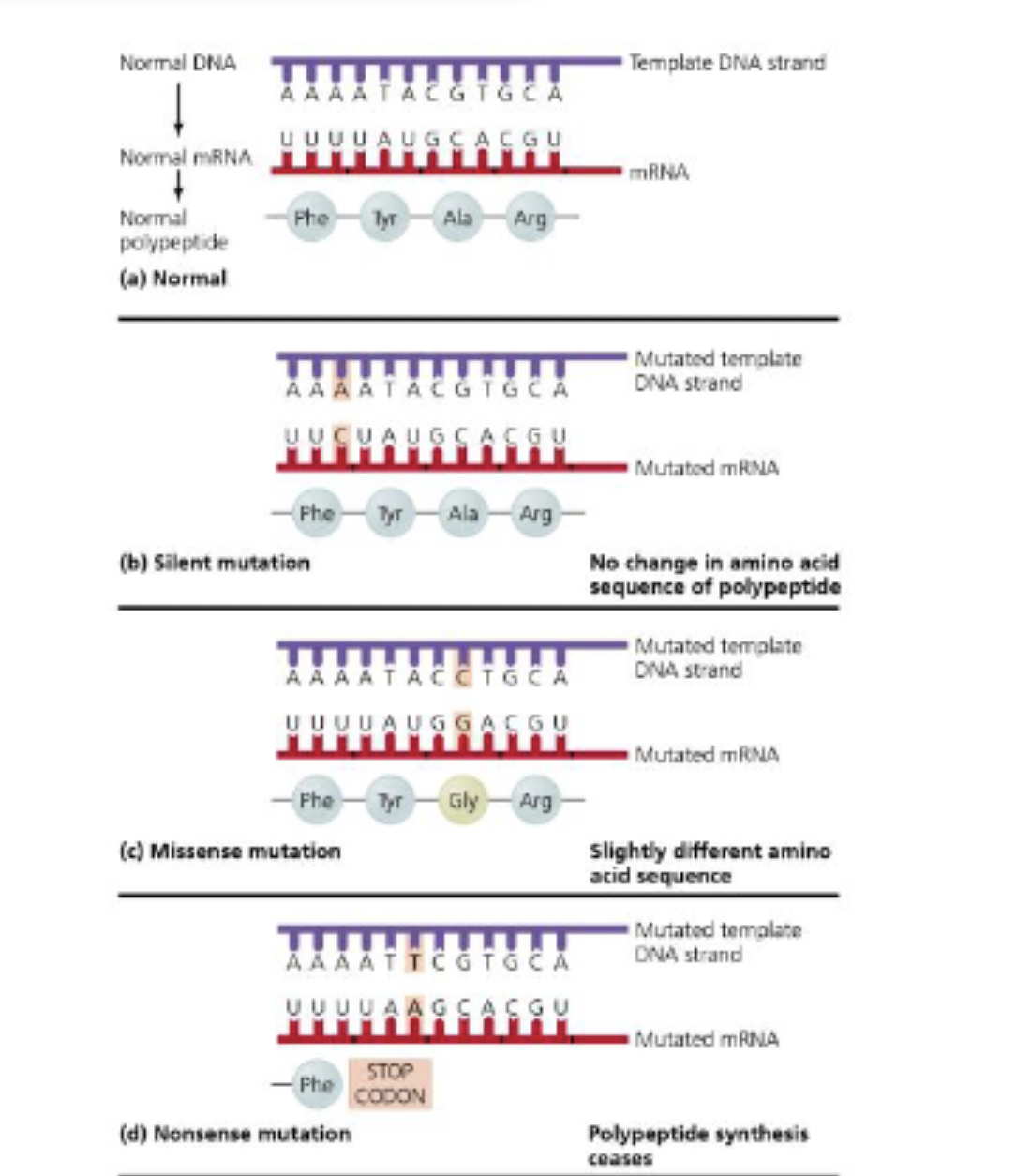
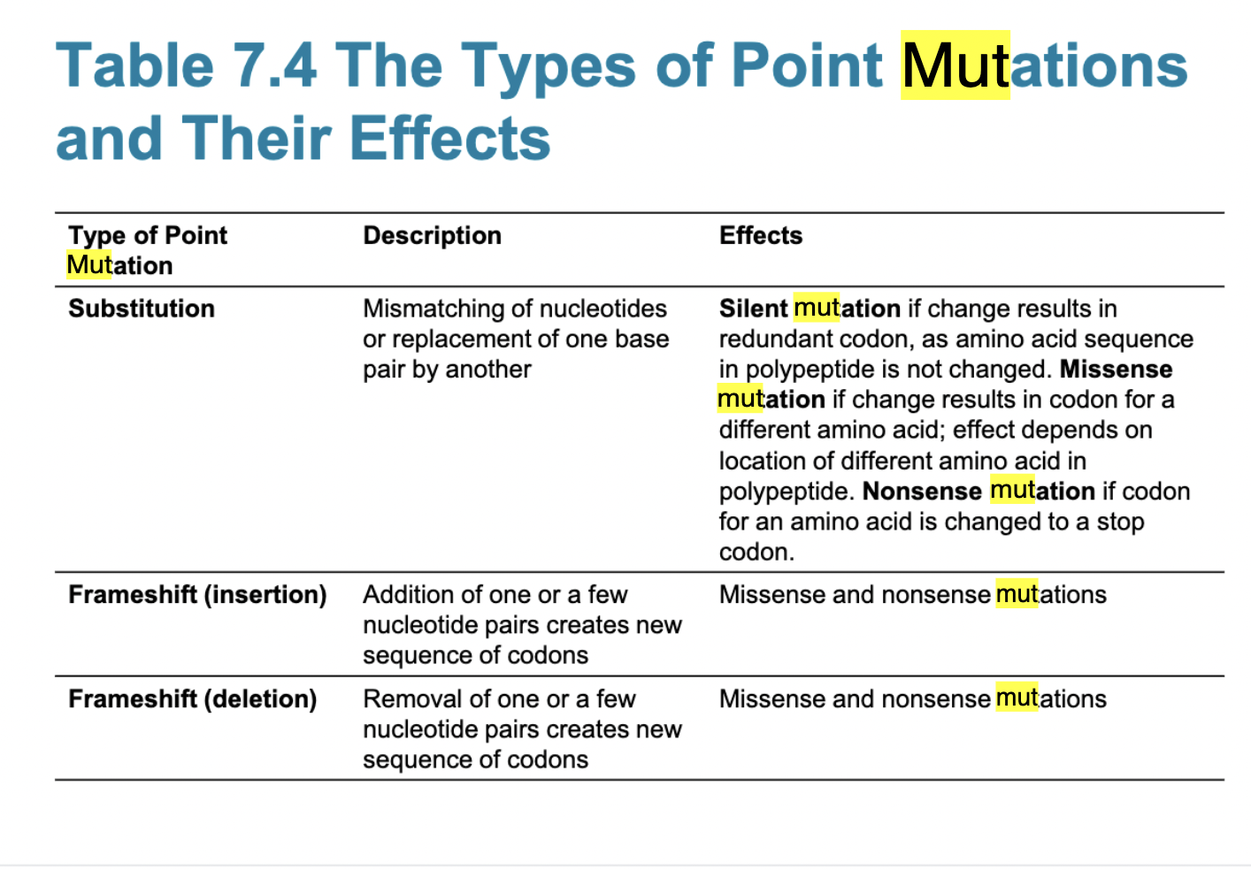
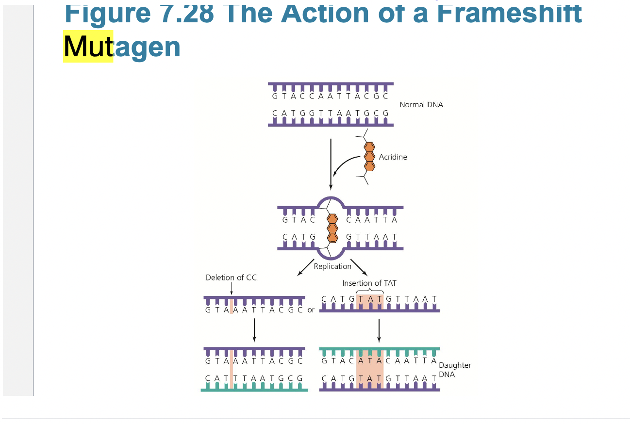
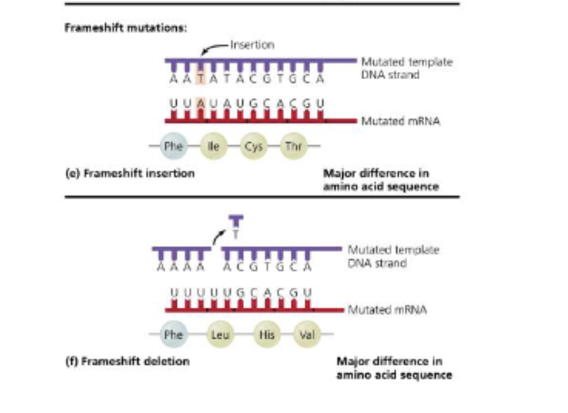
Types of DNA mutations
Types of Mutations
– Point mutations
▪ One base pair is affected
▪ ex: Substitutions and frameshift (insertion and deletion) mutations
– Gross mutations
▪ Include inversions, duplications, and transpositions
Mutagens increase the mutation rate by a factor of 10
to 1000 times.
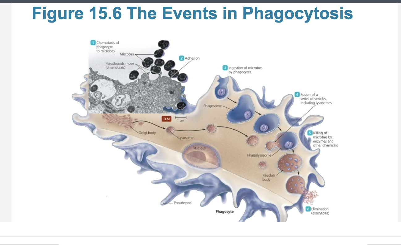
Steps of phagocytosis
1. Chemotaxis & Activation: The phagocyte detects chemical signals (e.g., cytokines, microbial components) and migrates to the site of infection or tissue damage. [1, 2, 3]
2. Recognition & Attachment: Receptors on the phagocyte's surface bind to the target. This process is highly enhanced by opsonization, where the target is coated with proteins like antibodies or complement factors. [1, 2, 3, 4]
3. Engulfment: The phagocyte extends parts of its cell membrane, known as pseudopods, around the target particle. These extensions eventually meet and fuse, enclosing the particle within a pouch. [1, 2, 3]
4. Phagosome Formation: The fused membrane pinches off inside the cell to form a specialized, membrane-bound vesicle called a phagosome. [1, 2]
5. Phagolysosome Formation: The phagosome travels into the cytoplasm and fuses with a lysosome, forming a phagolysosome. Lysosomes contain destructive enzymes and reactive oxygen species. [1, 2, 3, 4]
6. Destruction: The enzymes within the phagolysosome activate in an acidic environment, actively degrading and destroying the engulfed microbe or debris. [1, 2]
7. Elimination: The remaining indigestible waste is converted into a residual body, which is subsequently expelled from the cell through exocytosis. [1, 2, 3]
![<ul><li><p><span><strong>1. Chemotaxis & Activation:</strong> The phagocyte detects chemical signals (e.g., cytokines, microbial components) and migrates to the site of infection or tissue damage. [1, 2, 3]</span></p></li><li><p><span><strong>2. Recognition & Attachment:</strong> Receptors on the phagocyte's surface bind to the target. This process is highly enhanced by <strong>opsonization</strong>, where the target is coated with proteins like antibodies or complement factors. [1, 2, 3, 4]</span></p></li><li><p><span><strong>3. Engulfment:</strong> The phagocyte extends parts of its cell membrane, known as pseudopods, around the target particle. These extensions eventually meet and fuse, enclosing the particle within a pouch. [1, 2, 3]</span></p></li><li><p><span><strong>4. Phagosome Formation:</strong> The fused membrane pinches off inside the cell to form a specialized, membrane-bound vesicle called a <strong>phagosome</strong>. [1, 2]</span></p></li><li><p><span><strong>5. Phagolysosome Formation:</strong> The phagosome travels into the cytoplasm and fuses with a lysosome, forming a <strong>phagolysosome</strong>. Lysosomes contain destructive enzymes and reactive oxygen species. [1, 2, 3, 4]</span></p></li><li><p><span><strong>6. Destruction:</strong> The enzymes within the phagolysosome activate in an acidic environment, actively degrading and destroying the engulfed microbe or debris. [1, 2]</span></p></li><li><p><span><strong>7. Elimination:</strong> The remaining indigestible waste is converted into a residual body, which is subsequently expelled from the cell through exocytosis. [1, 2, 3]</span></p></li></ul><p></p>](https://assets.knowt.com/user-attachments/2cbddc14-9b2a-4938-9b92-35e9042f1cbe.png)
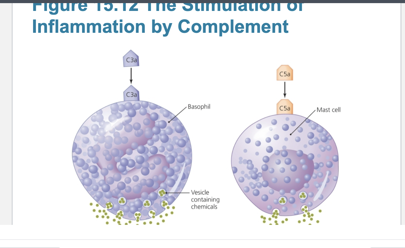
Hallmarks of inflammation
!!!redness, heat, swelling, and pain are hallmarks of inflammation
Inflammation
– Acute inflammation
▪ Develops quickly and is short lived
▪ Is typically beneficial
▪ Is important in the second line of defense
– Dilation and increased permeability of the blood vessels
– Migration of phagocytes
– Tissue repair
– Chronic inflammation
▪ Long-lasting
▪ Damage to tissues can cause disease
– Migration of phagocytes
▪ Neutrophils and monocytes delivered to site of infection
– Recruited by chemotactic factors
▪ Attach to receptors on blood vessels
▪ Squeeze between cells of vessel wall and enter site of
infection
– Tissue repair
▪ Delivery of nutrients and oxygen to site facilitates repair
▪ Some tissues cannot be repaired
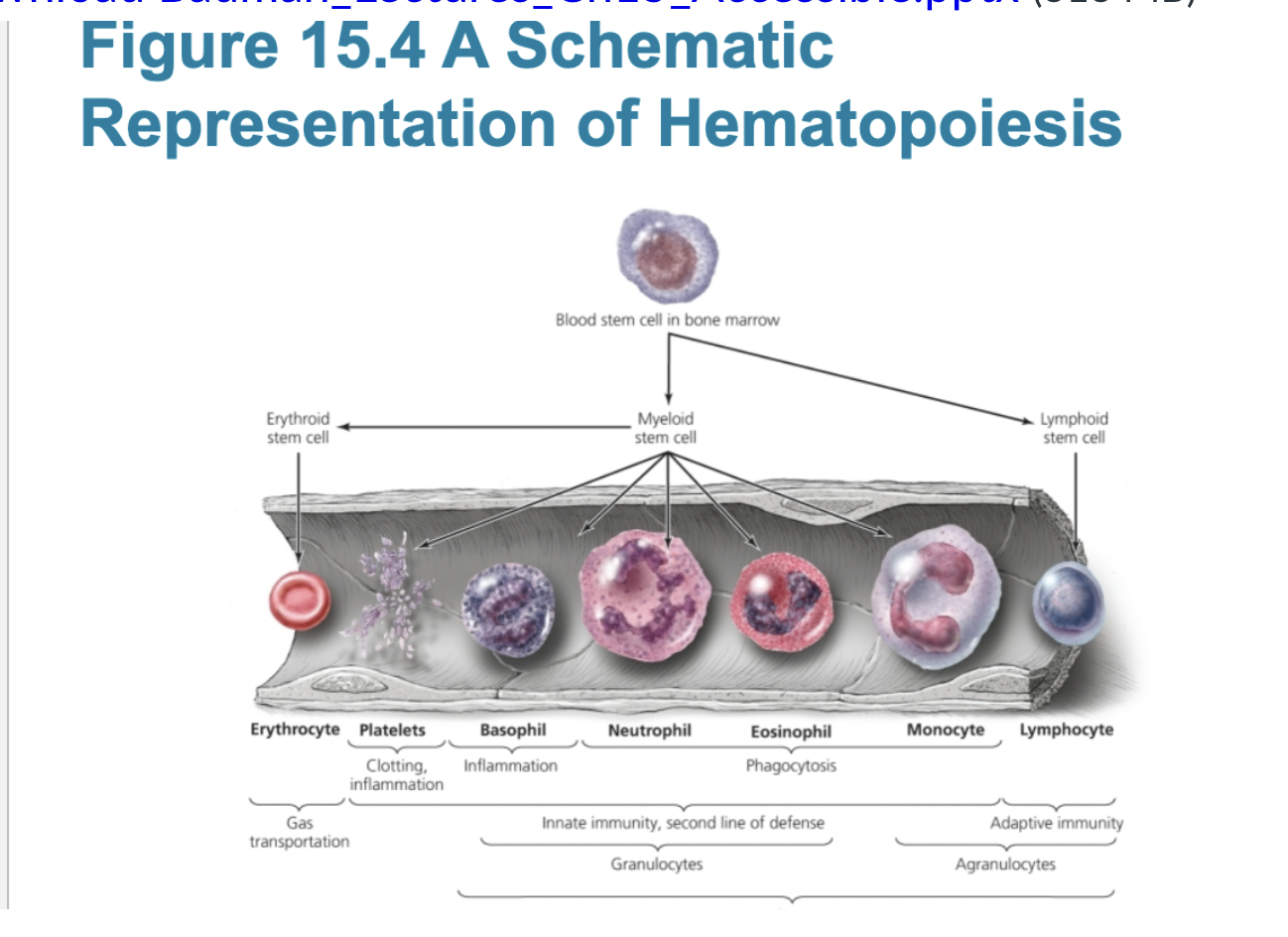
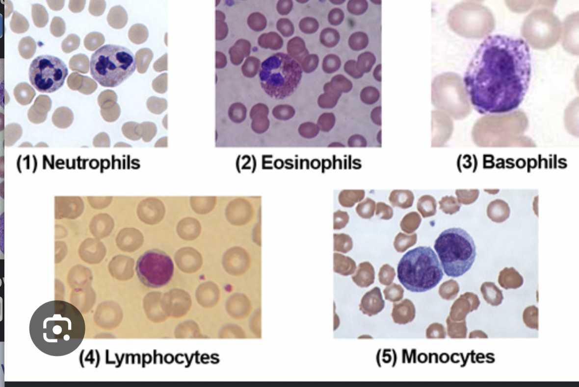
types of leukocytes and their function
Defensive blood cells: Leukocytes
▪ Granulocytes
– Contain large granules that stain different colors
– Three types
• Basophils—stain blue with basic dye methylene blue, release inflammatory chemicals, do not phagocytize
• Eosinophils—stain red/orange with acidic dye
eosin, phagocytize pathogens, capable of diapedesis, allergies or parasites
• Neutrophils—stain lilac with mix of acidic and
basic dyes, phagocytize pathogens, capable of diapedesis
▪ Agranulocytes
– Cytoplasm appears uniform under a light microscope
– Two types
• Lymphocytes
• Most involved in adaptive immunity
• Natural killer lymphocytes
• Monocytes
• Leave the blood and mature into macrophages
• Phagocytic cells that devour foreign objects
Differential white blood cell count can signal
disease
• Increased eosinophils indicate allergies or
parasitic worm infection
• Bacterial diseases often show increase in
leukocytes and neutrophils
• Viral infections show increase in
lymphocytes