gas exchange system
1/34
Earn XP
Description and Tags
Name | Mastery | Learn | Test | Matching | Spaced | Call with Kai | Chat |
|---|
No analytics yet
Send a link to your students to track their progress
35 Terms
Why do single-celled organisms not need exchange surfaces
Metabolic activity it low so low demand for oxygen for respiration
Large surface area to volume ratio
Why are exchange surfaces necessary for larger organisms
High metabolic activity
Smaller surface area to volume ratio
Larger distances between cells where oxygen is needed and the supply of oxygen
How does size of organism correlate to SA:V
The bigger the organism, the smaller the SA:V
What are the features of efficient exchange surfaces
large surface area - provides more surface area for diffusion to take place through (eg root hair cells)
Thin - short diffusion distance (eg epithelial cells of alveoli)
Good blood supply - maintains steep concentration gradient (eg dense capillary network around alveoli)
Good ventilation - maintains steep concentration gradient (eg fish gills)
Haemoglobin - binds to oxygen
Define gas exchange in mammals
Process whereby oxygen enters the blood capillaries in the alveoli and carbon dioxide leaves
What is the diaphragm and what is its role
A muscular sheet that contracts and relaxes to alter the pressure and helps control inhalation and exhalation
What is the role of the lungs
Contains alveoli for gas exchange
What is the role of cartilage
To keep airways open
What is the pleural membrane and fluid, what is its function
The membrane surrounding the lungs which lubricates the lungs to reduce friction during inhalation and exhalation.
What is the difference between cartilage in the bronchi and in the trachea
The trachea has C shaped rings due to its proximity to the oesophagus where the bronchi have interconnected cartilage plates instead of
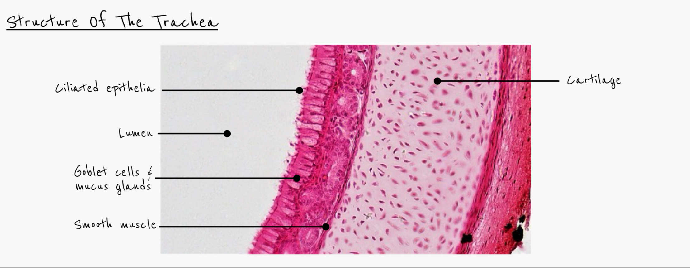
What is the structure and function of the trachea
carry clean, warm air from nose to chest
Supported by C-shaped rings of cartilage to stop trachea from collapsing but allowing food to move down oesophagus
Smooth muscle contracts to narrow lumen
Elastic fibres allow lumen to dilate
Lined with goblet cells (produce mucus to trap pathogens) and ciliated epithelial cells (move mucus to throat)
What is the structure and function of the bronchi
leads to left and right lungs
Supported by smaller rings of cartilage
Smooth muscle contracts to narrow lumen
Elastic fibres allow lumen to dilate
Lined with goblet cells and ciliated epithelial cells
What is structure and function bronchioles
Narrow tubes with columnar epithelial cells
Made from smooth muscle and elastic fibres so can contract and relax to control air flow
No cartilage or goblet cells present
What is the structure and function of the alveoli
site of gas exchange
Elastic fibres allow alveoli to stretch and recoil to return to original shape.
Have squamous epithelial cells, but no smooth muscle, cartilage or goblet cells
How are goblet cells adapted to their function
microvilli to increase the surface area
Lots of secretory vesicles to release mucus through exocytosis
Lots of rough endoplasmic reticulum to produce large quantities of mucin (protein in mucus)
Golgi to process the mucin
How does the respiratory system deal with external air
nasal passages are highly vascular so warm any cold air
Nasal hair traps any larger particles
Goblet cells secrete protective mucus which traps pathogens so macrophages roaming in mucus can engulf pathogen.
Ciliated epithelial cells waft mucus up and it is swallowed into stomach acid which kills the pathogens.
Explain the importance of ciliated epithelial cells and goblet cells in the trachea and bronchi
goblet cells secrete mucus
Dust particles and bacteria stick to the mucus
Cilia beat these particles up to the throat where they are swallowed
Stomach acid kills any pathogens
Keeps bronchial tree clean and defends against infection
Explain the importance of cartilage in the trachea and bronchi
Keeps trachea and bronchi open during pressure changes of ventilation
When food is passing down oesophagus, ring structure enables trachea to be flexible so neck can bend
Provides attachment for smooth muscles in trachea
Ring structure allows trachea to lengthen and shorten during ventilation movements.
Explain the importance of smooth muscle in the respiratory system
muscle contracts to enable trachea/bronchi/bronchioles to dilate or constrict
Changes diameter of airway, helping regulate airflow
Gives some elastic recoil after stretching, which helps move air out.
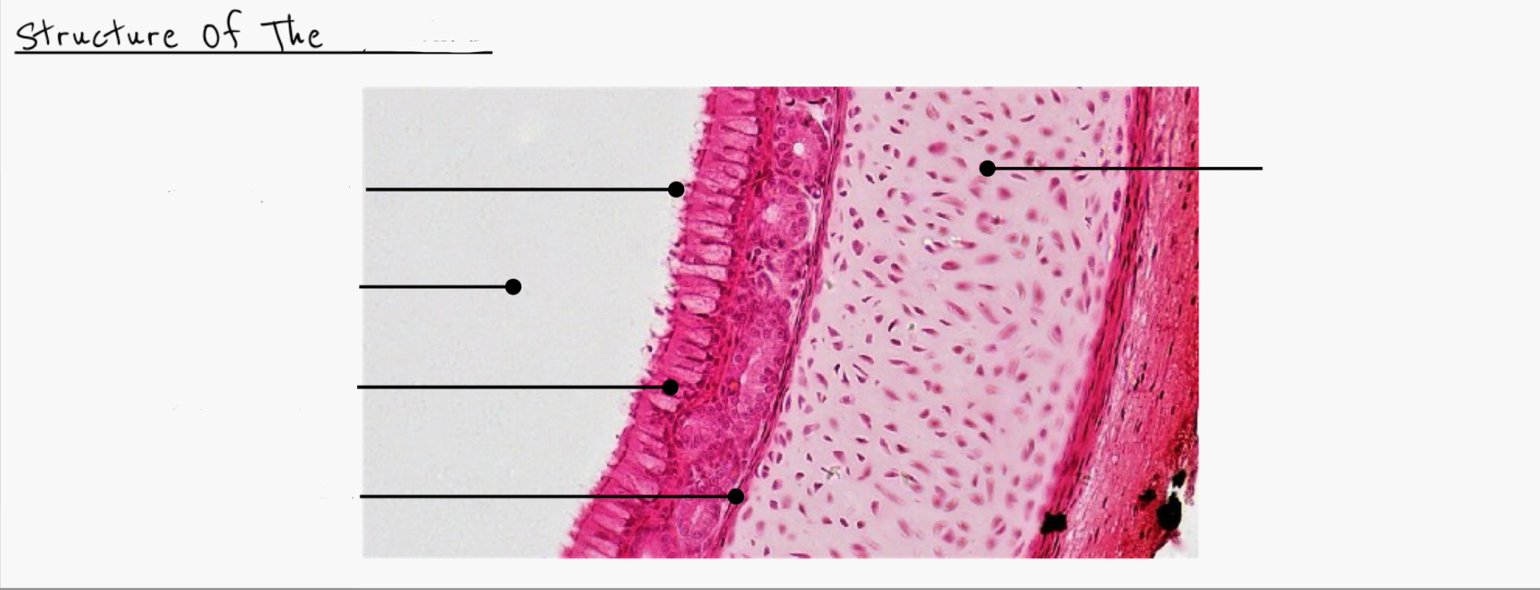
Name and label this structure
Trachea
Describe what happens in the alveoli
gas exchange
Oxygen diffuses from air to blood and carbon dioxide from blood to air
Oxygen binds to haemoglobin in red blood cells
Volume of alveoli increases during inspiration
Ventilation ensures concentration gradients of gases maintained
How are the alveoli adapted for gas exchange
very large surface area so large surface area to volume ratio
Thin squamous epithelial cells so short diffusion distance
Most, lined with tissue fluid so gases can dissolve - contains surfactants to lower surface tension and allow lung expansion
Good blood supply from capillaries and good ventilation maintaining steep concentration gradient.
Define ventilation
Inhalation and exhalation of air between the lungs and the outside
How are the alveolar walls adapted for enabling ventilation movements
Contains elastic fibres
Enables dimension changes due to ventilation
Elastic recoil helps to push air out of alveoli
Which structures contain cartilage
Trachea
Bronchi
Which structures contain smooth muscle
Trachea
Bronchi
Bronchioles
Which structures contain ciliated epithelial cells
Trachea
Bronchi
Which structures contain goblet cells
trachea
Bronhci
Which structures contain squamous epithelial cells
Alveoli
Which structures contain columnar epithelial cells
Bronchioles
Which structures contain capillaries
Alveoli
What is this
Trachea
What is this
Bronchi
What is this
Bronchioles
What is this
Alveoli