tanginamo pag to naalis nanaman
1/18
There's no tags or description
Looks like no tags are added yet.
Name | Mastery | Learn | Test | Matching | Spaced |
|---|
No study sessions yet.
19 Terms
Protozoans
They are “single-celled” organisms which are generally classified according to their ‘organelles of locomotion’
Unicellular, eukaryotic organisms
“Animal-like” protists
No cell wall
Additional info of protozoans
Organelles:
Nucleus
Nucleolus
Mitochondria
Locomotor Organelles
Two Regions of Cytoplasm:
Ectoplasm (outer)
Endoplasm (inner)
Contains at least one and some several nuclei
Some contain vacuoles
With special organs for locomotion
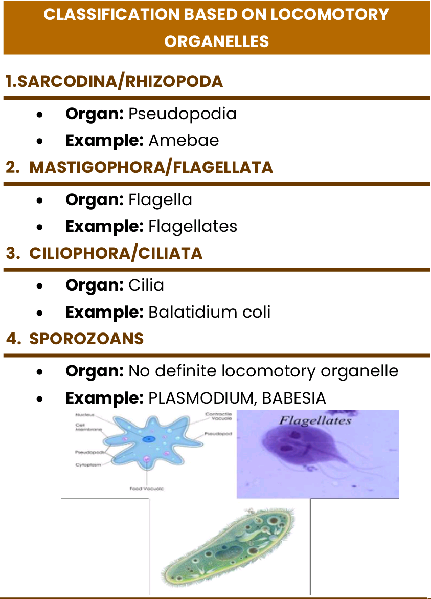
Class Sarcodina
• Have protoplasmic processes, or pseudopodia, for locomotion
• Possess in their life cycle the:
Trophozoite stage
Precystic stage
Cystic stage
Metacystic stage
• With cystic stage exc. for Entamoeba gingivalis (mouth)
• Inhabit the large intestine exc. for Entamoeba gingivalis
• Commensals exc. for Entamoeba histolytica (non-pathogenic)
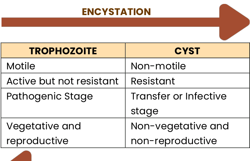
Cyst
• Non-motile
• Usually infective
• Found in formed feces
• Resistant to damage
• Usually smaller than trophozoite
• Best visualized using iodine
Trophozoite
Motile
Usually not infective
Found in liquid feces
Susceptible to damage; easily disintegrates
Usually larger than cyst
Best seen using permanent stains
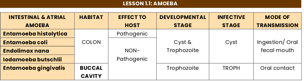
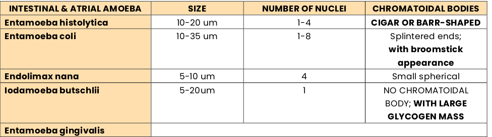
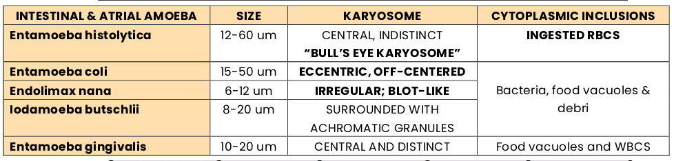
Entamoeba histolytica
12-60 um
central, indistinct “bull’s eye karyosome”
Ingested RBCs
Entamoeba coli
15-50 um
Eccentric, off-centered
Bacteria, food vacuoles, & debri
Endolimax nana
6-12 um
Irregular, “blot-like”
Bacteria, food vacuoles, & debri
Iodamoeba butschlii
8-20 um
Surrounded with achromatic granules
Bacteria, food vacuoles, & debri
Entamoeba gingivalis
10-20 um
Central and distinct
Food vacuoles and WBCs
Entamoeba histolytica (Intestinal Amebiasis)
Acute/Symptomatic
Acute/Symptomatic
Blood diarrhea, abdominal pain
Patient passes out ‘trophozoites’ in feces
Chronic/Asymptomatic
Stool consistency is ‘normal’
Patient are carriers
Patient passess ‘cyst’ in feces
Entamoeba histolytica (Extraintestinal Amebiasis)
• Trophozoite is ‘tissue invading’ forms:
Hepatic amebiasis
Pulmonary amebiasis
Cerebral amebiasis

Naegleria fowleri
Cyst size: 7-15 um
# of Nucleus: 1
Trophozoite:
Measures: 10-35 um
2 Forms:
Amoeboid Troph: Lobophodia
Flagellated Troph: 2 Flagella
Infective Stage: Amoeboid Trophozoite
Diagnostic Stage: Trophozoite in CSF & Brain tissues; flagellated forms occasionally in CSF
Cyst is ‘not seen’ in brain tissues
Cyst has ‘double wall’
Flagellated forms with 2 flagella and ‘rapidly motile’
Acanthamoeba spp.
Cyst Size: 10-25 um
# of Nucleus: 1
Trophozoite:
Measures: 15-45 um
With “Spine-like” Projections: Acanthopodia
Infective Stage: Cyst & Trophozoite
Diagnostic Stage: Cyst & Trophozoites in tissues;
Trophozoite has ‘spine-like’ process called “acanthophodia” (slowly motile”
Balamuthia mandrillaris
Isolated from soil and dust or from autopsy specimens of infected humans and animals
Infective Stage: Cyst & Trophozoite
Can Enter Thru: Nasal Passages & Ulcerated & Broken Skin
Cyst & Trophozoite in tissues
Cyst is 10-25 um with ‘double cyst wall’

Dientomoeba fragilis
Previously classified as an ‘amoeba’
Trophozoite is 5-15 um
With Hyaline “leaf-like” pseudopodia
Karyosome is composed of a cluster of 4-8 granules
Often referred to as “fragmented karyosome” or “tetrakaryosome”