Anatomy: Skeletal Landmarks: Skull
1/68
There's no tags or description
Looks like no tags are added yet.
Name | Mastery | Learn | Test | Matching | Spaced | Call with Kai | Chat |
|---|
No analytics yet
Send a link to your students to track their progress
69 Terms
Anterior; top and front part of the skull; Forehead bone
Frontal Bone
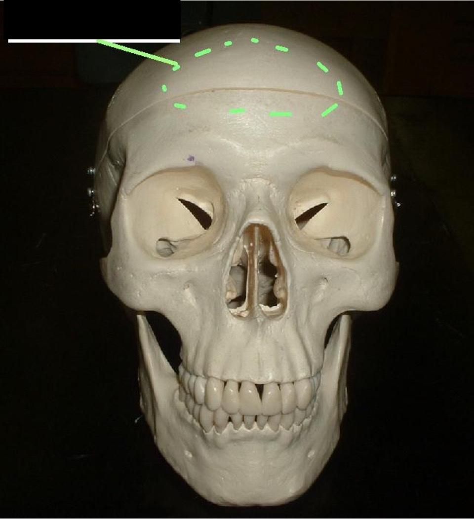
The connection between the frontal bone and parietal bone (between the front and the back)
Coronal Suture
middle, front, flat part of the frontal bone
Frontal Squama
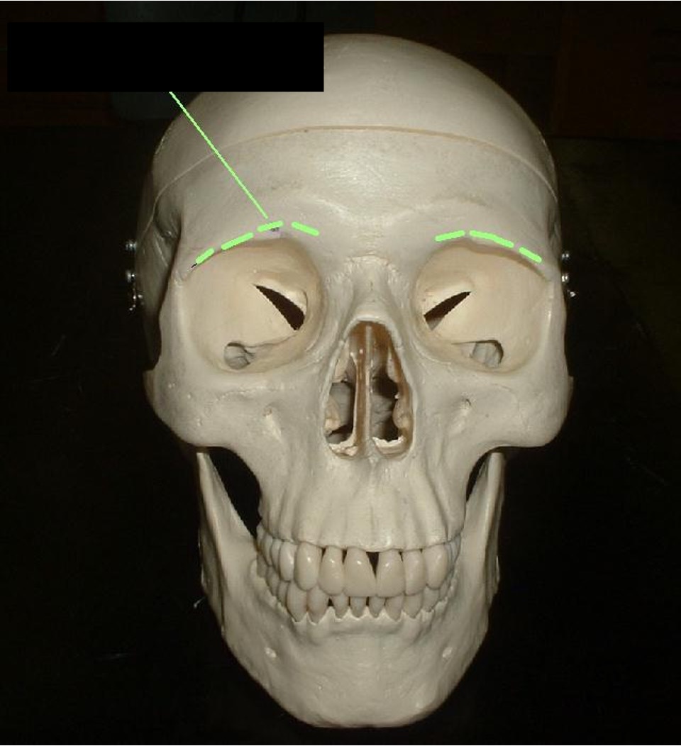
around the top of the eye socket; edge; eyebrow bone
Supraorbital Margin
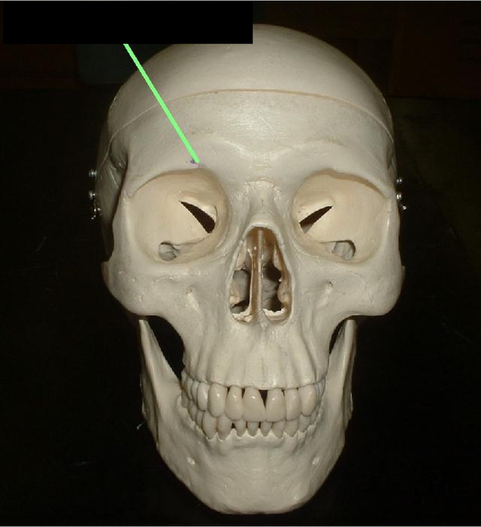
above the eye socket; a hole that is usually circular.
Supraorbital Foramen
lower forehead, above the nose, in between eyebrows. (you cannot see on the skull)
Frontal sinus
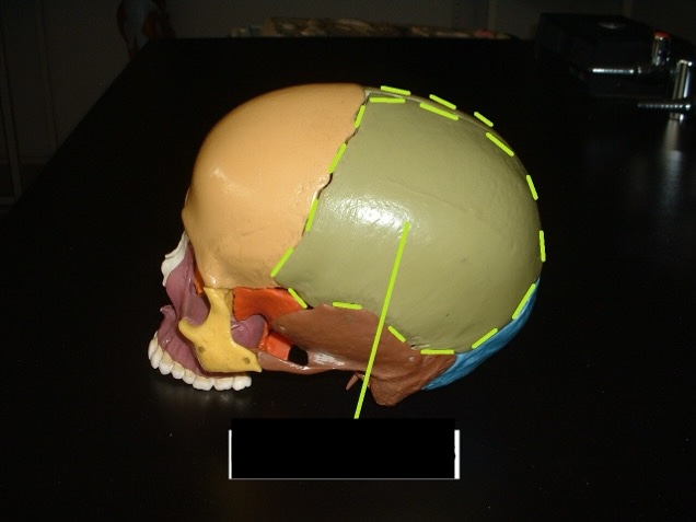
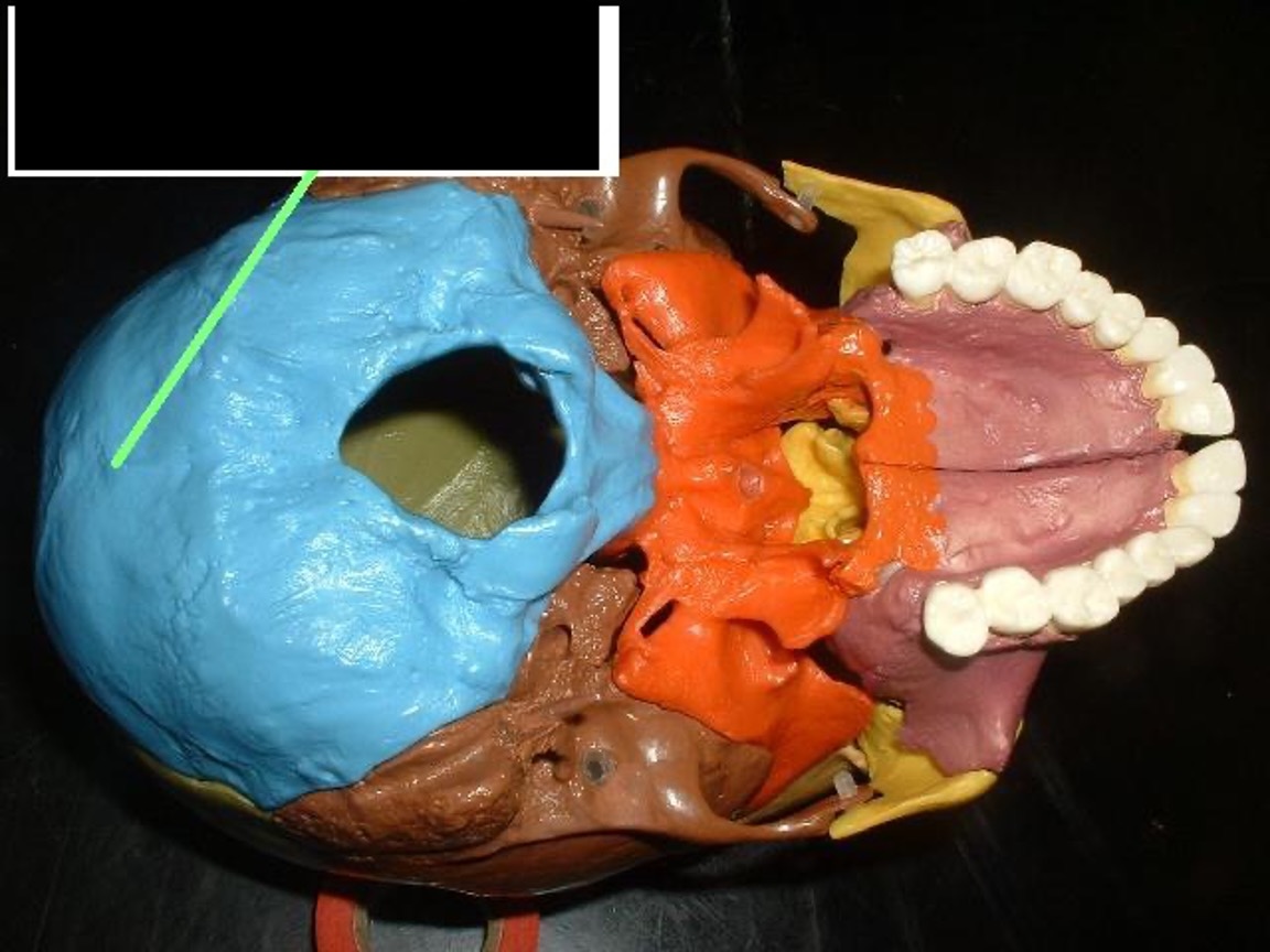
superior posterior; on top and behind the frontal bone but before the occipital bone.
Parietal Bones
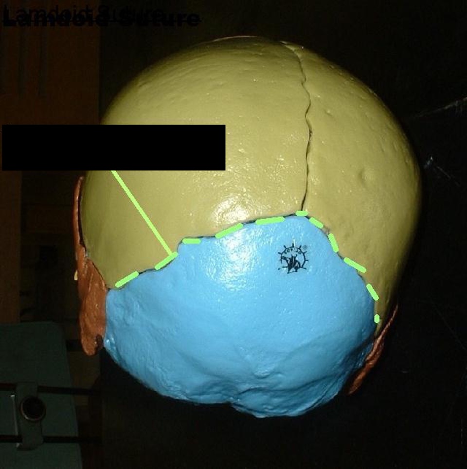
suture separating parietal and occipital bone. (looks like a lambda)
Lambdoid Suture
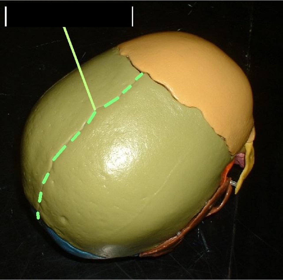
mid-sagittal suture separating the left and the right of the parietal bone.
Sagittal Suture

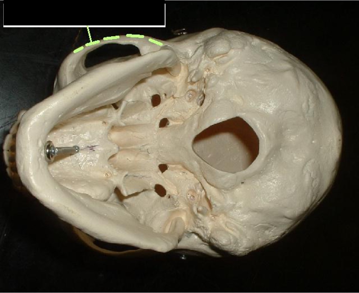
suture separating the parietal and temporal bones.
Squamous Suture
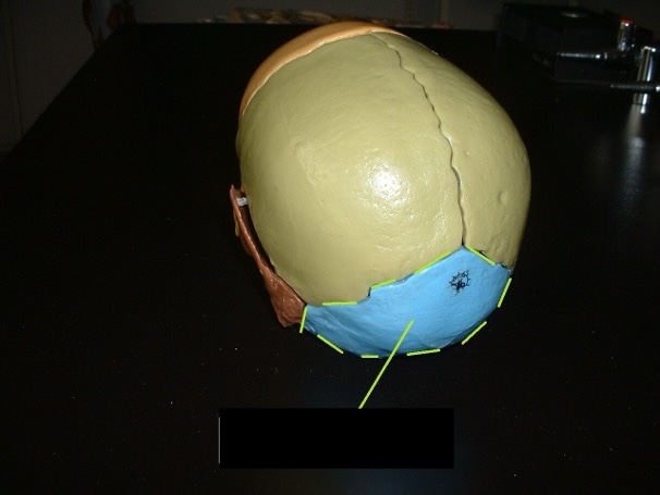
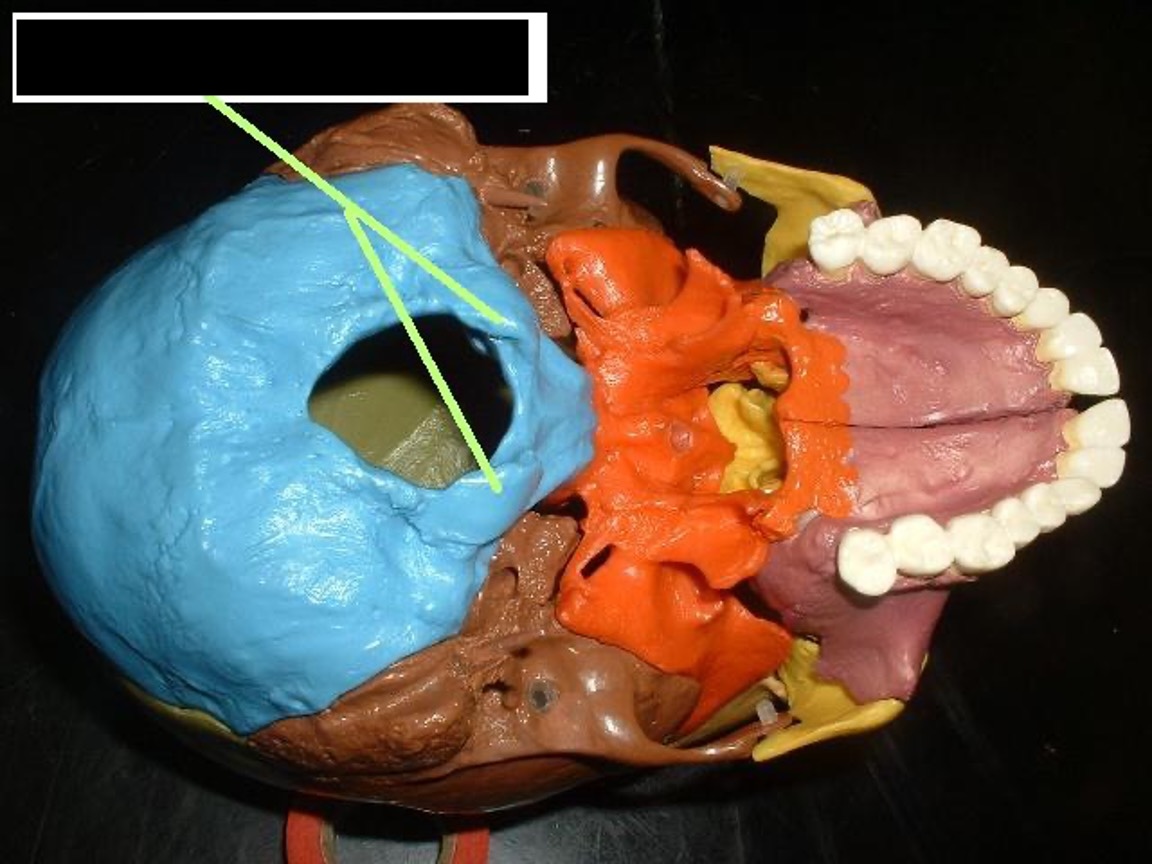
the back of the head; inferior, posterior. it has a hole in the bottom that the spinal cord goes through (foramen magnum).
Occipital Bone
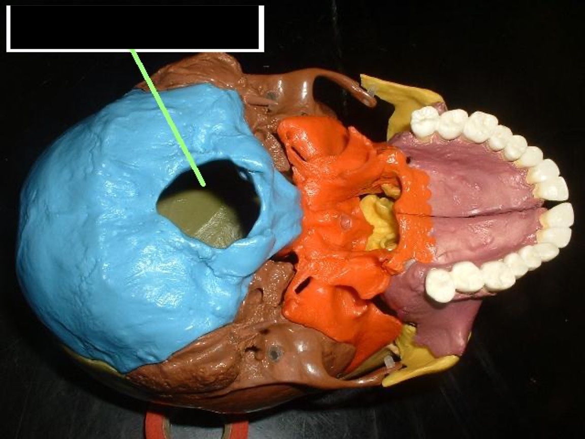
a hole in the bottom of the occipital bone for the spinal cord.
Foramen Magnum
bumps on the sides of the foramen magnum
Occipital Condyle
bump on the middle back of the head.
External Occipital Protuberance
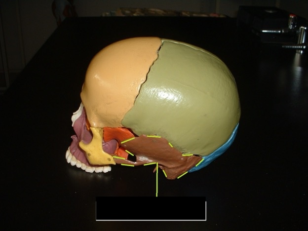
The bones on the side of the head
Temporal Bone

the flat part of the temporal bone
Temporal Squama

the part that connects the temporal bone to the zygomatic bone
Zygomatic Process
It looks like a handle off of the side of the head; it includes temporal bone, zygomatic process, and maxilla; it sticks out.
Zygomatic Arch
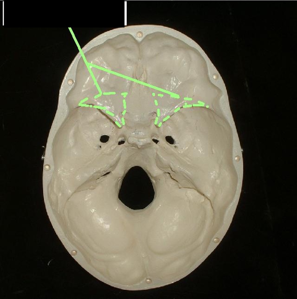
Diagonal rigid bone inside the top of the skull. (Think Cayman Islands Hell)
Petrous Portion
Left and right of the foramen magnum the circular hold closer to the jaw
Carotid Foramen
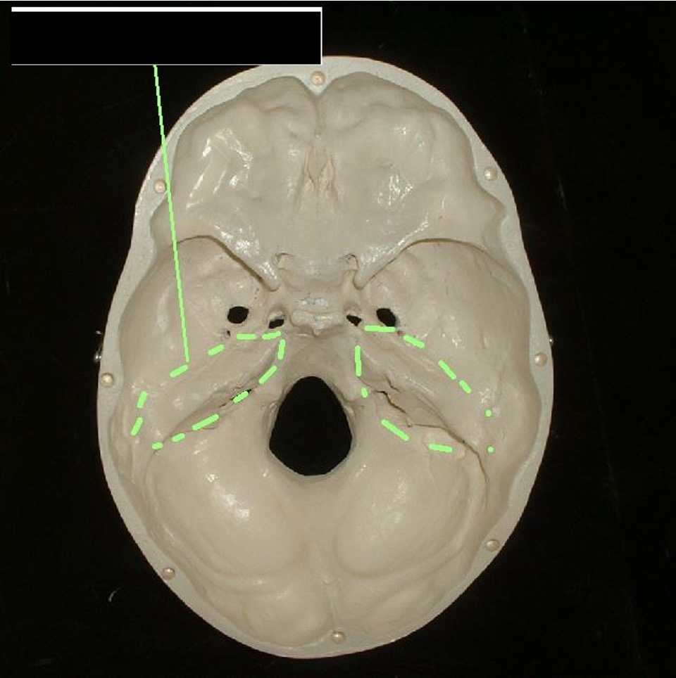
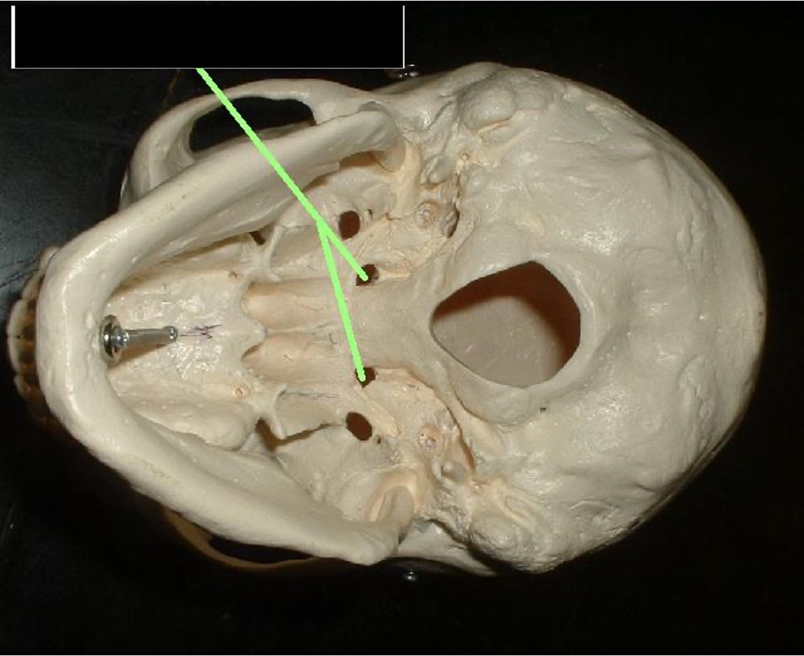
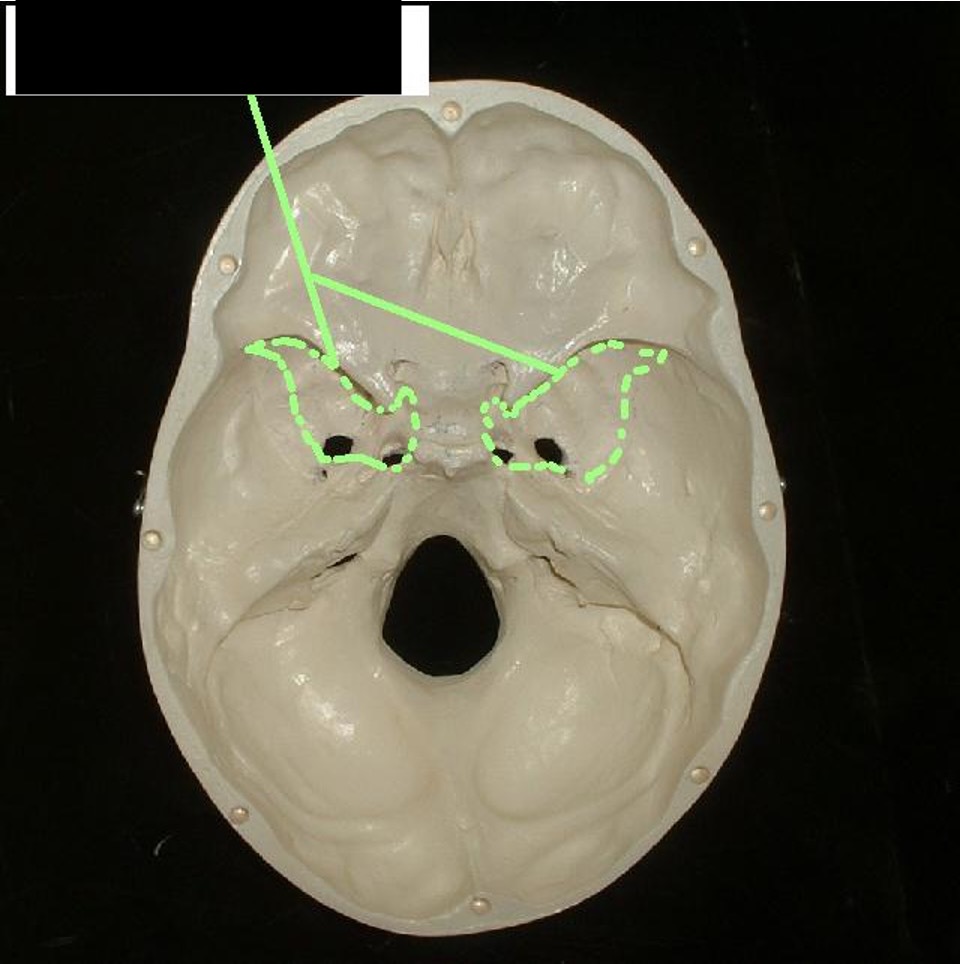
left and right of the foramen magnum; peanut (in shell shaped); behind the Carotid Foramen
Jugular Foramen
two holes that are close together in front of the carotid foramen
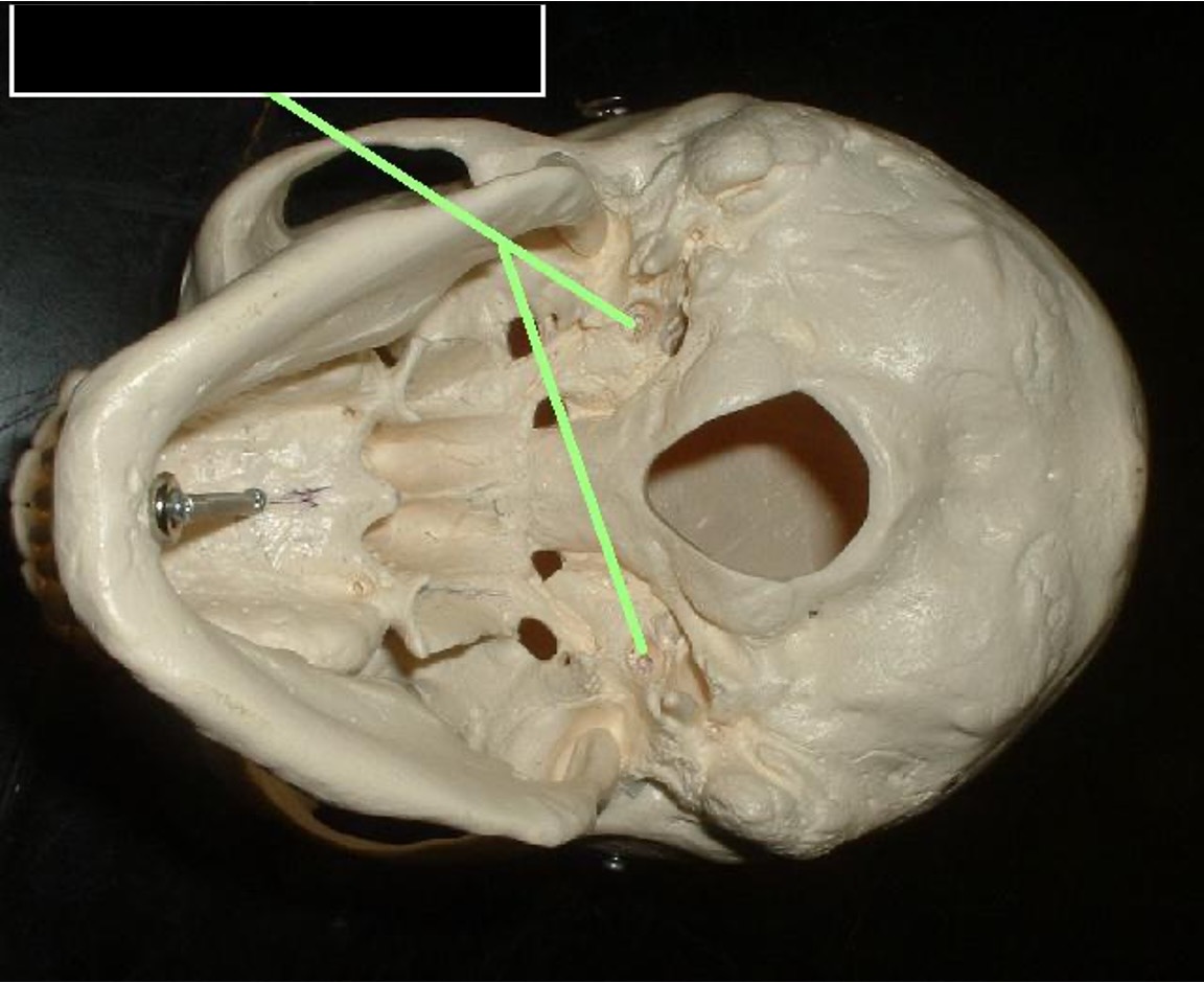
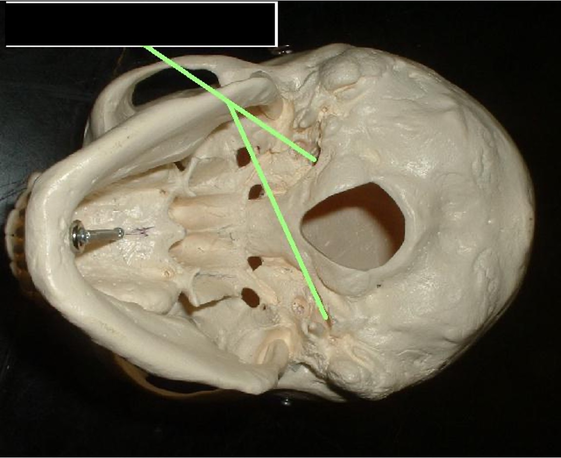
Foramen Lacerum
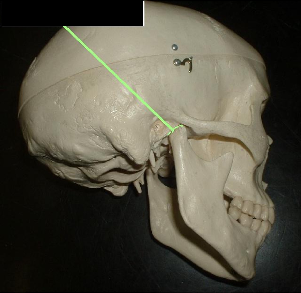
a dent that the mandible fits into; it is in front of the ear lobe.
Mandibular Fossa
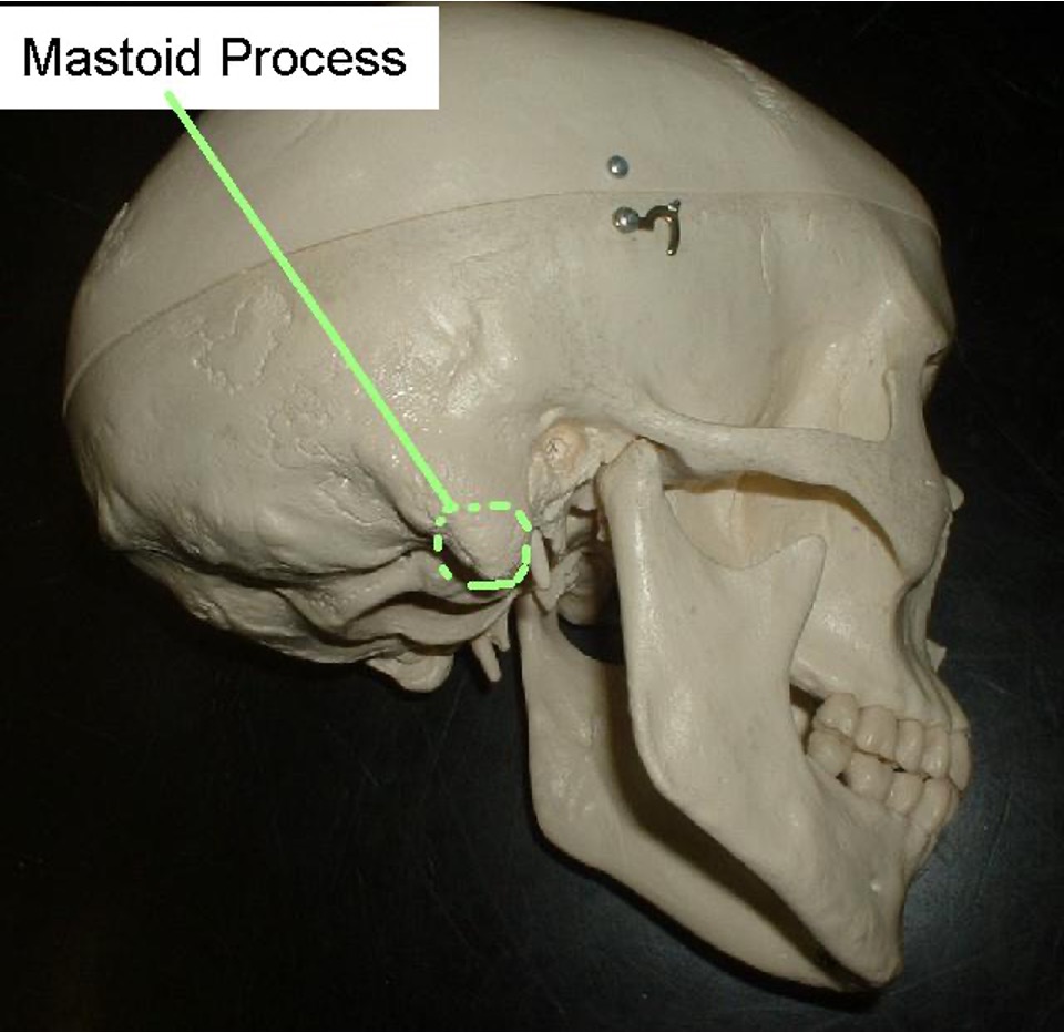
below the mastoid portion that is small, circular, and pinchable.
Mastoid Process
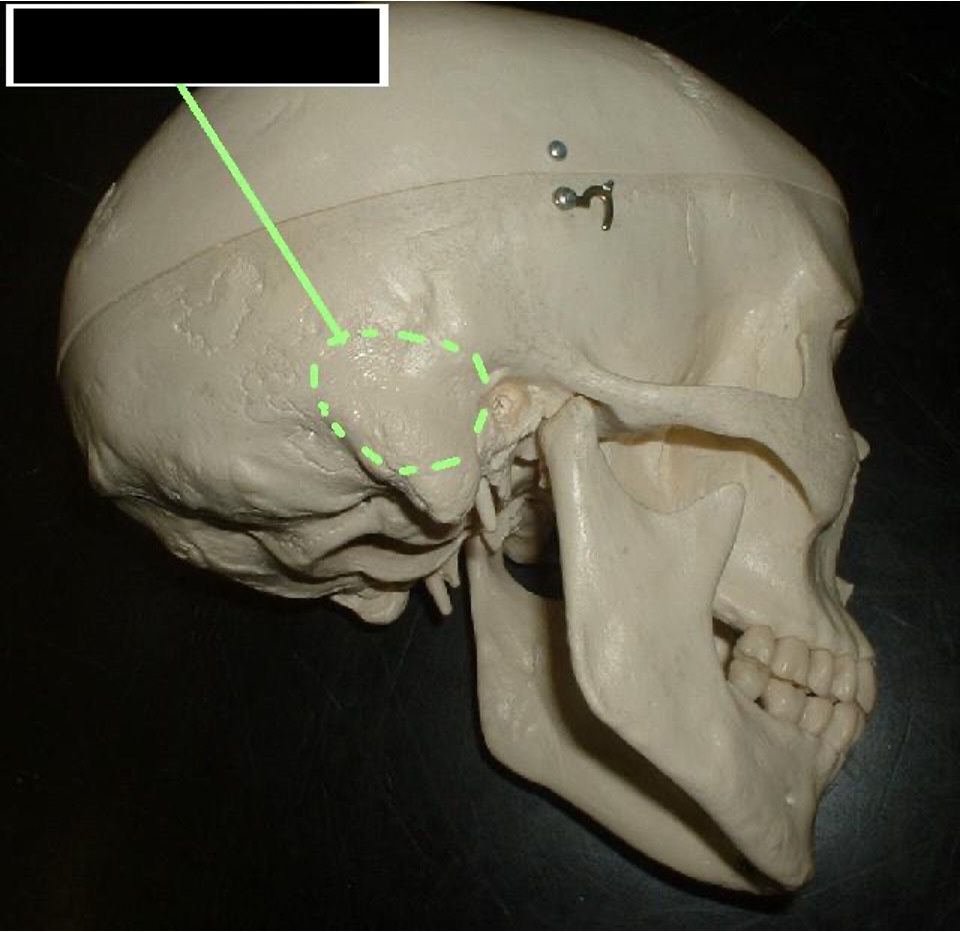
bump behind ear hole
Mastoid Portion
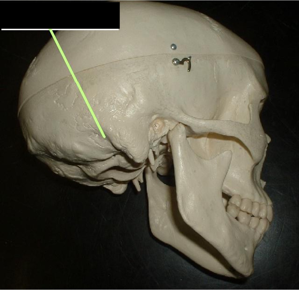
a hole behind the mastoid portion
Mastoid Foramen
the ear hole; it is in between the mastoid process and the zygomatic process
External Auditory Meatus

the back side of the petrous portion that runs into the ear hole
Internal Auditory Meatus
it looks like fangs on the inside of the mastoid process
Styloid Process
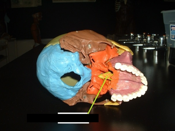
well surrounded by other bones; looks like a bat (when removed).
Sphenoid Bone
middle of the skull, view of the top of the skull; above petrous portion
Body of Sphenoid Bone
behind the eye socket; concave.
Greater Wings
little shelf above greater wings
Lesser Wings
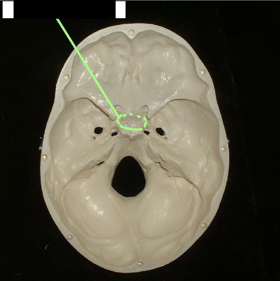
In between the two sides of the greater wings.
Sella Turcica
empty space inside the sphenoid bone.
Sphenoid Sinus
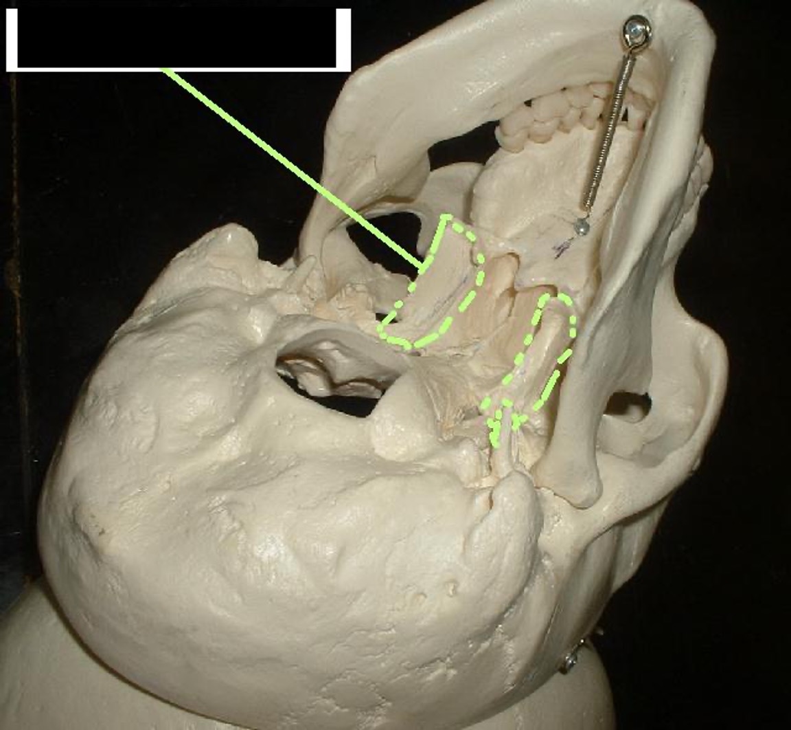
extends down and towards molars.
Pterygoid Process
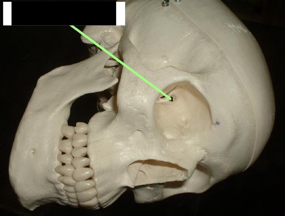
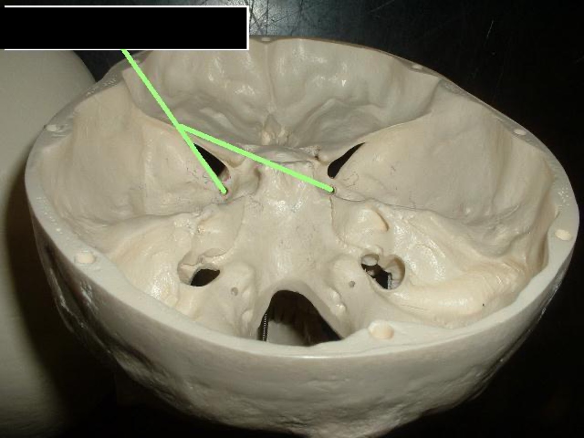
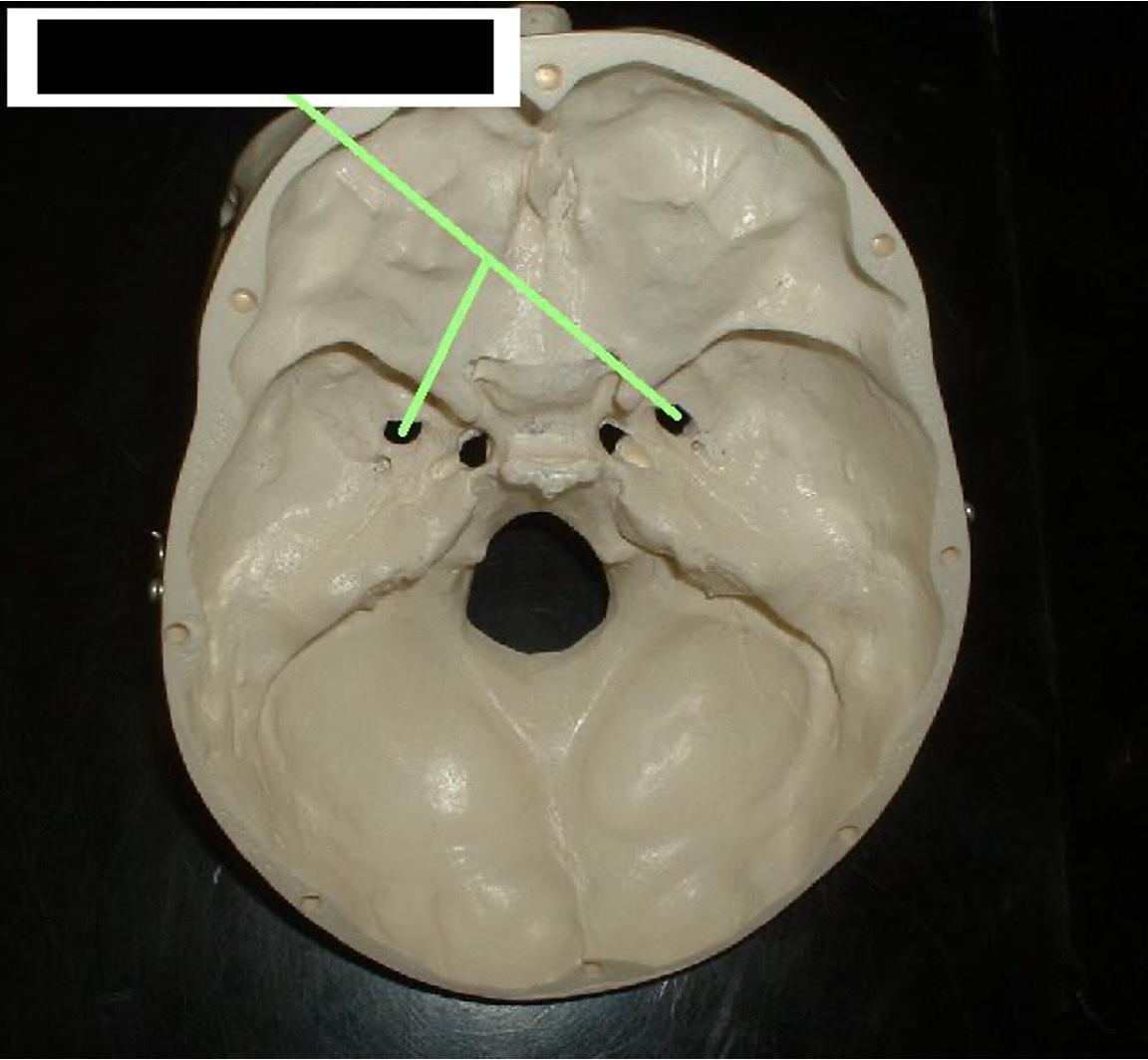
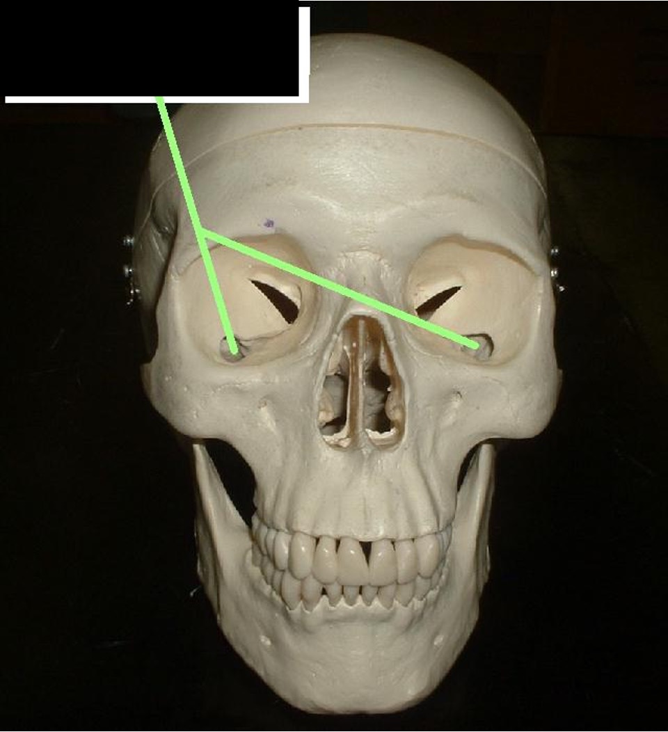
Inside of eye socket (circular) through lesser wings.
Optic Foramen
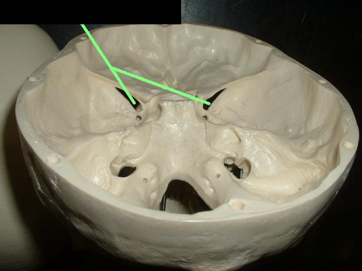
the crack beside the optic foramen
Superior Orbital Fissure
a round hole below the superior orbital fissure
Foramen Rotundum
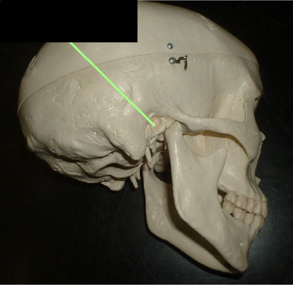
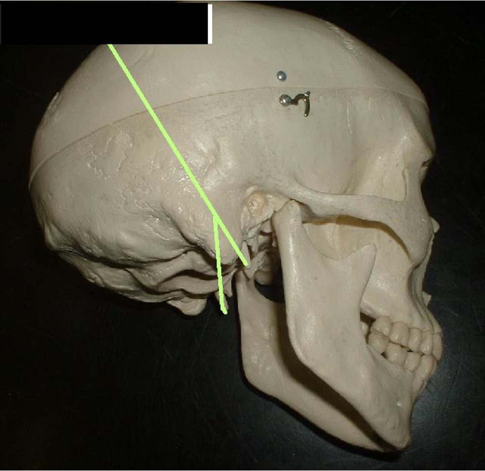
Oval-shaped hole that is about an inch behind the foramen rotundum
Foramen Ovale
a small hole that is next to the foramen ovale.
Foramen Spinosum
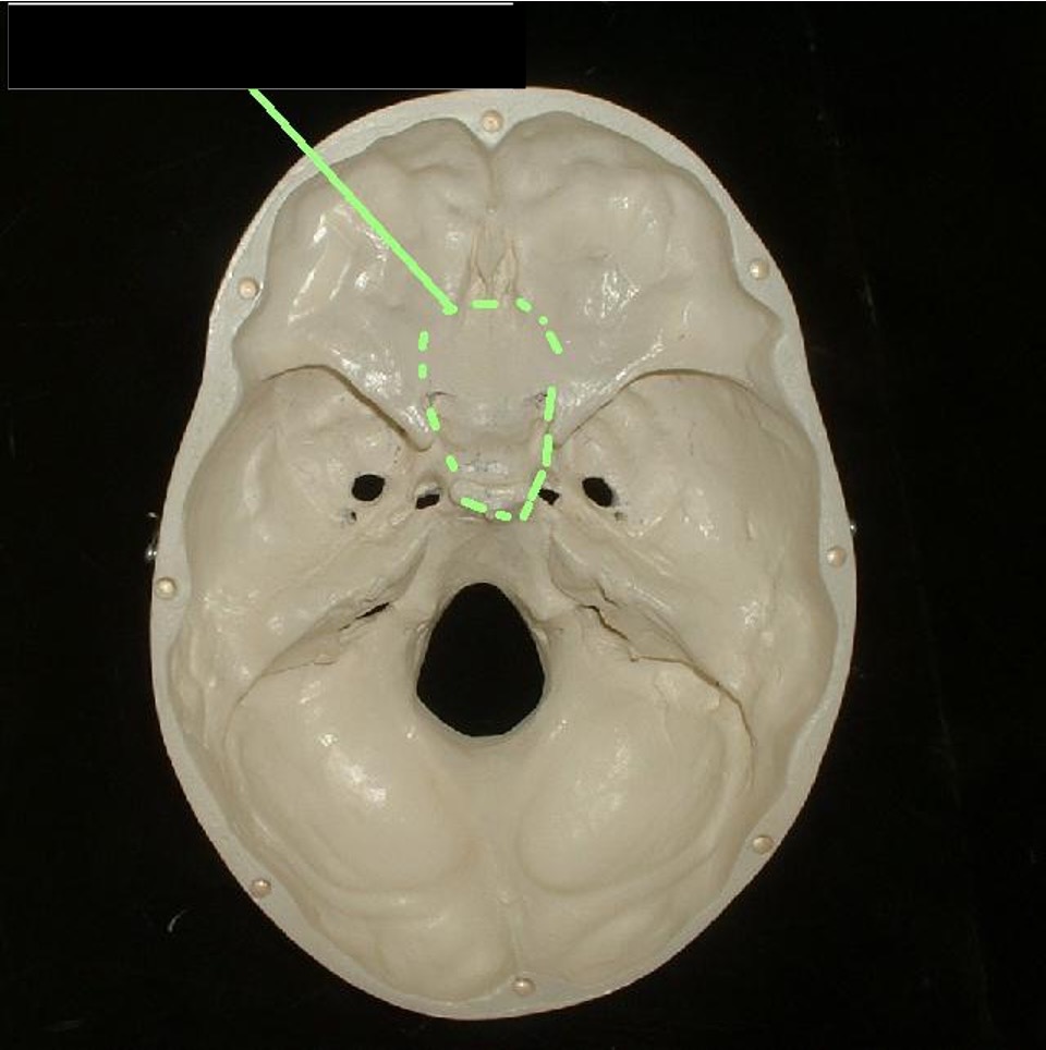
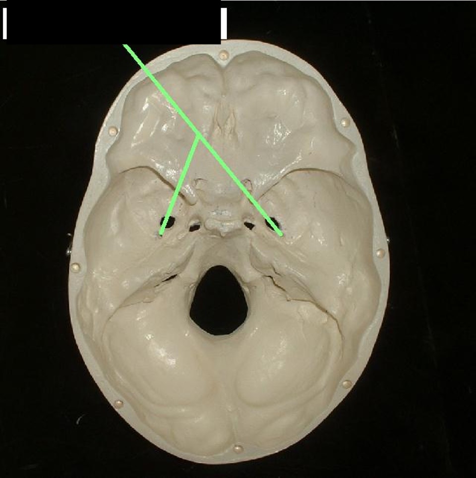
to the left and right of the Crista Galli (concave)
Cribriform Plate
middle of the nose; nose bone between the eyes.
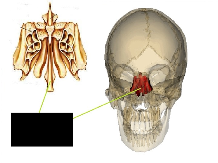
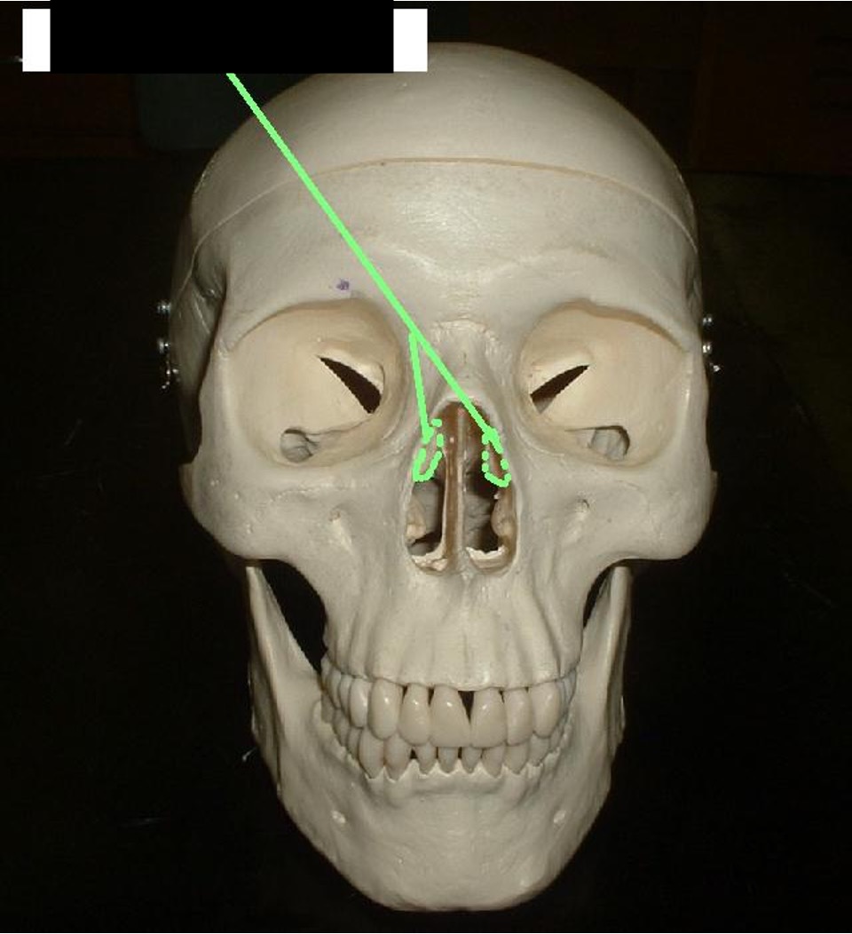
Ethmoid Bone
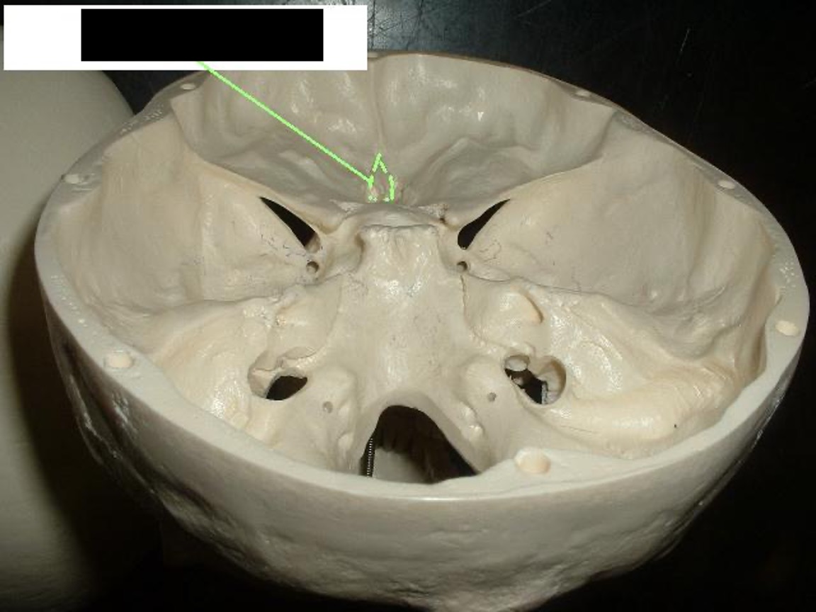
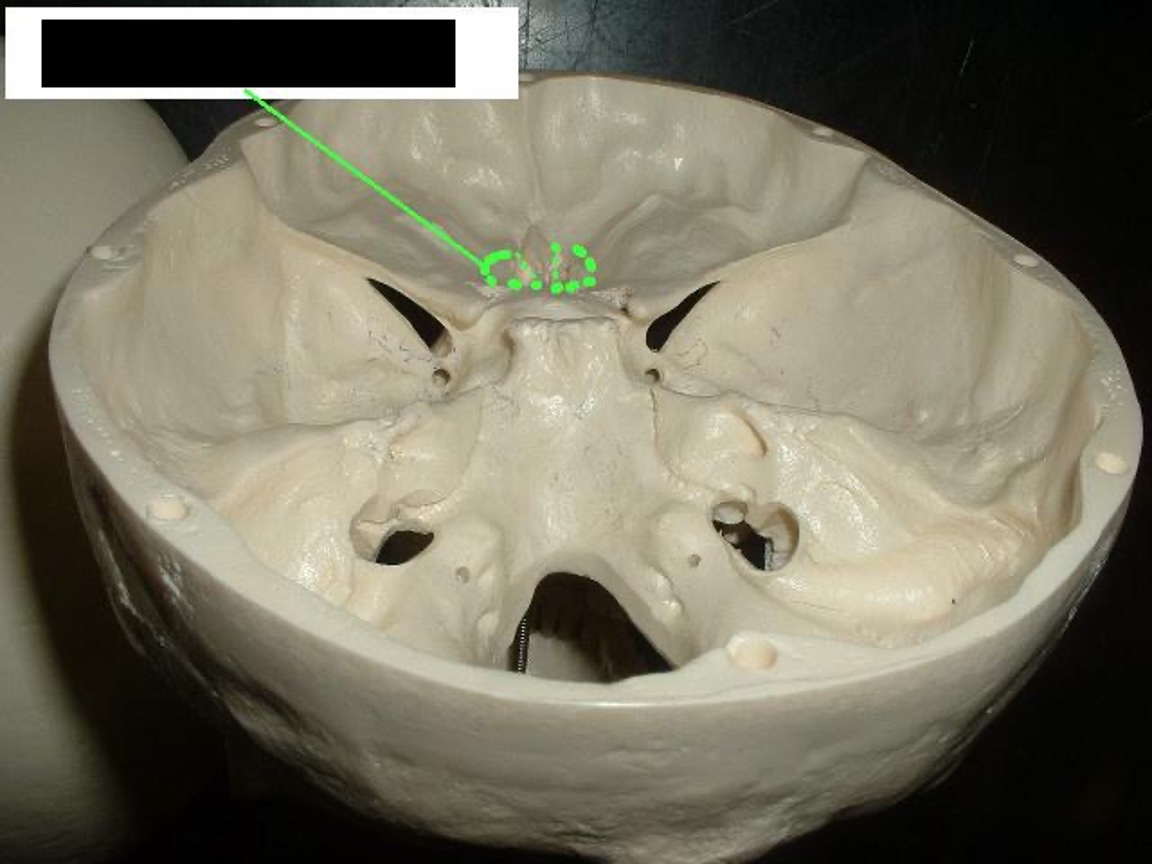
The sharp part in front of the “bat” sticks up in between the Cribiform Plate.
Crista Galli

extending vertically from the Cribiform Plate.
Perpendicular Plate
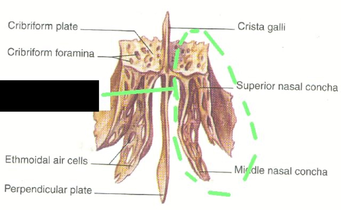
the two flaps on each side of the Perpendicular Plate.
Lateral Mass
one notch on both sides of the nose
Middle Turbinates
two bones that make up the top portion of your nose that do not connect in the middle. (bridge of your nose)
Nasal Bones
the upper jaw and the bottom part of the eye socket
Maxilla

the place where the teeth connect to the top jaw.
Alveolar Process (top Jaw)
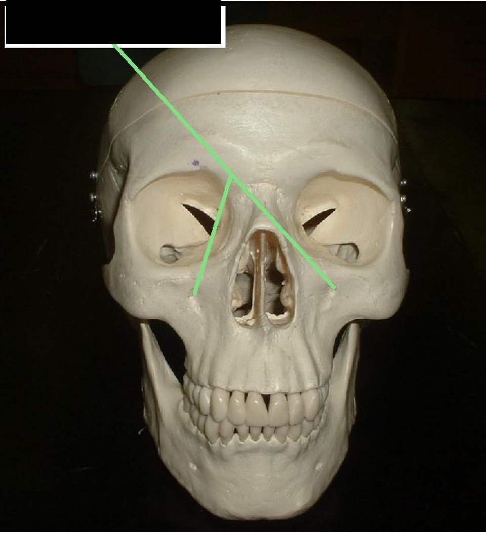
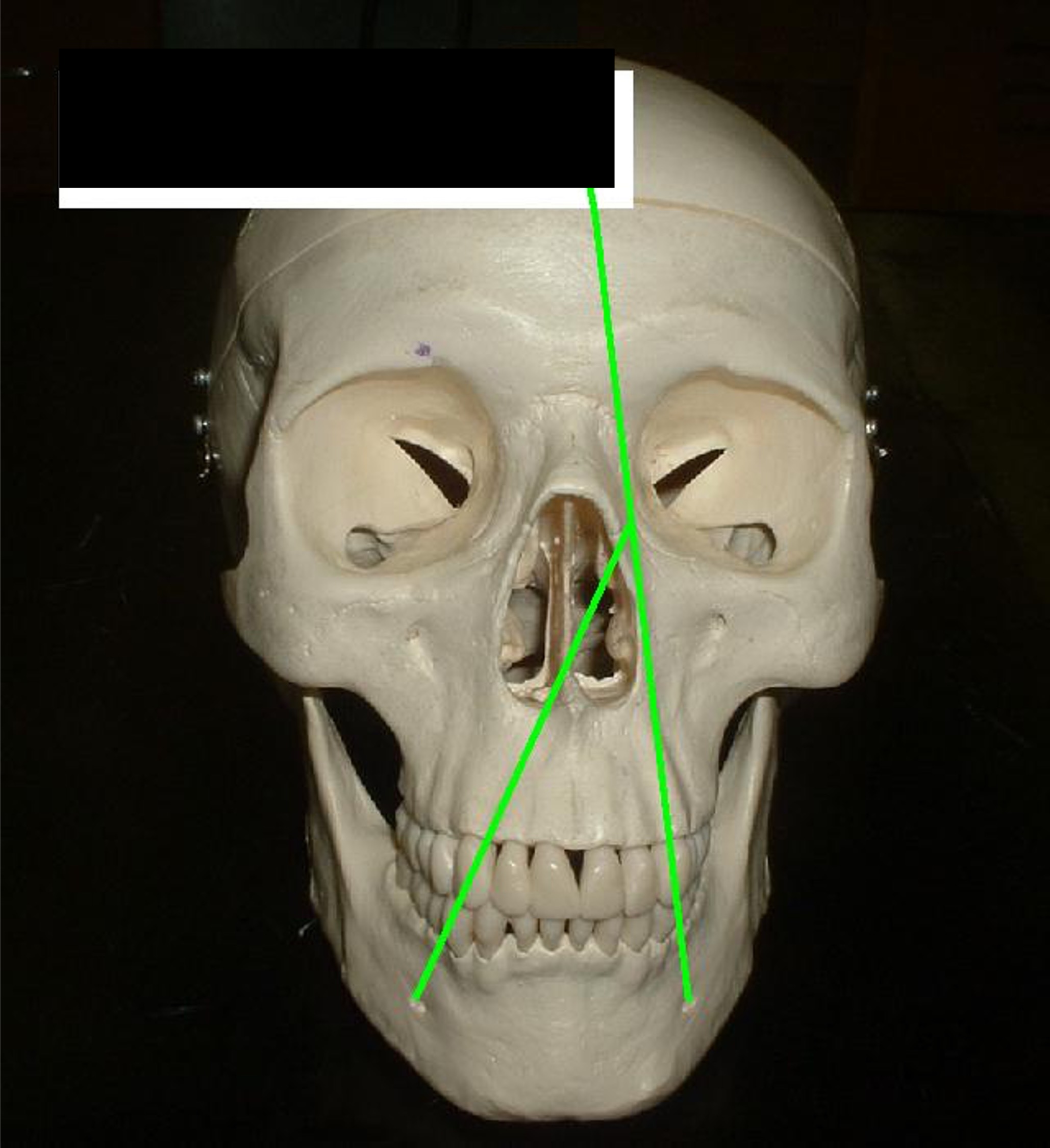
a hole below the eye socket (cheek area)
Infraorbital Foramen
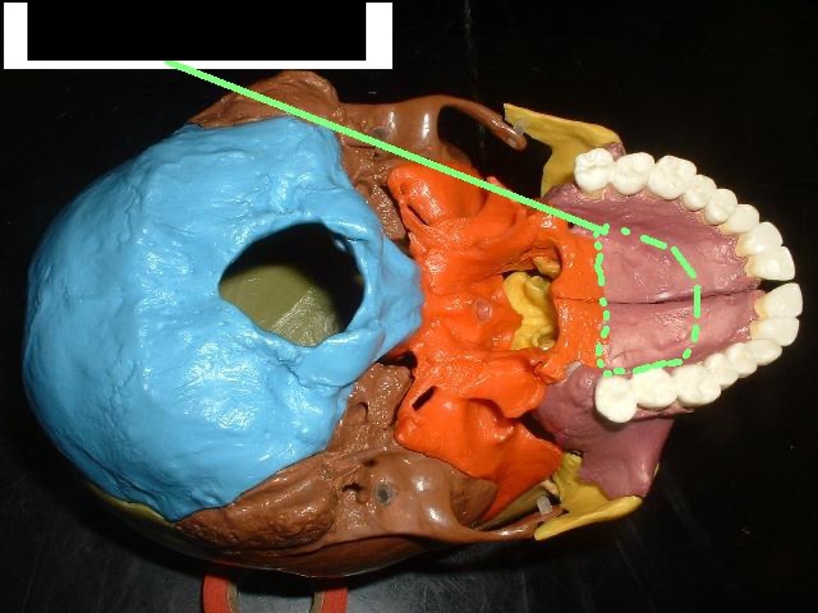
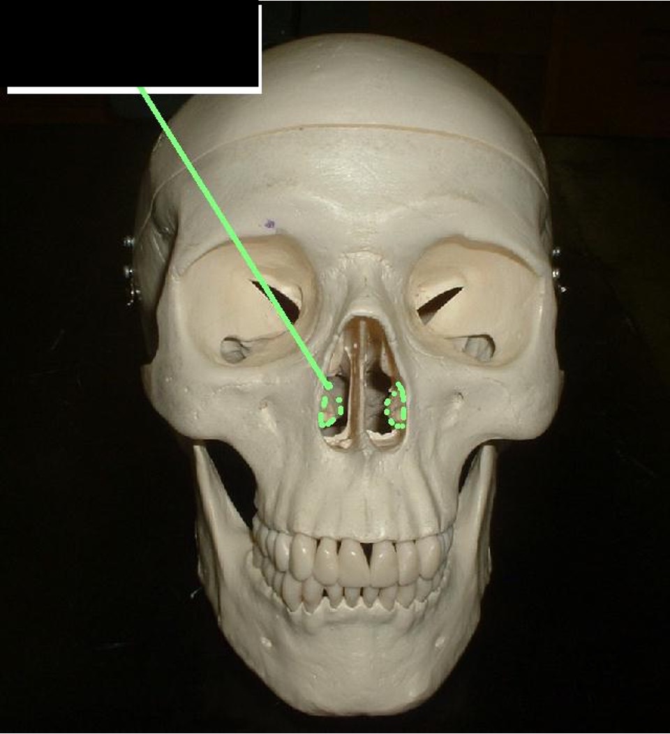
the front of the roof of the mouth
Palatine Process
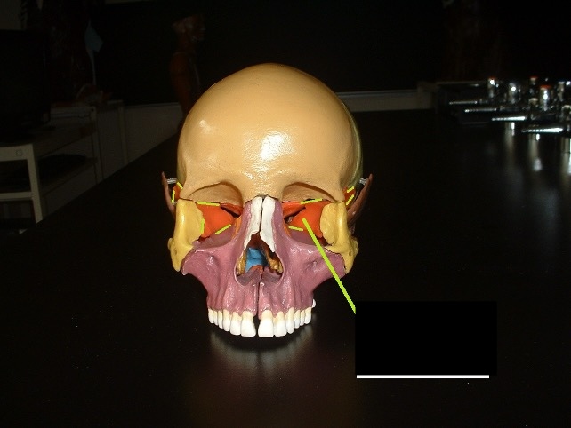
A teardrop-shaped crack in the lower eye socket
Inferior Orbital Fissure
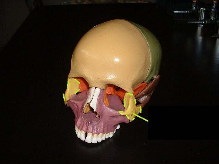
cheekbone and outer part of the eye socket
Zygomatic Bone
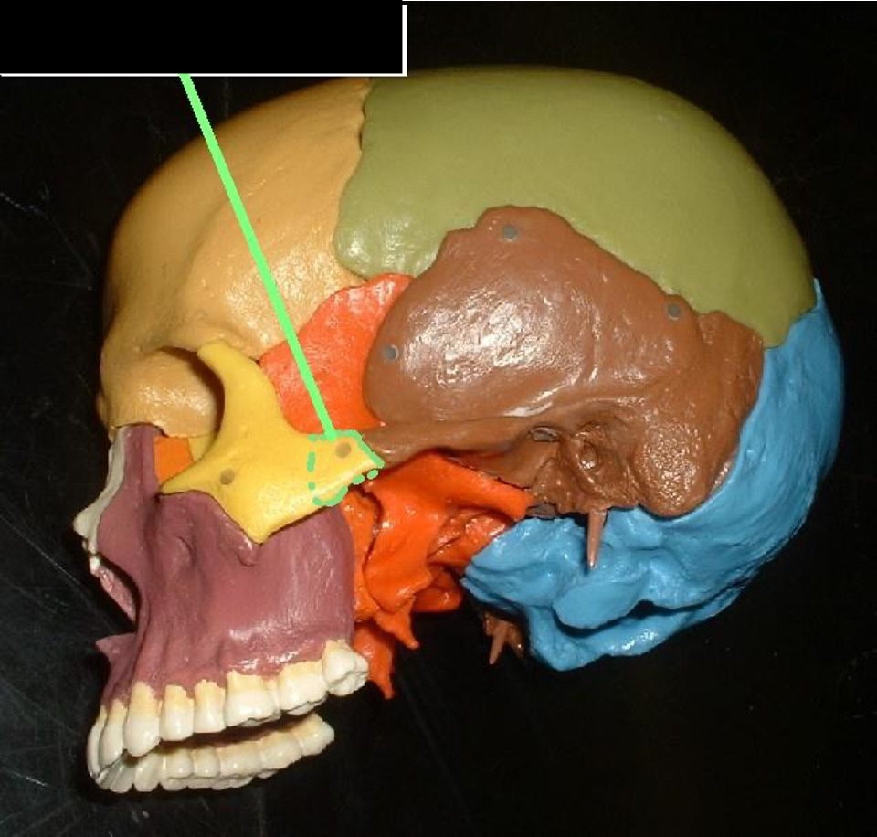
connects the Zygomatic bone to the temporal bone.
Temporal Process
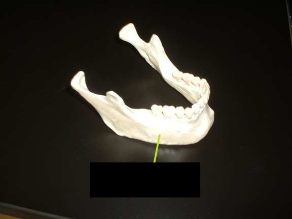
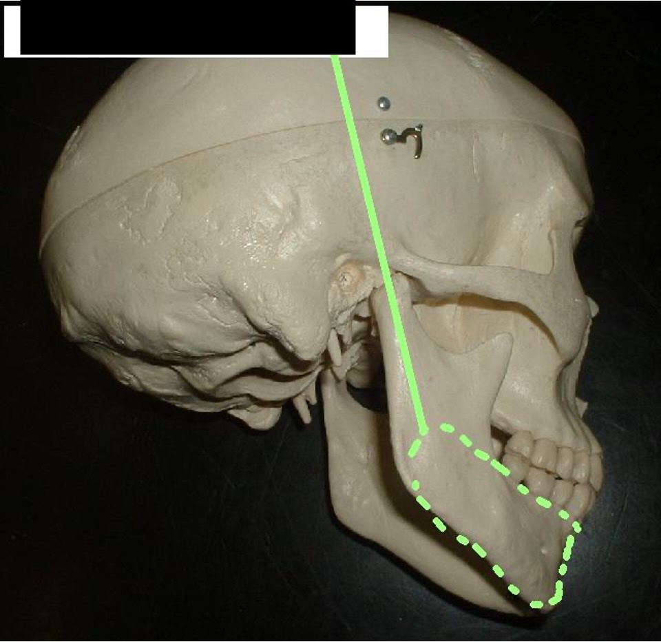
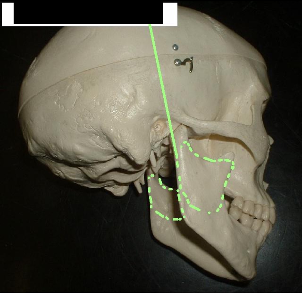
The lower jaw.
Mandible
the bottom of the mandible from a side view
Body of the Mandible
The part that runs up and down the sides of the Mandible.
Rami of Mandible
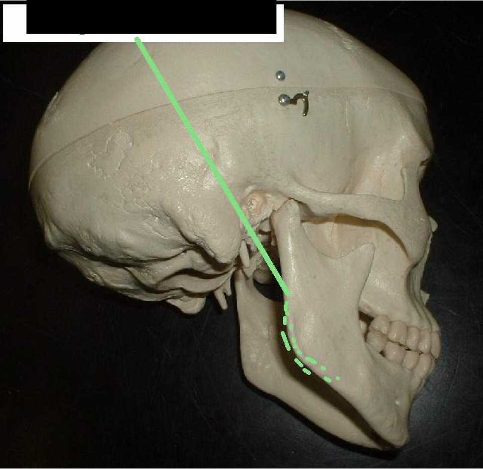
The jawline (where the jaw changes directions)
Angle of Mandible
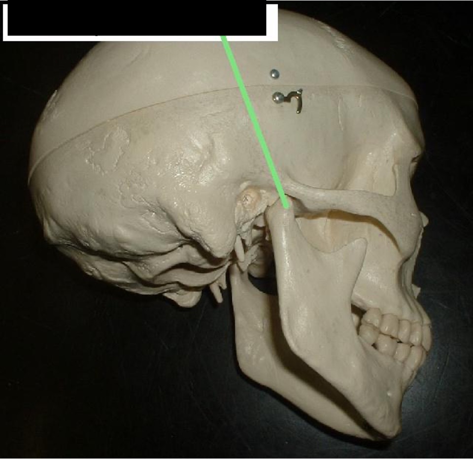
where the Mandible connects to the upper jaw.
Condylar Process
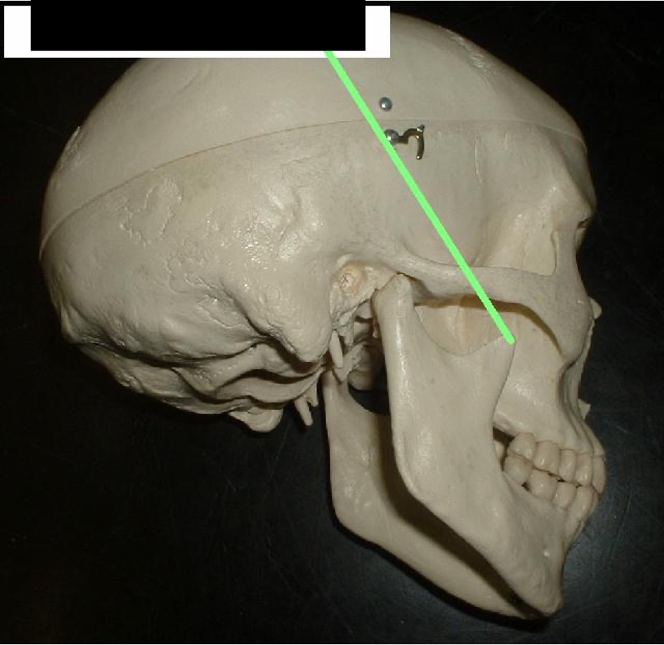
where the jaw moves up and down (with assistance from muscles)
Coronoid Process

A dent that is between the coronoid process and the condylar process.
Mandibular Notch
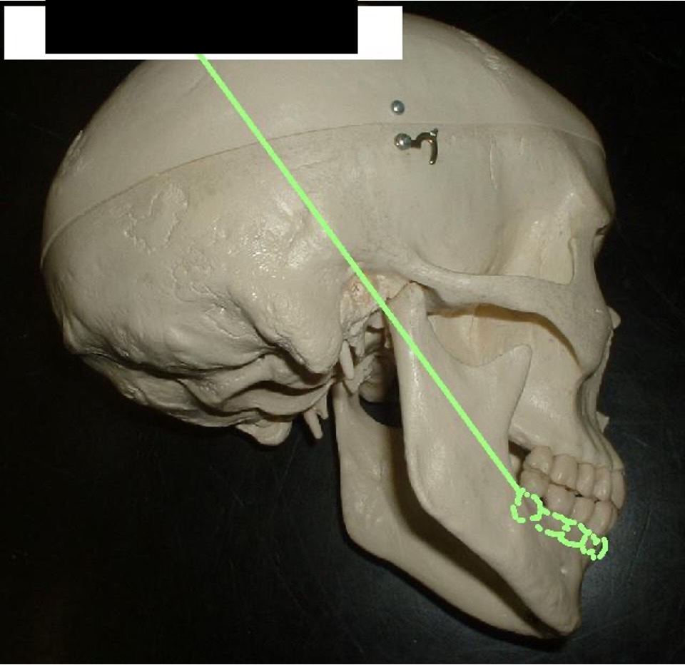
where the teeth connect to the lower jaw.
Alveolar Process (lower Jaw)
small holes (2) in the side of the chin (off center not side of the face)
Mental Foramen

triangle portion between the side of the nose and eye socket.
Lacrimal Bone
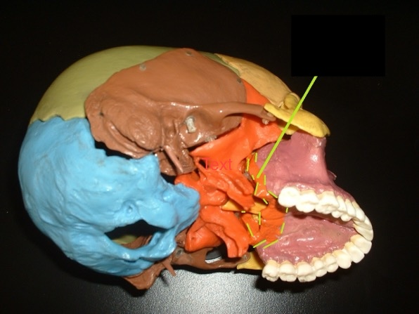
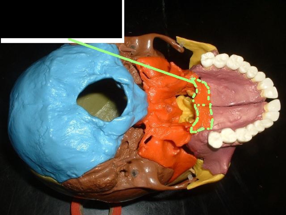
a boxy “U”-shaped bone behind the palatine process. The back of the roof of the mouth.
Palatine Bone
where the pterygoid process connects
Horizontal Plates of Palatine Bone
the bottom notches inside the nose underneath the Middle ______
Inferior Nasal Conchae
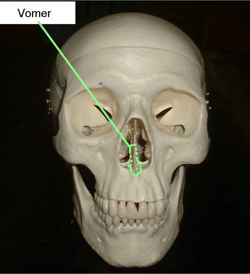
a flat triangular bone that divides the nose; the point of the Vomer sticks out of the lower jaw; If removed looks like a Dorito.
Vomer