Lecture 9: Bovine Infectious Infertility & Abortion
 Call Kai
 Learn
Learn Practice Test
 Spaced Repetition
 Match
Match Flashcards
 Knowt Play
1/39

There's no tags or description
Looks like no tags are added yet.
Name | Mastery | Learn | Test | Matching | Spaced | Call with Kai | Chat |
|---|
No analytics yet
Send a link to your students to track their progress
40 Terms
What is Tritrichomonas foetus?
a flagellate protozoa that primarily habituates the preputial crypts (older bulls more commonly), but can also be found in the vagina and uterus
What is the pathogenesis of tritrichomonas foetus?
• Coitus transmitted to female causing an established infection in the vagina which progresses to the uterus
• Does not prevent conception
• Causes fetal loss → Usually early embryonic death (EED)
• Bulls are primary reservoirs
How is tritrichomonas foetus diagnosed based on herd history?
• Reverse stair step
• Gradual increase in herd infertility
• Apparent conception then return to heats 60-90 days post service
• Prolonged interestrus intervals
• Early abortions - Often unobserved
• Abortions rarely past 150 days
• Postcoital pyometra*
How is tritrichomonas foetus diagnosed in males?
subclinical signs
samples: preputial scraping (pipette with aspiration) → cultured
lifelong infection in bulls → cull
one infected bull = infected herd
How is tritrichomonas foetus diagnosed in females?
clinical signs: repeat breeders - herd history
samples: fetal fluids (amnion, abomasum), uterine contents, cervico-vaginal mucus
How is trichomonas foetus treated?
• Test and Cull
• Sexual rest for cows for a minimum of 3 cycles → Then breed to uninfected bulls
• Quarantine or divide herd
• Use AI
• Use only culture negative or bulls less than 4 years old
• Keep fences in good repair
• Maintain a tight breeding season
• Vaccines – TrichGuard (Doesn’t prevent disease, but shortens recovery interval)
What is brucella abortus?
intracellular gram negative coccobacillus that was once the most important reproductive disease of cattle in the USA that causes abortion in last half of gestation
What is the pathogenesis of brucella abortus?
• Primary means of transmission is via mucus membranes to lymph nodes resulting in a bacteremia spreading to the uterus where it multiplies in chorioallantoic trophoblasts resulting in a fetal bacteremia and chorioallantoic necrosis eventually resulting in abortion
• Products of abortion are primary route of transmission
• Bulls can be unapparent carriers or have epididymitis/orchitis, however, is not commonly transmitted by coitus → has been transmitted by intrauterine deposition of frozen semen
What are the clinical signs of brucella abortus?
• ABORTION: Frequently after 5th month of gestation, RFM and metritis follow
• Some Brucella infected cows give birth to weak calves that die soon after birth
How is brucella abortus diagnosed?
Placentitis:
• Intercotyledonary areas may be dry, thickened, cracked
• Moroccan leather
Definitive Diagnosis requires isolation of the organism:
• Fetal tissues – lung, abomasum
• Uterine tissues
• Placenta
How is brucella abortus treated and controlled?
• Reportable disease in the USA
• No treatment for positives
• Routine serologic testing to ID infected herds
• Vaccination of heifers: Bangs Vaccination at 4-12 months of age, RB51 – antibodies do not interfere with testing
• Certified Brucellosis free herds are best source of new animals to assure you don’t introduce an infected animal
What is listeria monocytogenes?
a gram + coccobacillus found concentrated in rotting hay and improperly stored silage that causes encephalitis, abortion in the last trimester, and neonatal disease (predilection for fetoplacental tissues)
What is the pathogenesis of listeria monocytogenes?
• Ingestion – Replication in monocytes and macrophages – Placenta – Placentitis and fetal septicemia – Abortion
• Fetuses are usually retained in utero for a few days and undergo autolysis
What are the clinical signs of listeria monocytogenes?
• One of the few causes of abortion in cattle that you actually see sick infected cows before, during and after abortion
• Fever, Weight loss, RFM
How is listeria monocytogenes diagnosed?
Definitive diagnosis is from isolation of organism: Fetal tissues, Placenta
Lesions
Fetus: Autolyzed, Foci of necrosis in liver (similar to BHV-1)
Placenta: Pinpoint yellow, necrotic foci on tips of cotyledonary villi with focal or diffuse, intercotyledonary placentitis
How is listeria monocytogenes treated and prevented?
Treatment: Cow usually recovers
Prevention:
• Aimed at proper silage storage
• Clean up around old, wet hay rings
• Do not feed rotten material to pregnant animals
What is haemophilus somnus?
a common bacterial inhabitant of the vagina that can cause weak calves and stillbirhts more often than abortion
What two organisms are found in the “normal” female reproductive tract, but can cause granular vulvovaginitis, salpingitis (inflammation of fallopian tubes), and early and late term abortions (uncommon)?
mycoplasma and ureaplasma
What three viral diseases are known to cause infertility in cattle?
• Infectious Bovine Rhinotracheitis (IBR)
• Bovine Viral Disease (BVD)
• Bluetongue (BTV)
What is infectious bovine rhinotracheitis?
a latent bovine herpes virus -1 (BHV-1) with a venereal form causing infectious pustular vulvuvaginitis and balanoposthtitis and is the most frequently diagnosed cause of viral abortion in cattle in North America
How is the venereal form of IBR transmitted and what are the clinical signs?
• Transmission: Coitus, Instruments, Semen
• Pathogenesis and Clinical Signs: Pustules, nodules, ulcers, and/or erosions on vulva and penis
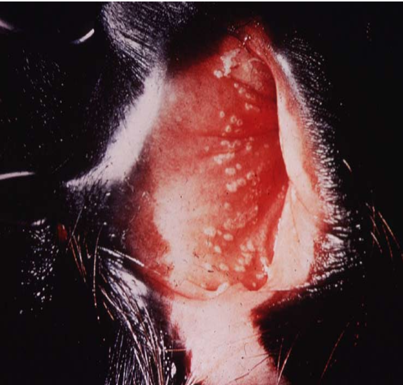
What is causing these pustules on this vulva?
venereal BHV (aka IBR)
How is the systemic form of IBR transmitted?
• Contracted from mucus membranes
• Virus shed in aborted fetus and respiratory fluids
What is the pathogenesis and clinical signs the systemic form of IBR?
• Naïve cow exposed – virus replication – fetal infections – abortion
• Abortion storms can occur with as many as 25-60% of cows in herd aborting
• Commonly abort between 4 and 8 months gestation
• Respiratory signs may or may not be present in cows
• Modified Live vaccine can cause abortions
How is IBR diagnosed?
Lesions are suggestive:
• Fetus: Autolyzed, Foci of necrosis in liver and other organs
• Immunohistochemistry (IHC) and microscopic exam confirms: Intranuclear inclusion bodies, Confirmed with Viral Isolation
How is IBR treated, prevented, and controlled?
Venereal: Lesions spontaneously resolve in 1-2 weeks
Control and Prevention
Biosecurity
Vaccination – Follow manufacturer’s recommendation
AI – use negative semen
What is BVDV?
bovine viral diarrhea virus = single stranded enveloped RNA virus that exists as different genotypes (BVDV1 and BVDV2) and as different biotypes (cytopathic or noncytopathic - all persistently infected calves)
How is BVDV transmitted vertically?
vertical:
Important because this is where creation of the PI occurs – primary source of BVD spread
• Variety of calf disease manifestations subject to time of gestational exposure
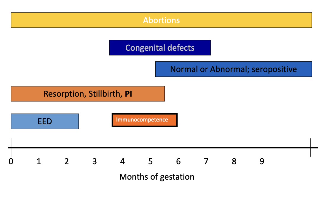
What congenital defects are associated with in utero infections of BVDV?
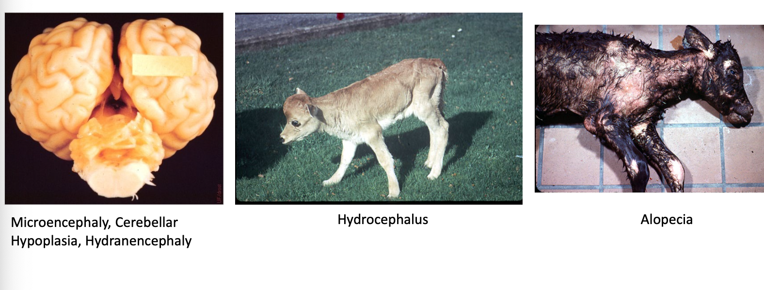
How is BVDV transmitted horizontally?
PI:
• Primary source of spreading virus
• Consistently shedding high numbers of virus particles
• 0.2-4% prevalence
Acutely infected animal:
• Usually exposed to PI
• May shed virus from 2 days to 2 weeks
Where do PI BVDV calves come from?
• PI cows – PI calves
• Fetus infected in utero prior to Day 120-150
What are the clincal features of BVDV?
• May be ill-thrift
• Intermittent diarrhea
• Chronic respiratory disease
• But, most show no clinical signs
How is BVDV diagnosed?
• Remains virus neutralization negative, tests 14 days apart
• Remains virus isolation positive for 14 days or longer
• Virus Isolation (VI) $: Buffy coat, serum, tissue; Gold Standard; Remember maternal antibodies
• Immunohistochemistry (IHC) $: Presence of Ag in ear notch sample, Maternal antibodies do not interfere
• PCR : Can run on pooled samples, Specificity low = False +, Re-test
• ELISA $: Like VI
What are the economic impacts of BVDV?
Reproductive:
• Decreased conception
• Abortions
• Stillborn
• Weak calves
• Ovarian pathology
Production:
• Decreased weight gain
• Respiratory
• Decreased immune status
• Decreased milk production
How is BVDV controlled?
biosecurity: closed herd, don’t buy from stockyard, quarantine and test new entries
vaccines
implement BVDV control strategy
PI animals are reservoirs for BVDV
control programs should aim for detection and elimination of PIs
monitoring and surveillance programs prevent the return of PI animals
What is bluetongue virus?
orbivirus transmitted by culicoides affecting cattle and sheep causing teratogenesis (cerebral malformations, weak calves, stillborns) and less commonly, abortions
How are cattle infected with aspergillus fumigatus?
transmission propagated in winter/spring during increased confinement of cattle in norhtern hemisphere → ingestion → hematogenous spread → placentitis → aboriton (causes around 70% of fungal abortions in cattle)
What lesions are cause by aspergillus fumigatus?
• Placenta: Necrosis of cotyledons and thickening of intercotyledonary space
• Fetus: Skin has mycotic plaques in ~25% of cases, Look like ringworm lesions
How is asperigllus fumigatus diagnosed?
fungal hyphae on microscope or histopath
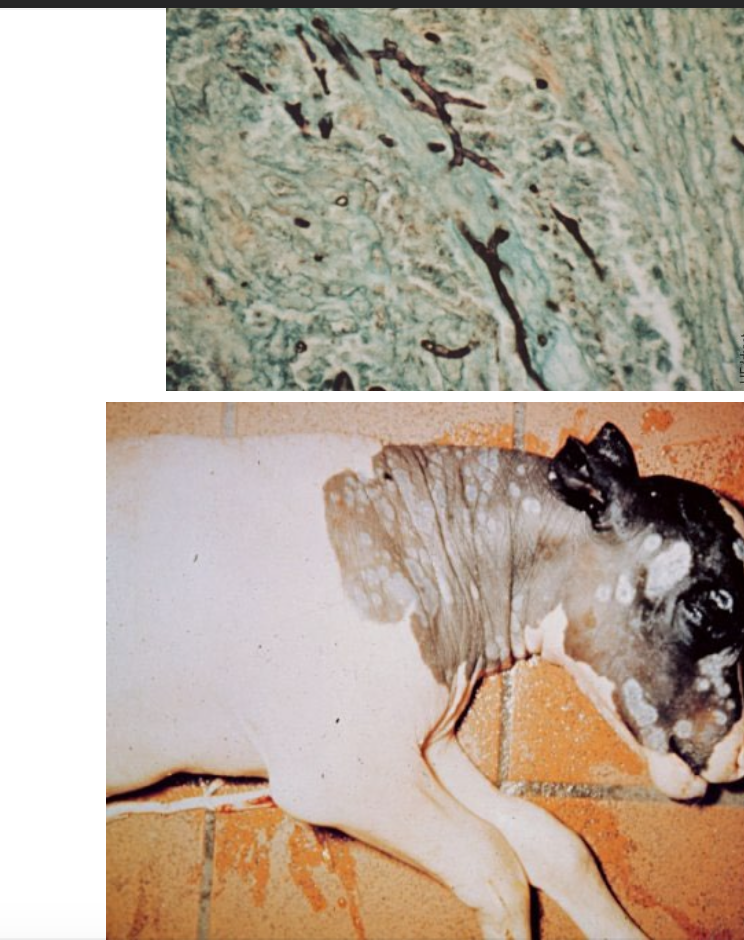
What etiologic agent has infected this fetus?
aspergillus fumigatus (note the fungal hyphae in the top photo and the mycotic plaques on the fetus)