Cog Neuro Exam 1
 Call Kai
 Learn
Learn Practice Test
 Spaced Repetition
 Match
Match Flashcards
 Knowt Play
1/205

There's no tags or description
Looks like no tags are added yet.
Name | Mastery | Learn | Test | Matching | Spaced | Call with Kai | Chat |
|---|
No analytics yet
Send a link to your students to track their progress
206 Terms
Top-down Processing
A cognitive process where higher-level functions guide perception and interpretation of sensory information, influenced by expectations and prior knowledge
Bottom-up Processing
Perception is built from the smallest pieces of sensory information without the influence of prior knowledge or expectations.
Transcranial Magnetic Stimulation (TMS) Characterization
Has poor spatial and temporal resolution but is useful for understanding causal relationships in brain function. It creates a temporary virtual lesion, which is semi-invasive.
Electroencephalography (EEG) Characterization
Has excellent temporal resolution, poor spatial resolution, and is non-invasive.
Magnetoencephalography (MEG) Characterization
Has excellent temporal resolution, good spatial resolution, and is non-invasive. Similar to EEG but with better spatial resolution. Great for very shallow, surface level brain areas
Computerized Axial Tomography (CAT) Characterization
Has low spatial resolution, poor tissue contrast, and emits radiation. However, it’s fast, safe, and cheap.
Positron Emission Tomography (PET) Characterization
Has poor spatial and temporal resolution, is somewhat invasive, but gives unique data.
Magnetic Resonance Imaging (MRI) Characterization
Has excellent spatial resolution, decent temporal resolution, and is non-invasive. However, the magnet can be dangerous.
Functional MRI (fMRI) Characterization
Has excellent spatial resolution, decent temporal resolution, and is non-invasive. However, the magnet can be dangerous.
Cellular Recording Characterization
Excellent spatial and temporal resolution, but incredibly invasive
Superior or Dorsal
Top of brain
Inferior or Ventral
Bottom of brain
Anterior or Rostral
Front of brain
Posterior or Caudal
Back of brain
Medial
Towards middle of brain
Lateral
Towards side of brain
Sagittal Slice
Divides brain into left and right
Axial Slice
Divides brain into top and bottom (superior and inferior)
Coronal Slice
Divides brain into front and back (anterior and posterior)
Ipsilateral
On the same side
Contralateral
On the opposite side
Proximal
Toward the point where limb attaches to the body
Distal
Toward the end of the limb
Gray Matter
Tissue that contains unmyelinated cell bodies and dendrites on the outer layer of the brain, responsible for thinking and decision making.
White Matter
Tissue that contains myelinated axons and nerve fibers in deeper parts of the brain, responsible for carrying infromation over long distances.
Forebrain (Prosencephalon)
Divided into telencephalon and diencephalon
Telencephalon
Cerebral hemispheres: cerebral cortex, subcortical white matter, basal ganglia, and basal forebrain nuclei
Diencephalon
Thalamus and hypothalamus
Midbrain (mesencephalon)
Surivival-related conscious behaviors - periaqueductal gray, reticular formation, locus coeruleus, and substantia nigra
Hindbrain (rhombencephalon)
Divided into metencephalon and myelencephalon
Metencephalon
Pons and cerebellum
Myelencephalon
Medulla
Peripheral Nervous System
Connects spinal cord to the rest of the body, composed of somatic (outer) and autonomic (inner) nervous system.
Somatic Nervous System
Directs and processes information from external stimuli and uses it to guide the body’s movements - somatosensory inputs and motor outputs.
Autonomic Nervous System
Directs activity of internal (visceral) organs in response to signals from the internal environment - visceral inputs, sympathatic & parasympathetic outputs.
Sympathetic Nervous System
Fight or flight response - quickened heartbeat, increased respiration and blood pressure, slowed digestive tract, and circulation shift from digestive organs to muscles.
Parasympathetic Nervous System
Rest and regenerate response - slowed heart rate, decreased respiration and blood pressure, relaxed muscle tone, and blood flow shift to stomach and digestive organs.
Sensory Input
Enters spinal cord through dorsal nerve root at the back of the spinal cord
Motor Output
Exits spinal cord through ventral nerve root at the front of the spinal cord
Sensory Neurons
Collect information from inside and outside the body
Motor Neurons
Carry signals to muscles for movement.
Central Nervous System
Brain and spinal cord - central pattern generators in the spinal cord contain a combination of excitatory and inhibitory neurons that balance together to form simple movements
Cerebrum
Seperated into left and right hemispheres, initiates and coordinates movement, regulates temperature, handles thinking, speech, memory, learning, judgement, and senses. Divided into four lobes.
Frontal Lobe
High-order cognitive processsing, decision-making, control of voluntary movement, and perception of sensory stimuli. Contains Broca’s area - key role in speech.
Parietal Lobe
Processes sensory information like pain and touch, helps people understand spacial relationships and identify objects. Contains Wernicke’s area - key role in understanding spoken language.
Temporal Lobe
Auditory processing, memory formation, speech, musical rhythm, hearing, and vision.
Occipital Lobe
Visual processing - color perception, visualspatial processing, facial recogntition, and memory formation.
Cerebellum
Responsible for highly coordinated, fine-tuned movements, predicts outcome of motor actions, in addition to language, memory, attention, and emotion. Comprised of neurons and cerebral cortex.
Lateral Cotricospinal Tract
80% of corticospinal axons cross over at the medulla and form this pathway, 10% cross over when exiting the spinal cord, and the rest remain on the same side
Brainstem
Involuntary and automatic functions - composed of midbrain, medulla oblongata, and pons
Medulla Oblongata
Responsible for involuntary functions like breathing and heart rate, as well as reflexive activities like coughing and swallowing
Pons
Responsible for cerebellum-cortex relay, sleep, arousal, and sensory information
Periaqueductal Gray
Cluster of nerve cells that manage pain relief; fight, flight, or freeze responses; and basic bodily functions
Reticular Formation
Responsible for consciousness
Locus Coeruleus
Responsible for alertness and arousal
Substantia Nigra
Responsible for motor control and reward learning
Hypothalamus
Regulates autonomic responses and behaviors, releases hormones via pituitary gland
Thalamus
Relays sensory signals to cerebral cortex, conveys brain and motor signals to the body
Cerebral Cortex
Outer layer of the brain, responsible for reasoning, emotion, thought, memory, language, and consciousness
Basal Ganglia
Initiating and maintaining activity, especially in the motor cortex, also involved in habit and reward processing. Contains striatum (caudate nucleus and putamen), and globus pallidus
Straitum
Made up of caudate nucleus and putamen
Caudate Nucleus
Comet-shaped structure, motor and behavioral functions
Putamen
Sits within the “C” of the caudate, learning and motor control
Globus pallidus
Oval structure, regulates voluntary movement
Limbic System
Emotions, memory, and basic drives, shaped like a fist in the middle of the forebrain. Integrates internal and external representations. Mamillary bodies and fornix relay information between the hippocampus and other brain areas. Comprised of amygdala and hippocampus.
Amygdala
Emotional evaluation and learning, links external sensory inputs to emotional states
Hippocampus
Learning, memory, episodic memory, and spatial navigation
Fornix
C-shaped pathway of white matter connects the hippocampus to mamillary bodies and the thalamus
Mammillary Bodies
A pair of nuclei in the hypothalamus, helps with memory consolidation, spacial orientation, and connects hippocampus to anterior thalamic nucleus
Neuronal Anatomy
Soma, nucleus, dendrites, axon, axon terminals, and terminal branches
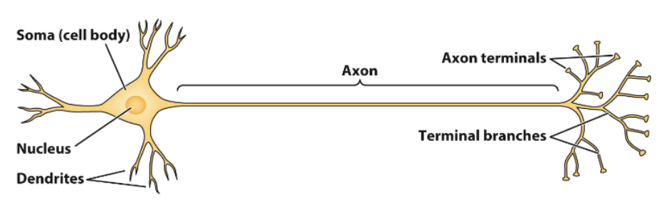
Soma
Integrates signals from dendrites and generates signal that travels down axon
Dendrites
Collects thousands of incoming signals
Axon
Conducts signals rapidly across long distances
Axon Terminals
Where messages are sent to a neighbor neuron
Synapse
Junction where axon terminals are in close proximity to the dendrites and somas of other cells
Resting Membrane Potential
The electrical charge difference across a cell’s plasma membrane when the cell is at rest. The inside of the cell is more negative than the inside (the cell is polarized).
Ion Channels
Allow ions to pass through a non-permeable cell membrane, generating resting membrane potential, action potentials, and synaptic responses.
Excitatory Postsynaptic Receptors (EPSPs)
Postive ions flow through a receptor into the cell, causing depolarization and increasing the chance of firing an action potential
Inhibitory Postsynaptic Potentials (IPSPs)
The binding of neurotransmitters makes the inside of the cell more negative from negative ions flowing into the cell or from positive ions flowing out of the cell, making it harder for the neuron to fire
Action Potential
Sudden change in electrical properties of a neuron’s membrane in an axon, composed of several phases, sodium and potassium play key roles.
Sodium Potassium Pump
Pumping sodium out and potassium into the cell creates a membrane potential
Postsynaptic Receptors
Receptors on the receiving end of a synapse that bind to neurotransmitters
Synaptic Cleft
Space between pre and post synaptic cells
Neurotransmitter Degradation
Enzymes come through and break down the neurotransmitter in the synaptic cleft
Neurotransmitter Diffusion
The neurotransmitter floats away from the synapses and gets lost
Neurotransmitter Reuptake
The neurotransmitter is taken back into the pre-synaptic cell to be recycled and released again
Polarized Cell
Greater concentration of positive ions outside the cell, the inside is negative relative to the outside
Depolarized Cell
Greater concentration of negative ions outside the cell, the inside is positive relative to the outside
Temporal Summation
Signals that arrive to the soma at the same time will add up once they reach the soma, leading to an action potential
Spatial Summation
Signals that arrive on different branches of the dendrites converge at the soma
EPSP + IPSP
If an EPSP and IPSP arrive at a different location at the same time, they will cancel each other’s effect at the soma
Action Potential Stages
Resting state, depolarization, action potential, repolarization, refractory period, return to resting state. Depolarization must hit a threshold to cause an action potential
Cell at Rest
High concentration of Na+ on the outside and lower on the inside; low concentration of K+ on the outside and higher on the inside
Voltage-Gated Ion Channels
Selectively pass Na+ and only open at particular voltages across the membrane, driven by concentration and electrical gradient. This depolarization triggers the opening of K+ channels, causing K+ to rush out the cell and repolarize the cell (inside becomes more negative).
Refractory Period
After an action potential, Na+ channels are more resistant to opening and need time to reopen, so the action potential can only travel forward towards the axon terminal
Glial cells
Non-neuronal cells that support, nourish, and protect neurons
Oligodendrocytes
Myelinate axons, called Schwann cells in the peripheral nervous system
Microglia
Immune responses, inflammatory responses, and debris removal
Ependymal Cells
Line ventricles
Astrocytes
Form blood-brain barrier, provide nutrients to neurons from blood, structural support, repair to central nervous system