BSC2086L E.4 Heart Anatomy
0.0(0)
Card Sorting
1/43
Study Analytics
Name | Mastery | Learn | Test | Matching | Spaced |
|---|
No study sessions yet.
44 Terms
1
New cards
apex
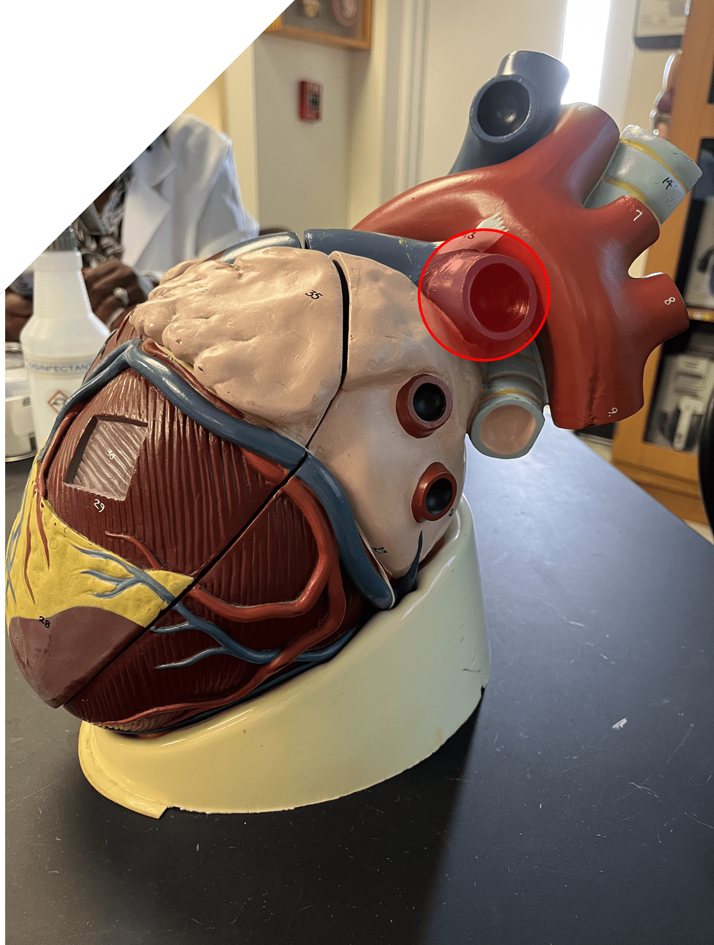
• The slightly pointed end of the heart.
• Located left of the medial line.
• Located left of the medial line.
2
New cards
base
• The broader side of the heart connected to the great vessels.
• Located beneath the second rib.
• Located beneath the second rib.
3
New cards
right atrium
• The superior, right chamber of the heart.
• Receives deoxygenated blood from the inferior and superior venae cavae.
• Pumps blood to the right ventricle.
• Receives deoxygenated blood from the inferior and superior venae cavae.
• Pumps blood to the right ventricle.
4
New cards
left atrium
• The superior, left chamber of the heart.
• Receives oxygenated blood from the pulmonary veins.
• Pumps blood to the left ventricle.
• Receives oxygenated blood from the pulmonary veins.
• Pumps blood to the left ventricle.
5
New cards
right ventricle
• The inferior, right chamber of the heart.
• Receives deoxygenated blood from the right atrium.
• Pumps blood into the pulmonary trunk.
• Receives deoxygenated blood from the right atrium.
• Pumps blood into the pulmonary trunk.
6
New cards
left ventricle
• The inferior, left chamber of the heart.
• Receives oxygenated blood from the left atrium.
• Pumps blood into the aorta.
• Receives oxygenated blood from the left atrium.
• Pumps blood into the aorta.
7
New cards
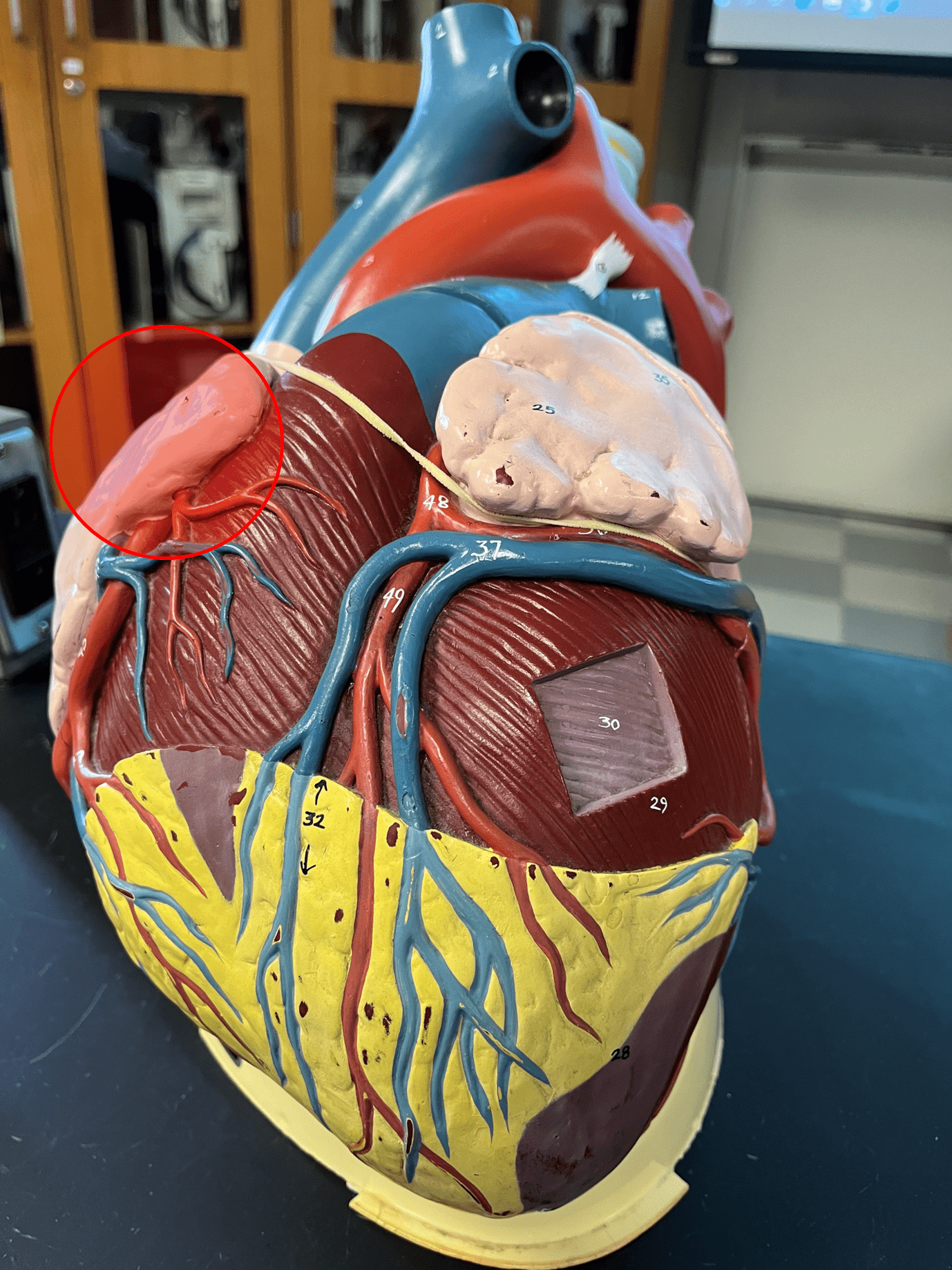
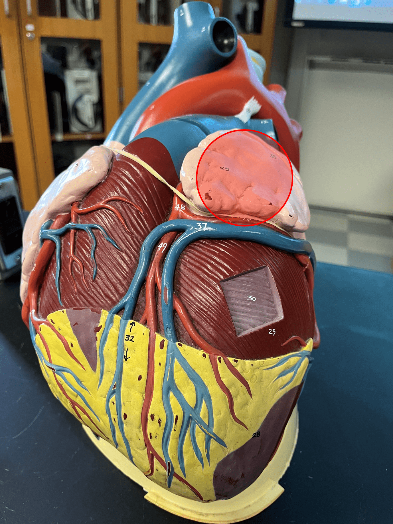
right auricle
A sac contained within and increasing the volume of the right atrium.
8
New cards
left auricle
A sac contained within and increasing the volume of the left atrium.
9
New cards
coronary sulcus
• Sulcus dividing the atria and ventricles of the heart.
• Contains the coronary sinus.
• Also known as the atrioventricular groove.
• Contains the coronary sinus.
• Also known as the atrioventricular groove.
10
New cards
anterior interventricular sulcus
• Sulcus dividing the right and left ventricles of the heart on the anterior side of the heart.
• Contains coronary blood vessels.
• Contains coronary blood vessels.
11
New cards
posterior interventricular sulcus
• Sulcus dividing the right and left ventricles of the heart on the posterior side of the heart.
• Contains coronary blood vessels.
• Contains coronary blood vessels.
12
New cards
aorta
• A great vessel of the heart.
• Carries oxygenated blood to the systemic circuit.
• Divided into ascending, arch-shaped, and descending portions.
• Carries oxygenated blood to the systemic circuit.
• Divided into ascending, arch-shaped, and descending portions.
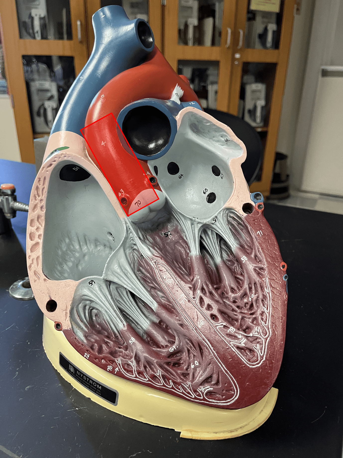
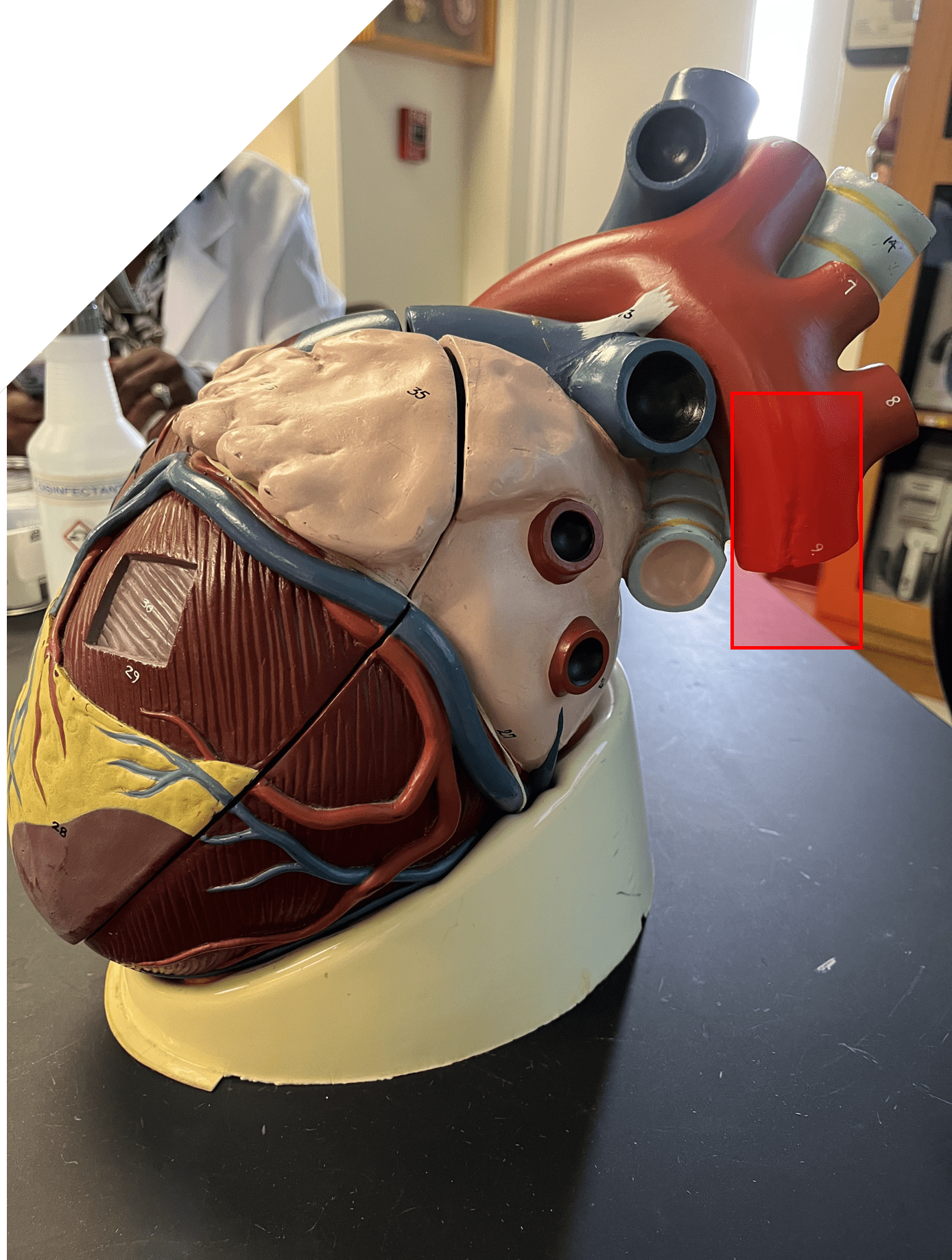
13
New cards
ascending aorta
• The part of the aorta arising from the base of the heart.
• Continues on as the aortic arch.
• Continues on as the aortic arch.
14
New cards
aortic arch
• The part of the aorta that inverts downwards.
• Continues on as the descending aorta.
• Continues on as the descending aorta.
15
New cards
descending aorta
The part of the aorta that descends posteriorly.
16
New cards
superior vena cava
• A great vessel of the heart.
• Carries deoxygenated blood to the right atrium from areas superior to the heart.
• Abbreviated SVC.
• Carries deoxygenated blood to the right atrium from areas superior to the heart.
• Abbreviated SVC.
17
New cards
inferior vena cava
• A great vessel of the heart (not shown).
• Carries deoxygenated blood to the right atrium from areas inferior to the heart.
• Abbreviated IVC.
• Carries deoxygenated blood to the right atrium from areas inferior to the heart.
• Abbreviated IVC.
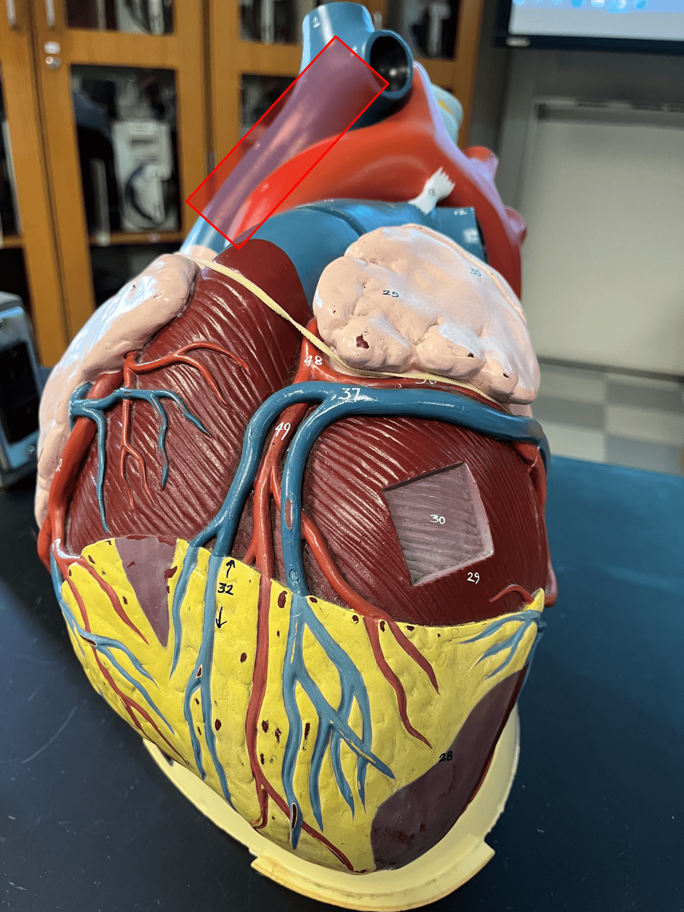
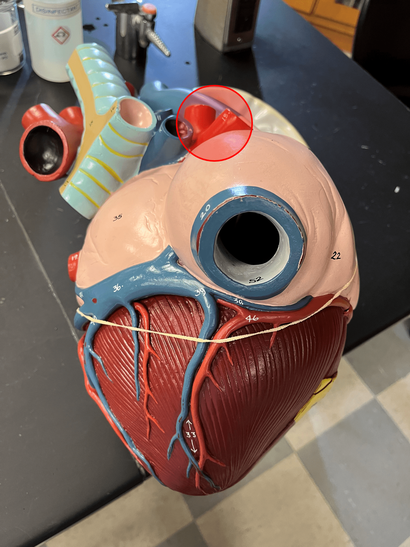
18
New cards
pulmonary trunk
• A great vessel of the heart.
• Carries deoxygenated blood from the right ventricle and branches off into the pulmonary circuit.
• Carries deoxygenated blood from the right ventricle and branches off into the pulmonary circuit.
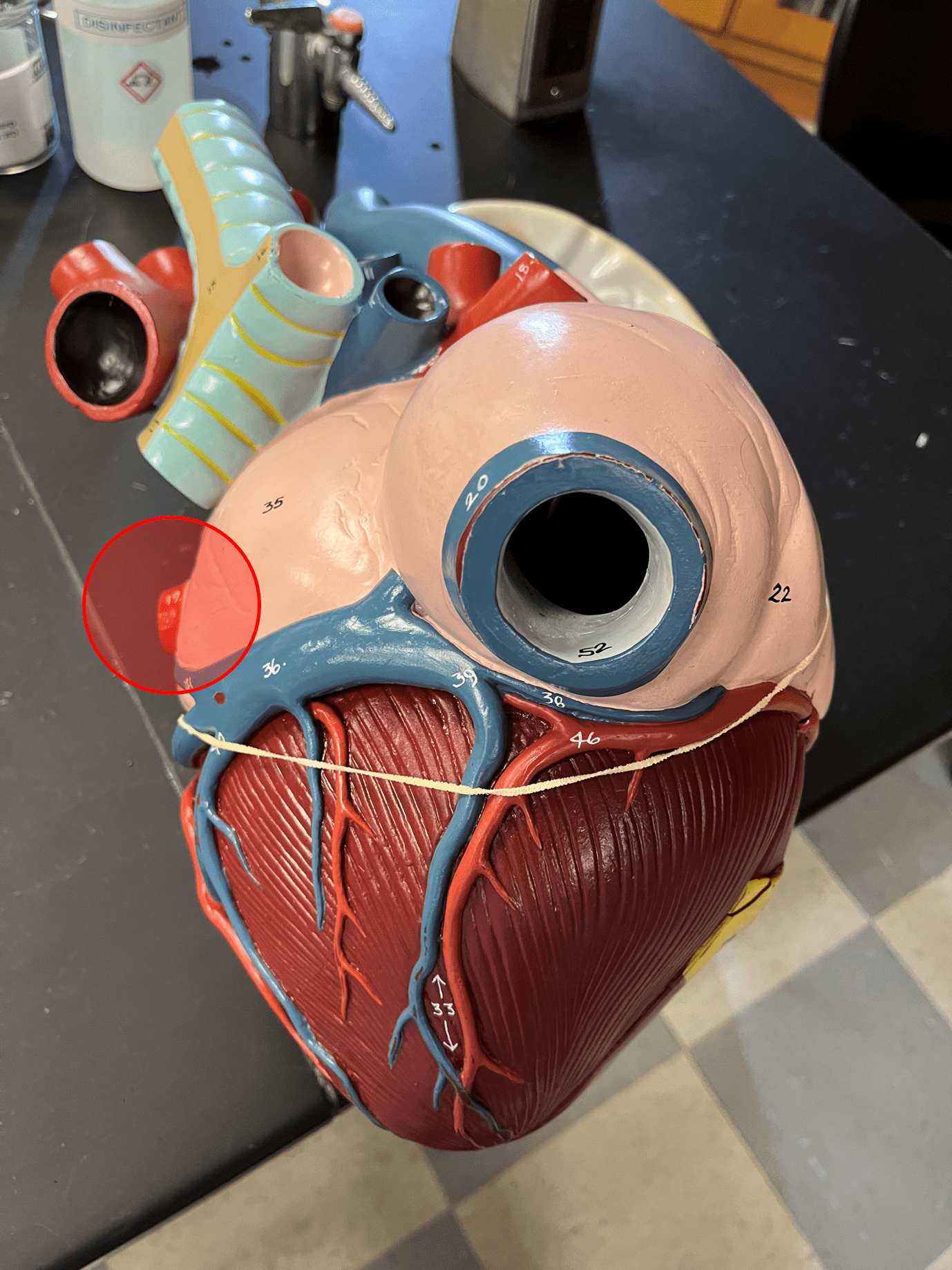
19
New cards
right pulmonary artery
• Carries deoxygenated blood to the right lung.
• Branches off from the pulmonary trunk.
• Branches off from the pulmonary trunk.
20
New cards
left pulmonary artery
• Carries deoxygenated blood to the left lung.
• Branches off from the pulmonary trunk.
• Branches off from the pulmonary trunk.
21
New cards
right pulmonary veins
• A pair of veins located posterior to the right side of the heart.
• Carries oxygenated blood from the right lung to the left atrium.
• Carries oxygenated blood from the right lung to the left atrium.
22
New cards
left pulmonary veins
• A pair of veins located posterior to the left side of the heart.
• Carries oxygenated blood from the left lung to the left atrium.
• Carries oxygenated blood from the left lung to the left atrium.
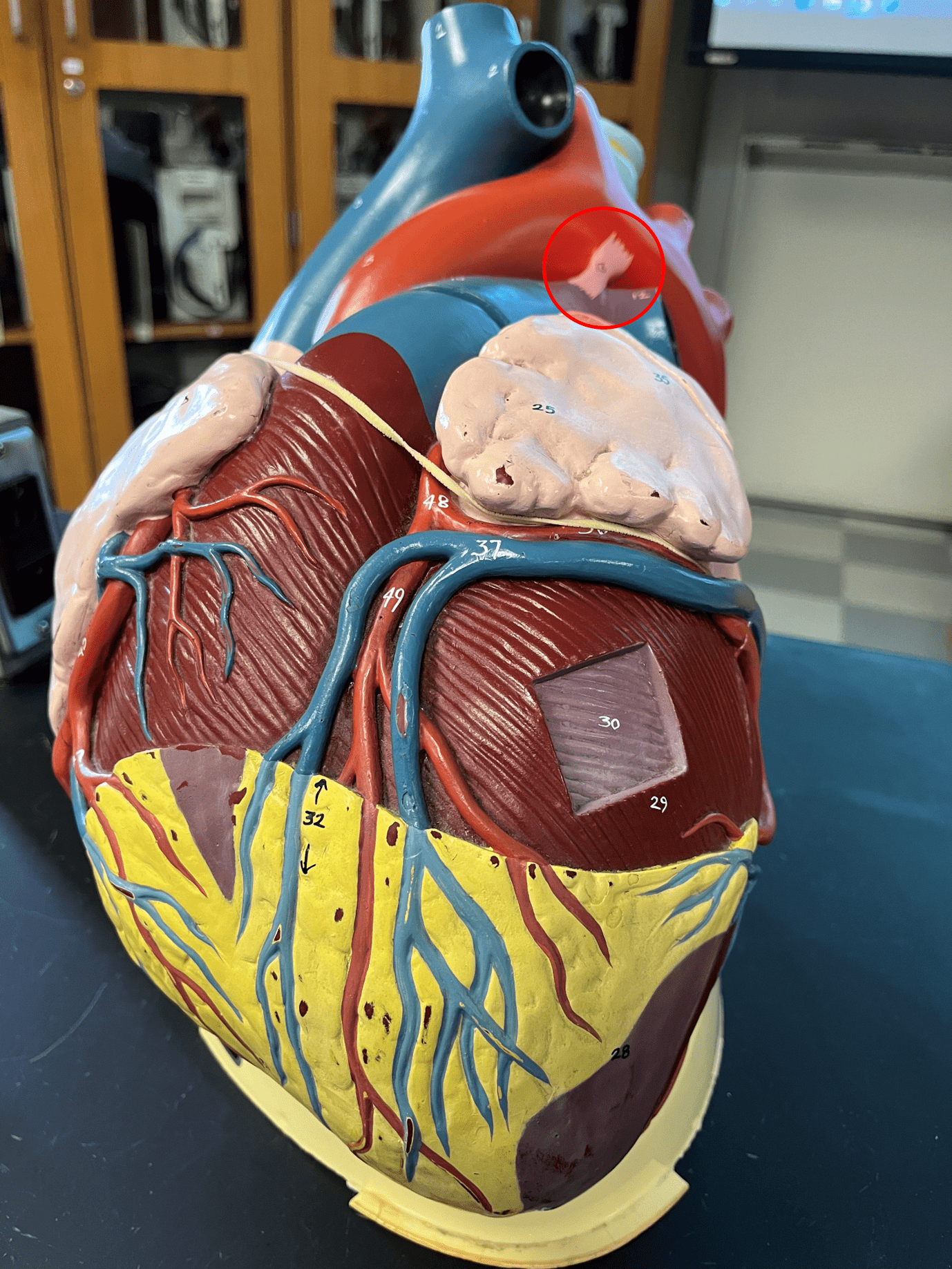
23
New cards
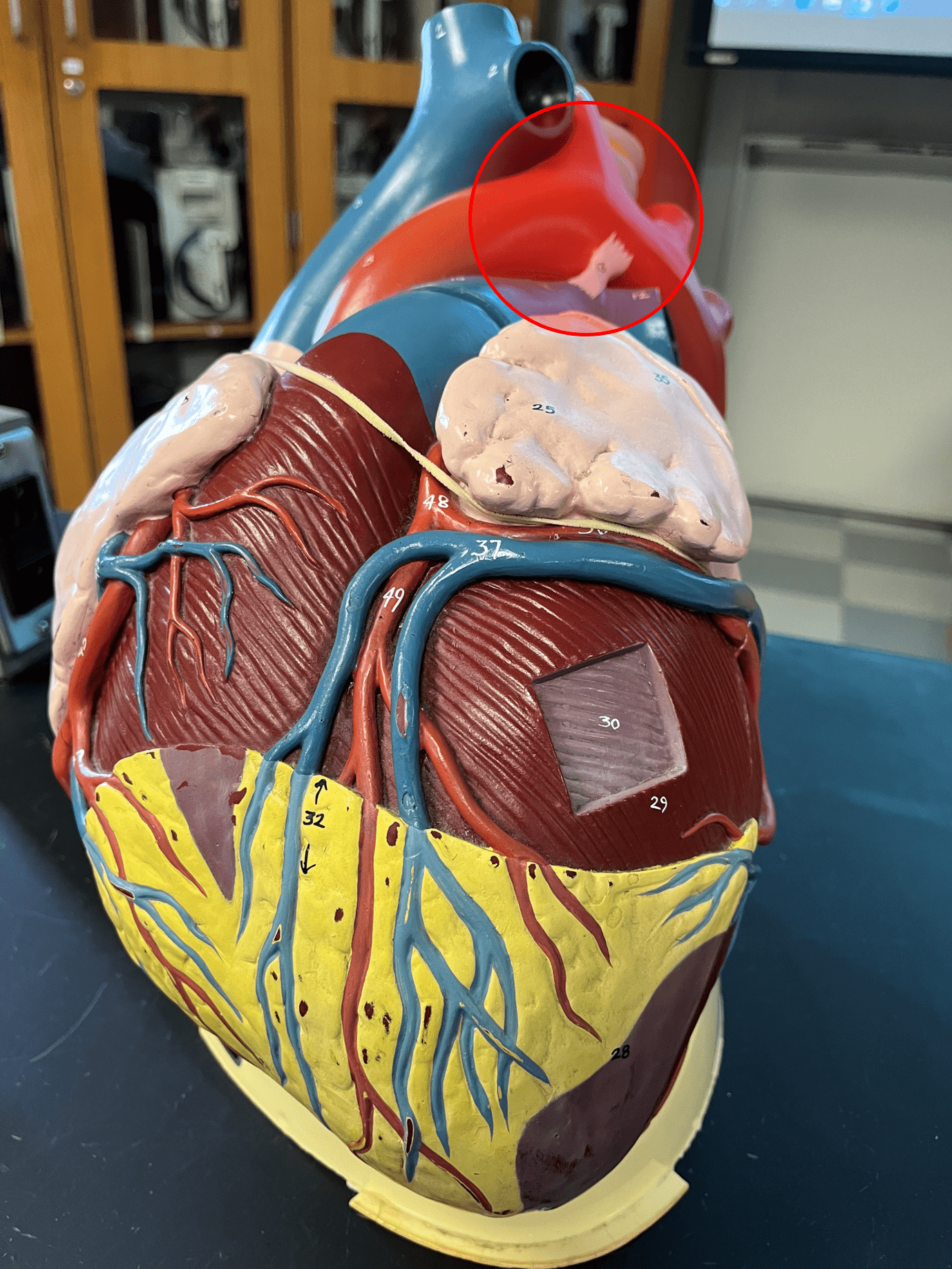
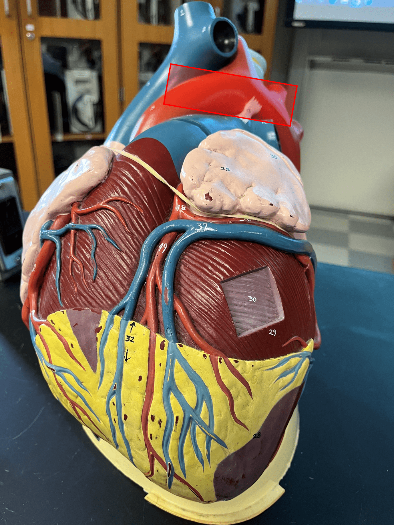
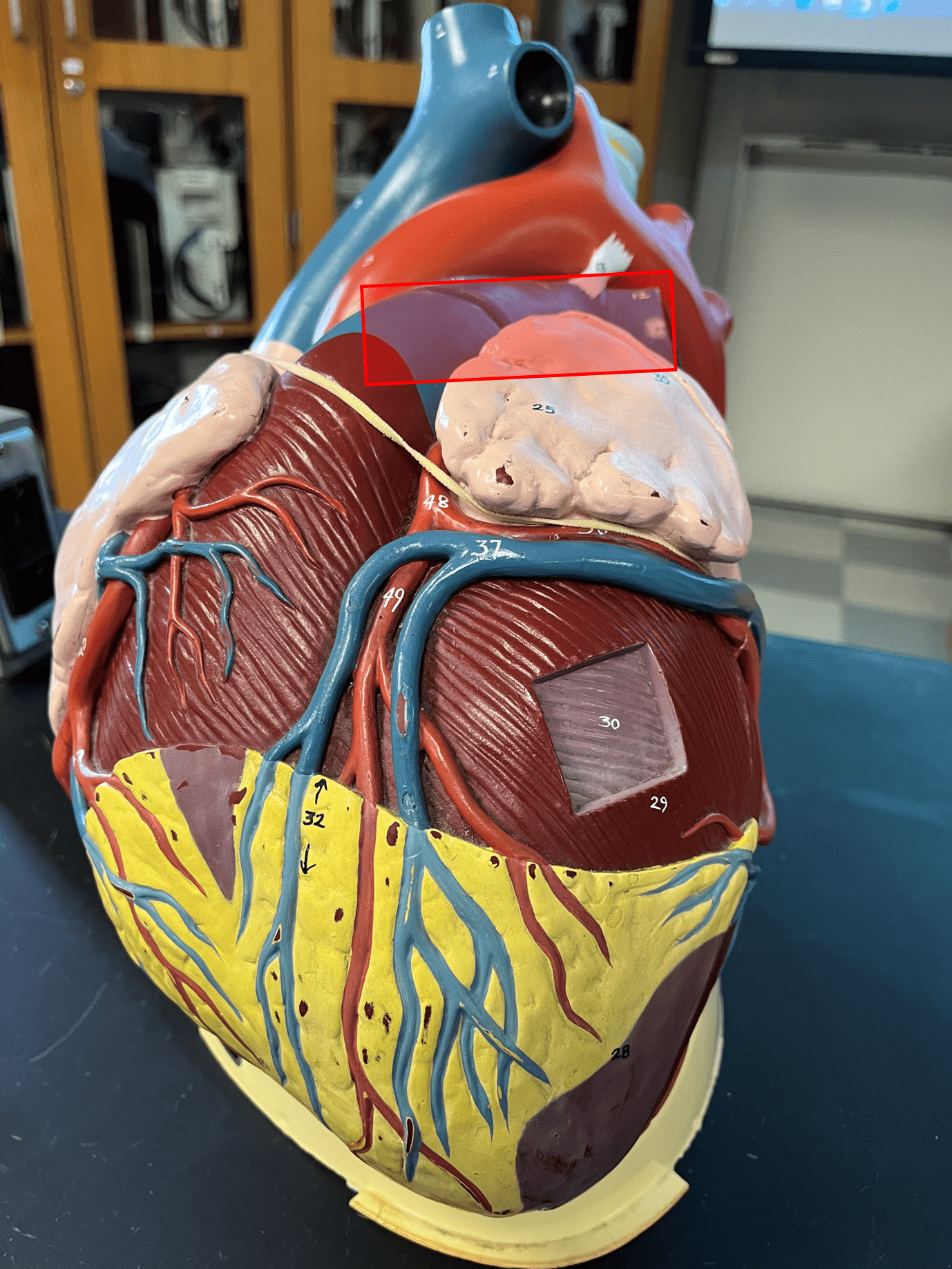
ligamentum arteriosum
• A small band of connective tissue connecting the pulmonary trunk and aorta.
• Vestigial structure in adults.
• Vestigial structure in adults.
24
New cards
interatrial septum
Separates the right and left atria of the heart (not shown).

25
New cards
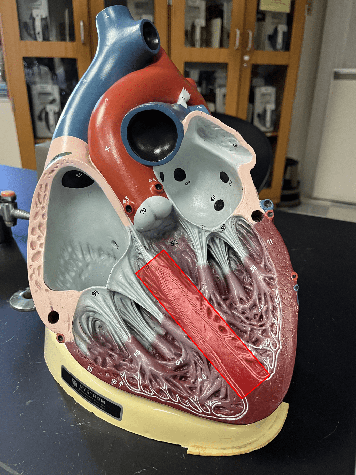
interventricular septum
Separates the right and left ventricles of the heart.
26
New cards
tricuspid valve
• An atrioventricular valve.
• Controls the flow of blood from the right atrium to the right ventricle.
• Also known as the right atrioventricular (AV) valve.
• Controls the flow of blood from the right atrium to the right ventricle.
• Also known as the right atrioventricular (AV) valve.
27
New cards
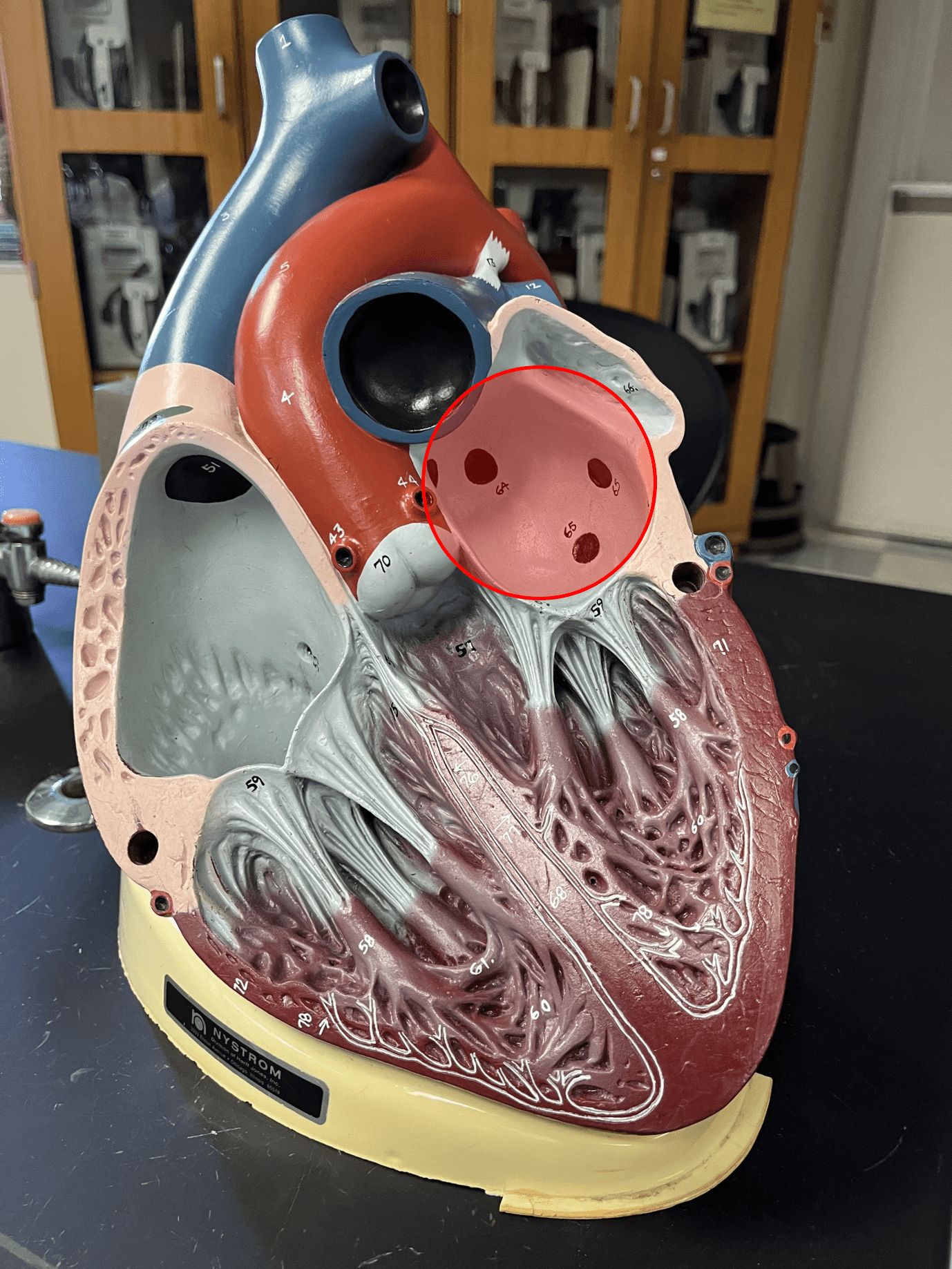
mitral valve
• An atrioventricular valve.
• Controls the flow of blood from the left atrium to the left ventricle.
• Also known as the left atrioventricular (AV) valve or the bicuspid valve.
• Controls the flow of blood from the left atrium to the left ventricle.
• Also known as the left atrioventricular (AV) valve or the bicuspid valve.
28
New cards
pulmonary valve
• A semilunar valve (not shown).
• Controls the flow of blood from the right ventricle to the pulmonary trunk.
• Also known as the right semilunar (SL) valve.
• Controls the flow of blood from the right ventricle to the pulmonary trunk.
• Also known as the right semilunar (SL) valve.
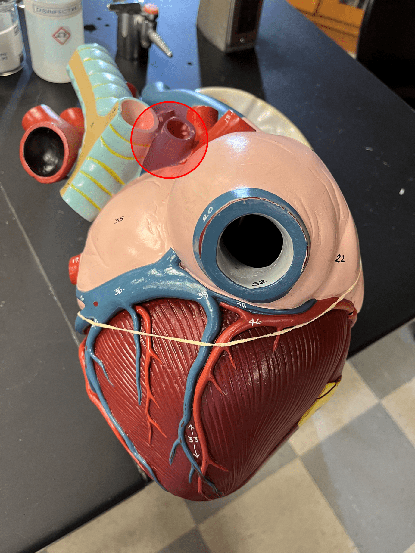
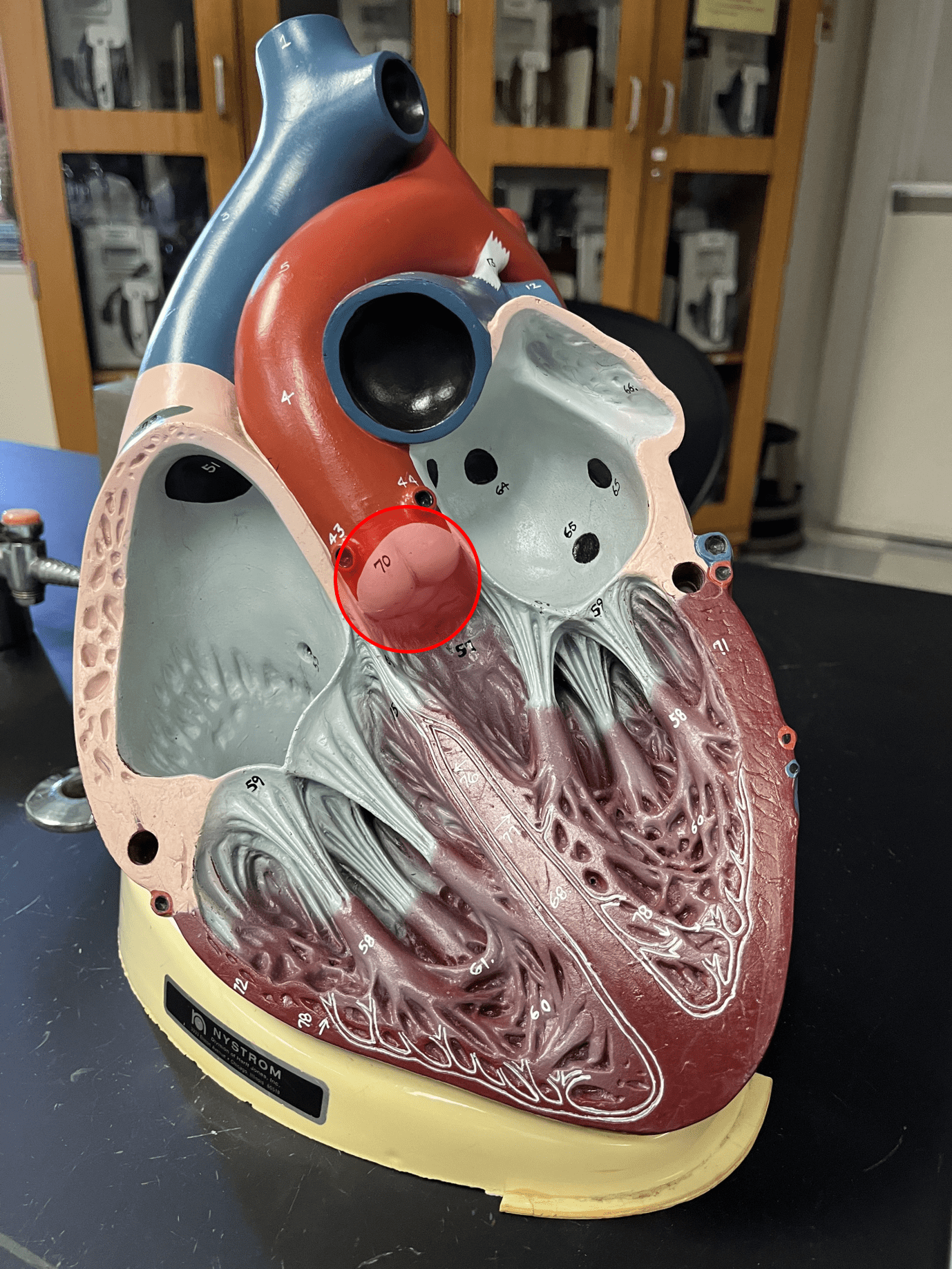
29
New cards
aortic valve
• A semilunar valve.
• Controls the flow of blood from the left ventricle to the ascending aorta.
• Also known as the left semilunar (SL) valve.
• Controls the flow of blood from the left ventricle to the ascending aorta.
• Also known as the left semilunar (SL) valve.
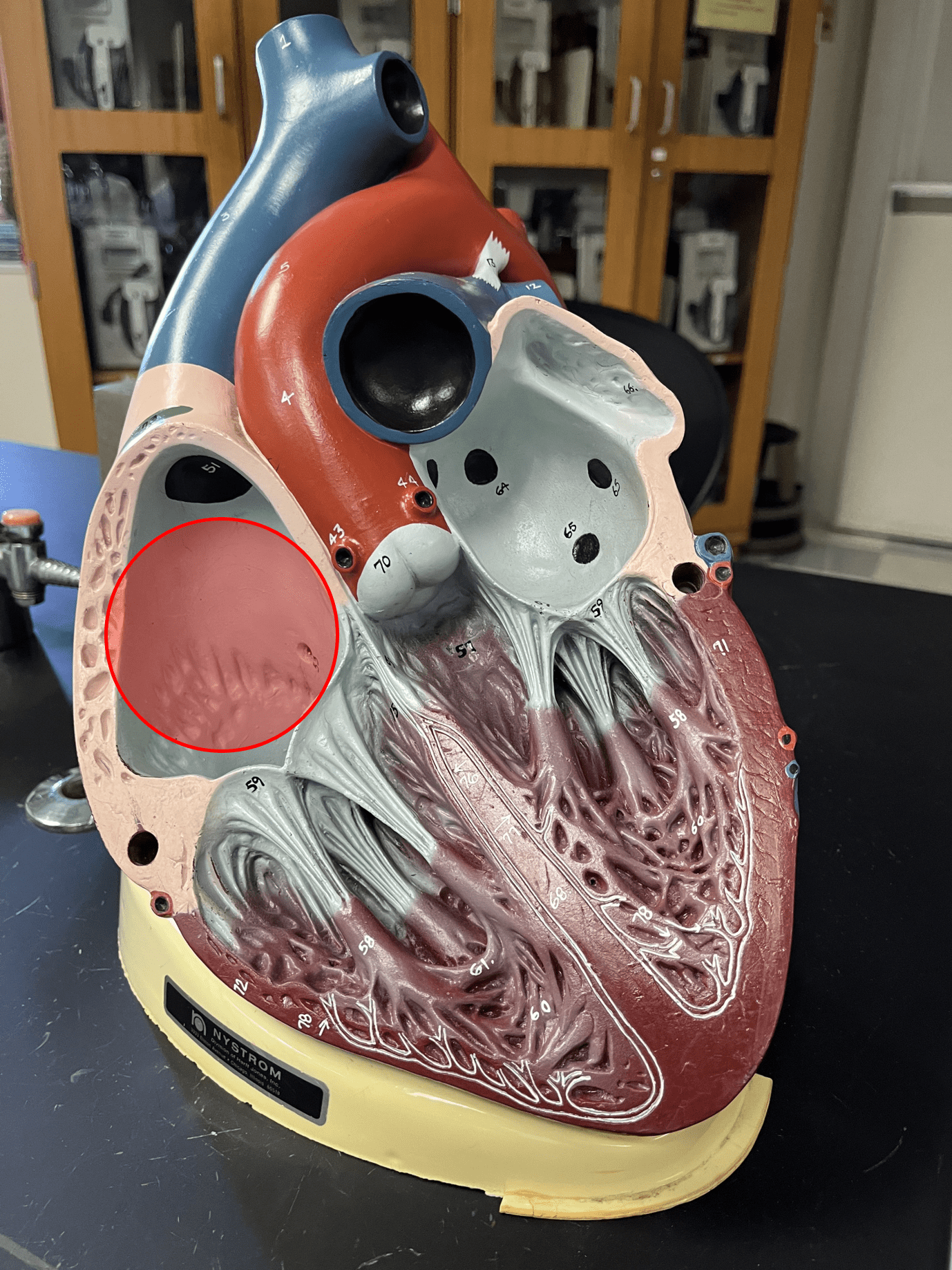
30
New cards
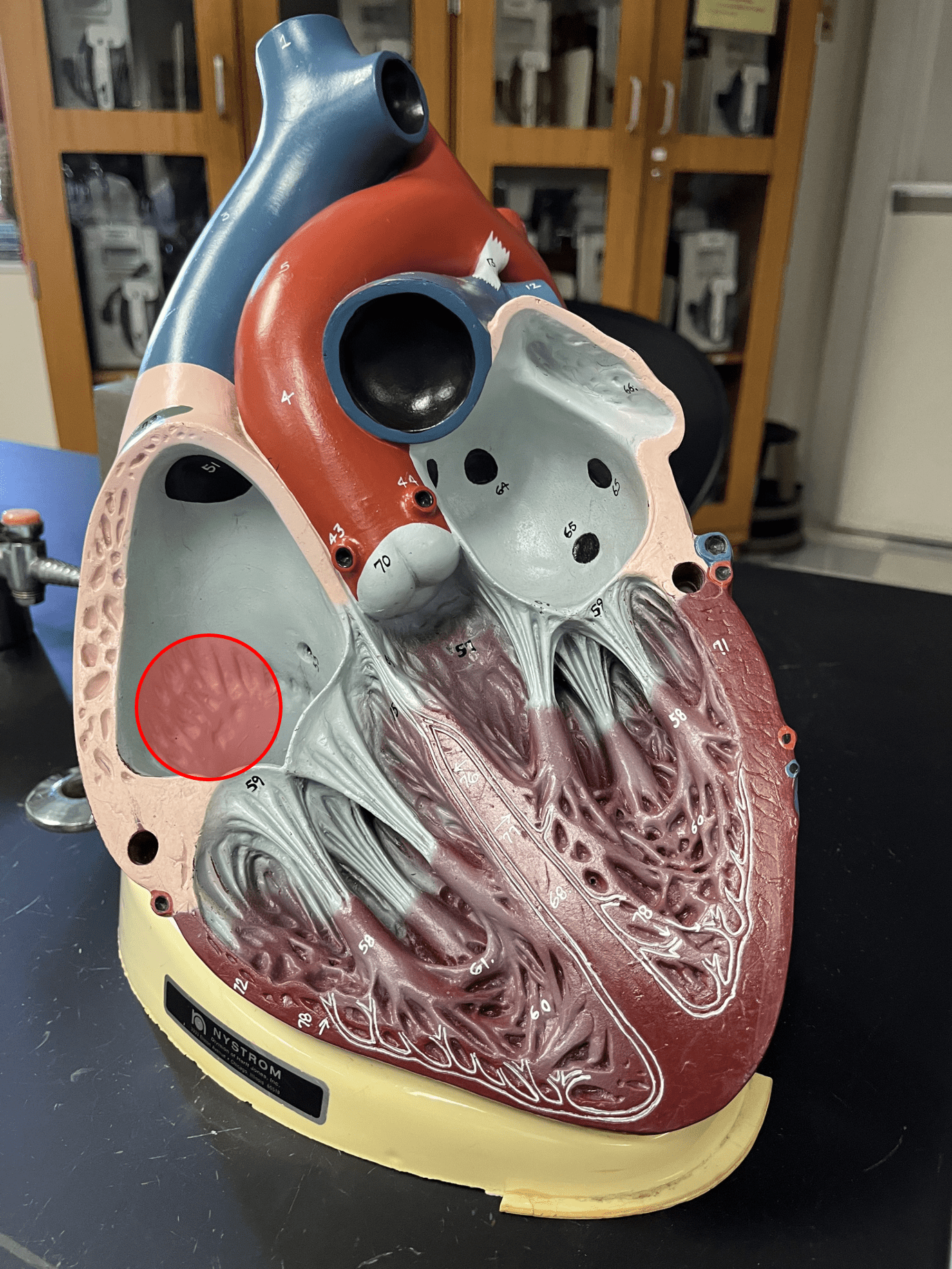
pectinate muscles
• Parallel arrangement of cardiac muscle bundles appearing as ridges.
• Located on the anterior side of the interior wall of the right atrium.
• Located on the anterior side of the interior wall of the right atrium.
31
New cards
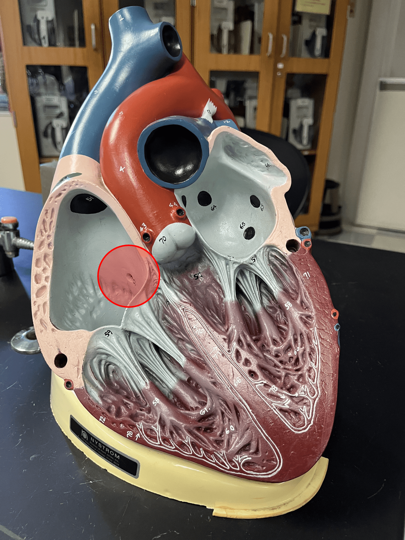
fossa ovalis
• A slight oval depression where the foramen ovale of the fetus was.
• Located in the interior wall of the right atrium.
• Located in the interior wall of the right atrium.
32
New cards
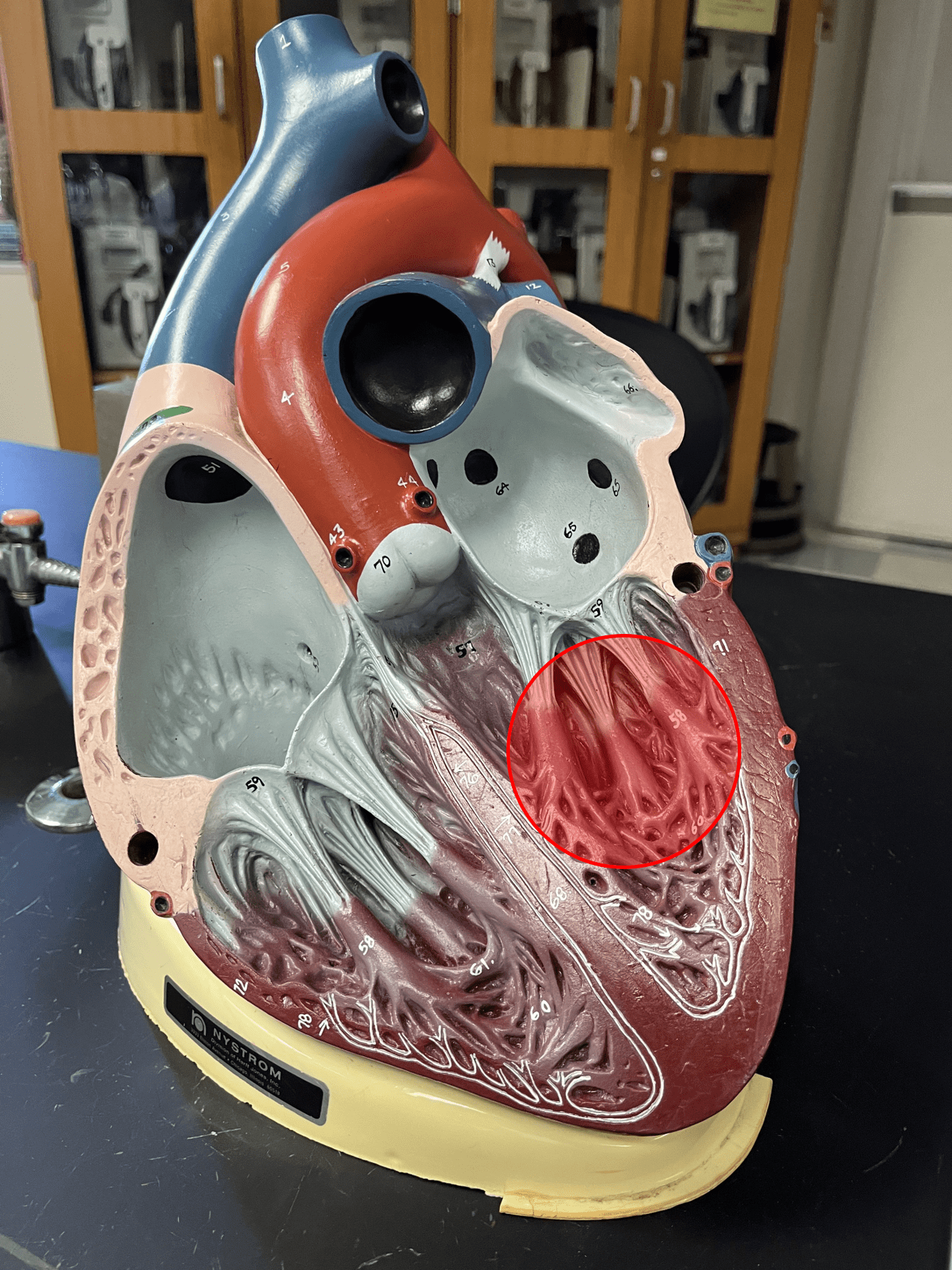
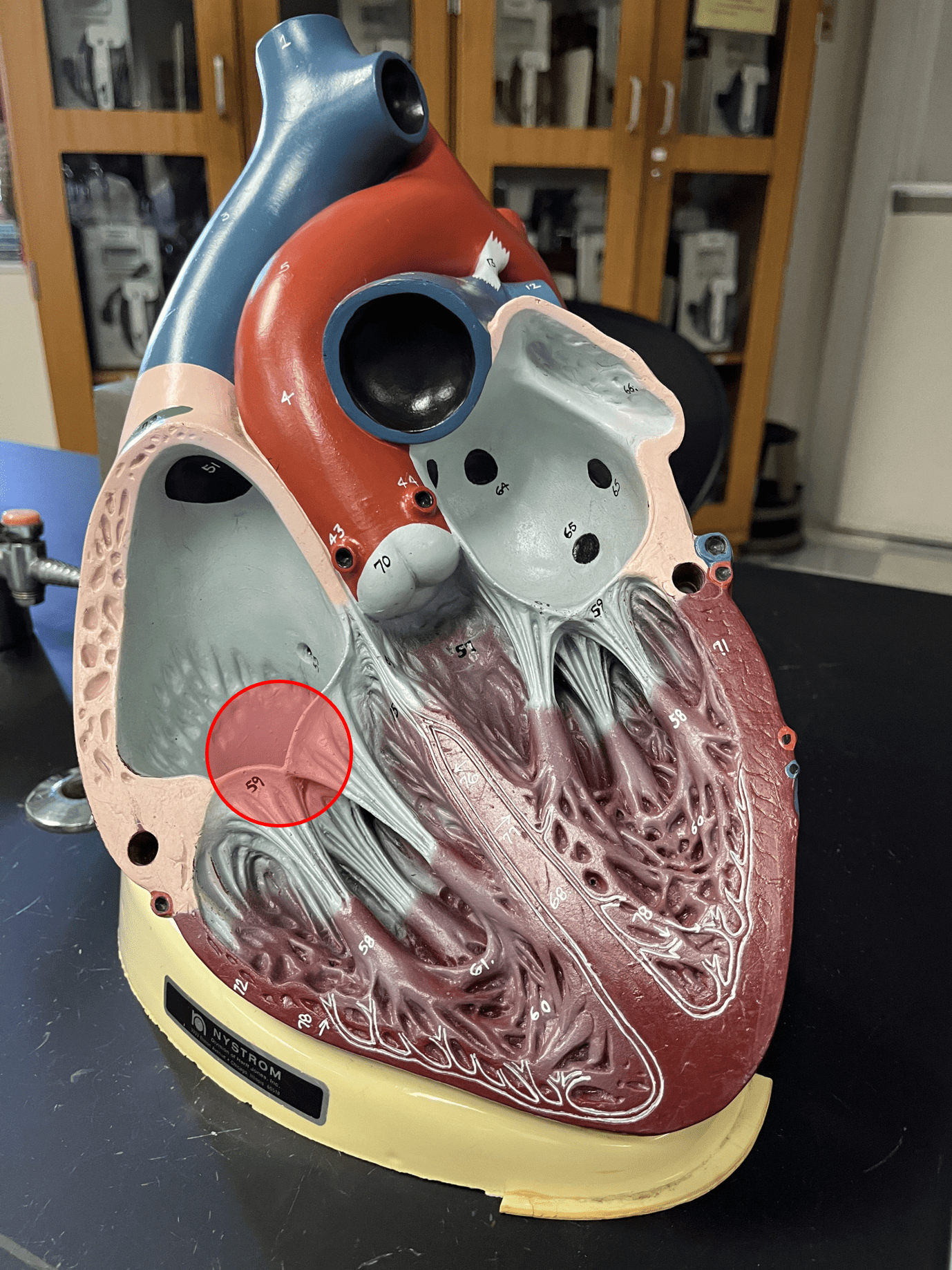
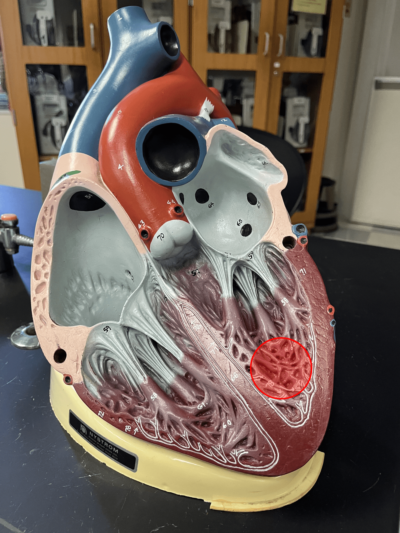
trabeculae carneae
• Prominent ridges of cardiac muscle.
• Located in the ventricles of the heart.
• Located in the ventricles of the heart.
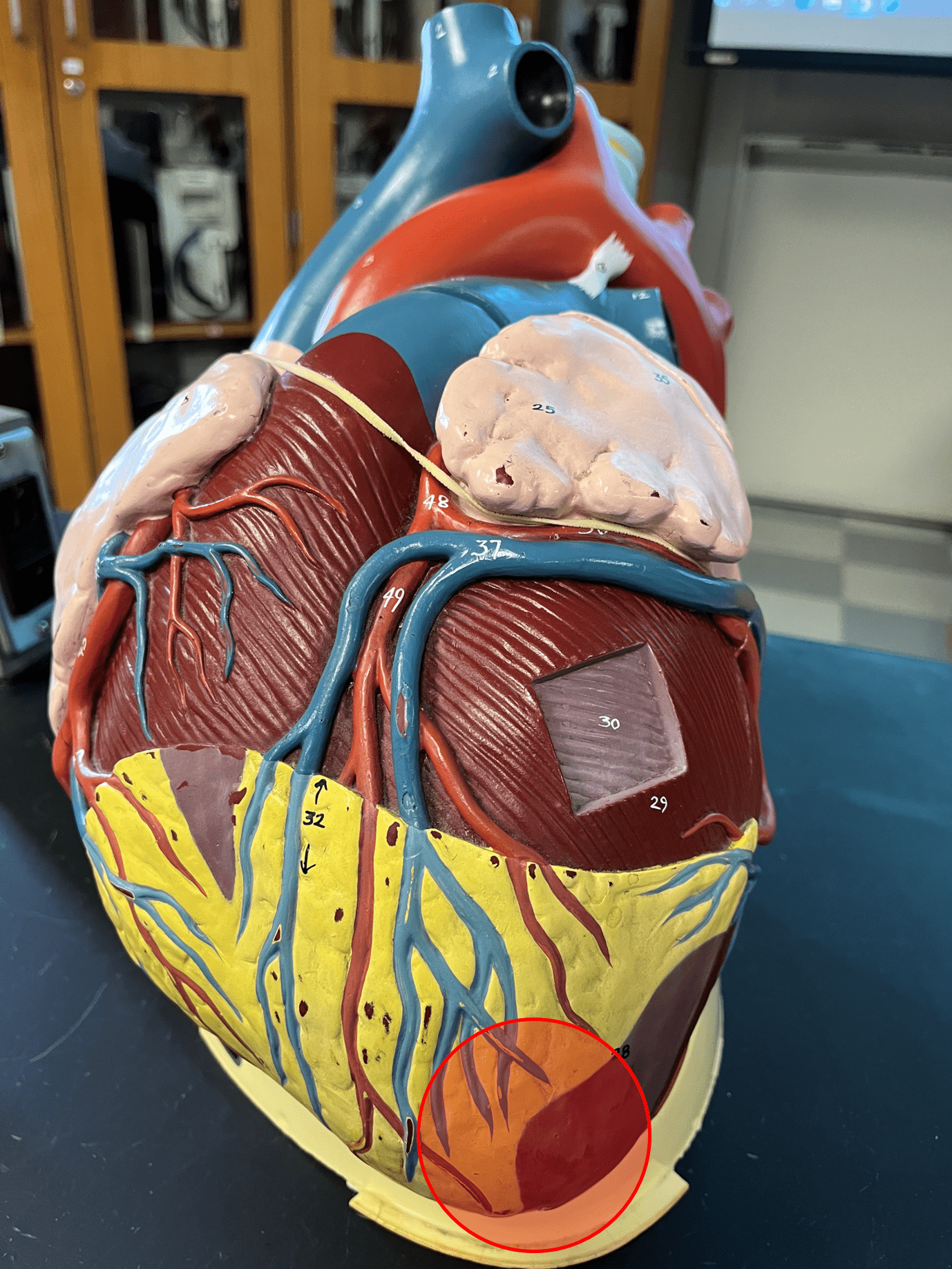
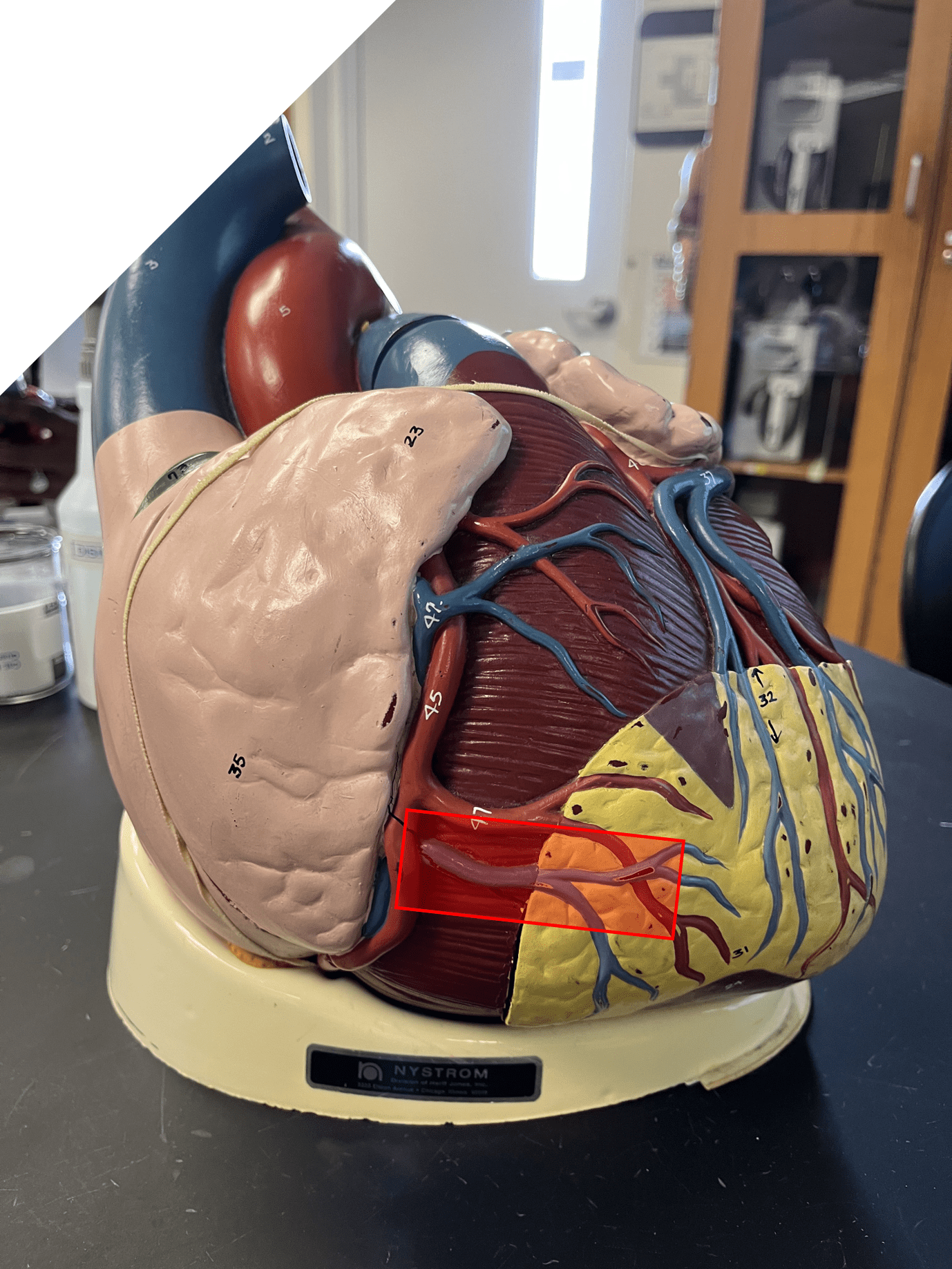
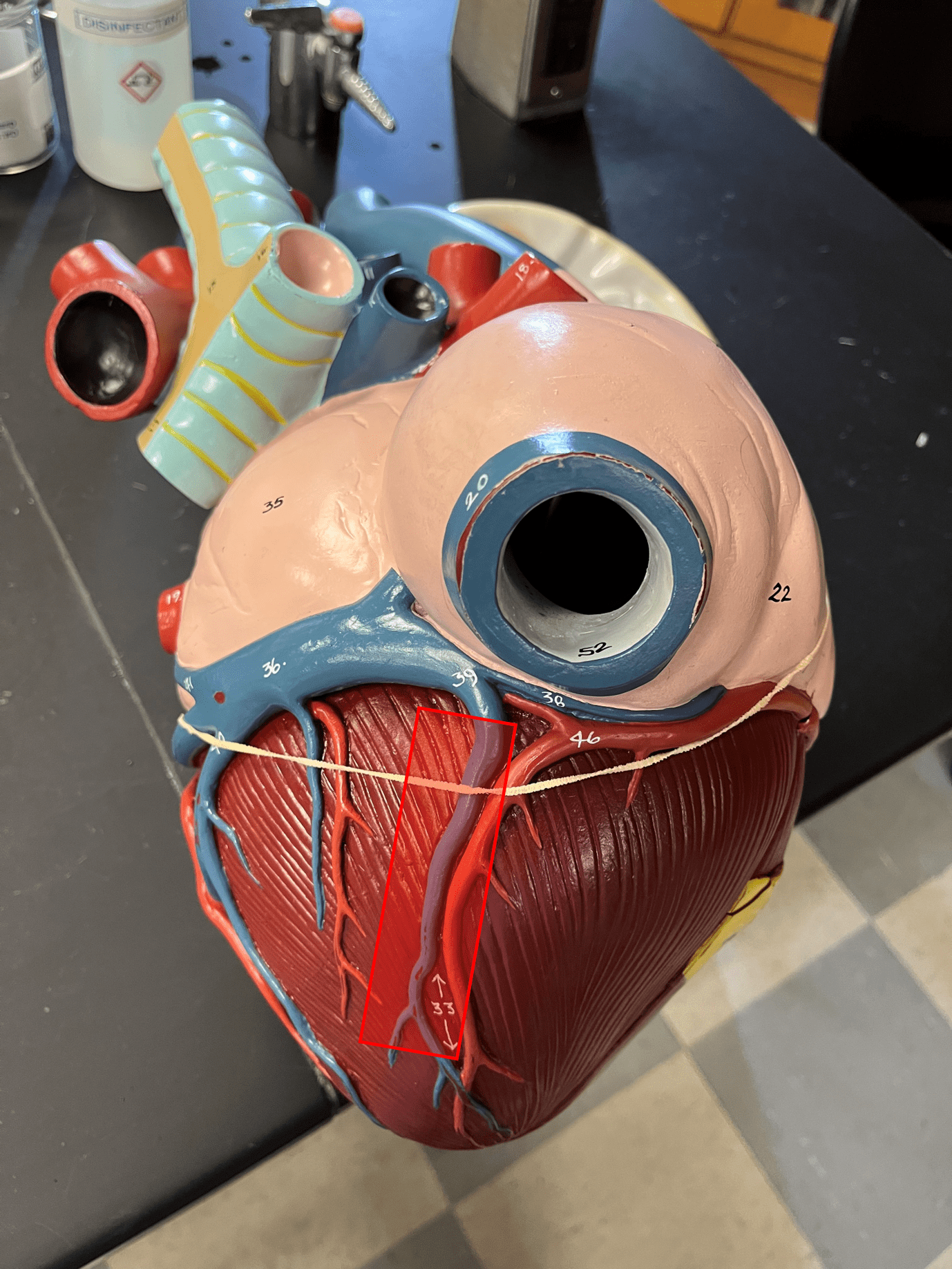
33
New cards
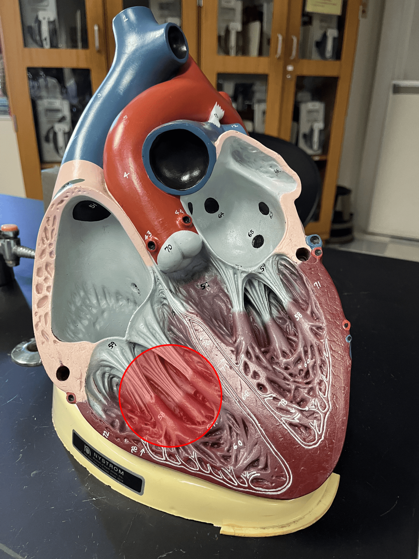
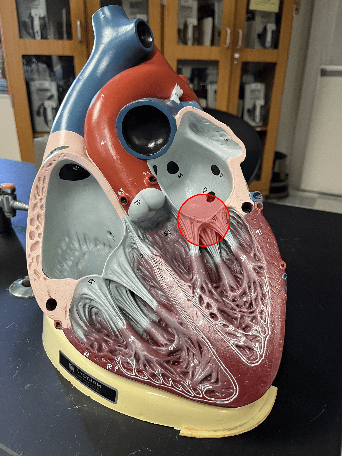
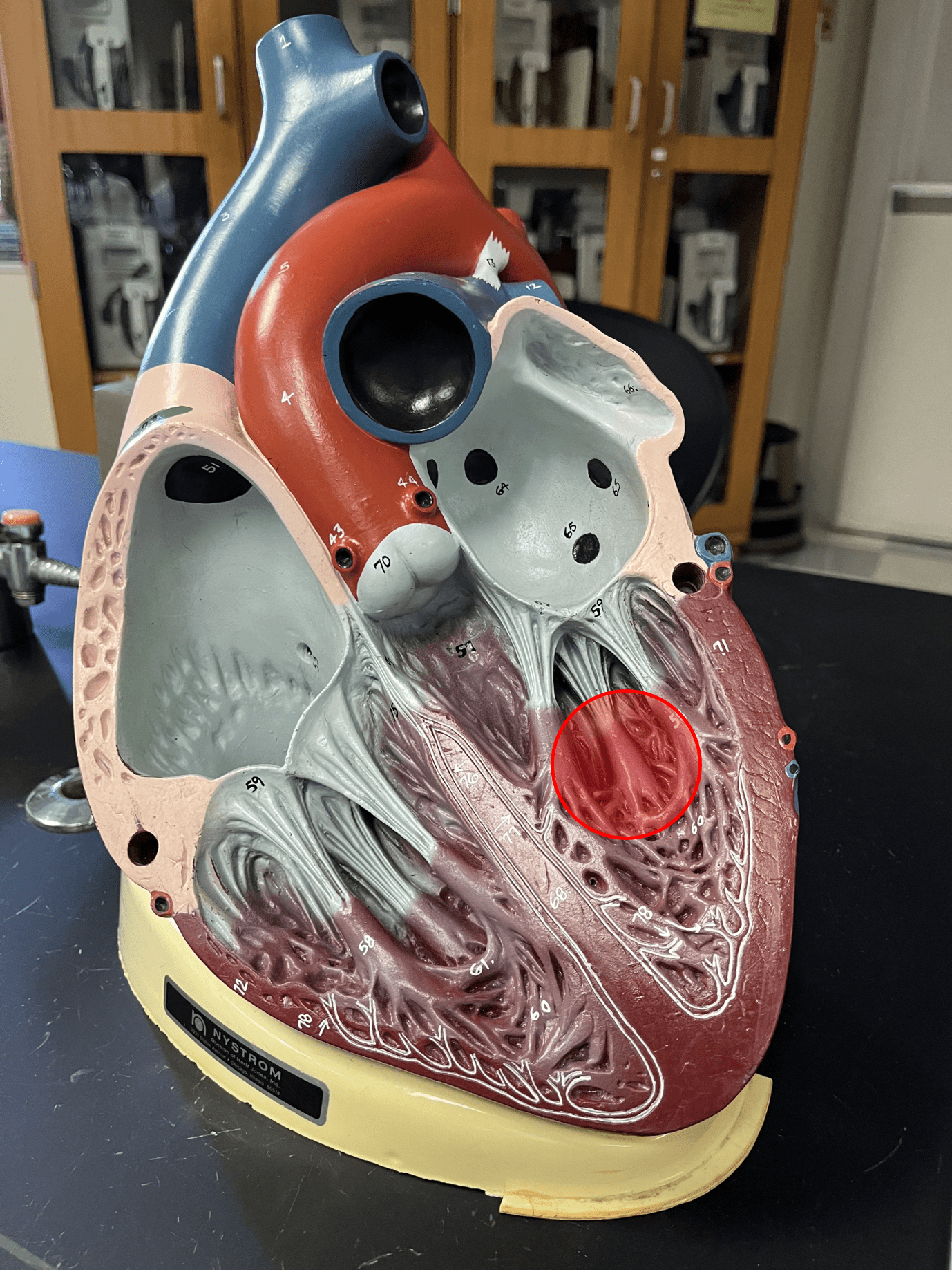
papillary muscle
• Cardiac muscle anchoring the chordae tendineae.
• Located in the ventricles of the heart.
• Located in the ventricles of the heart.
34
New cards
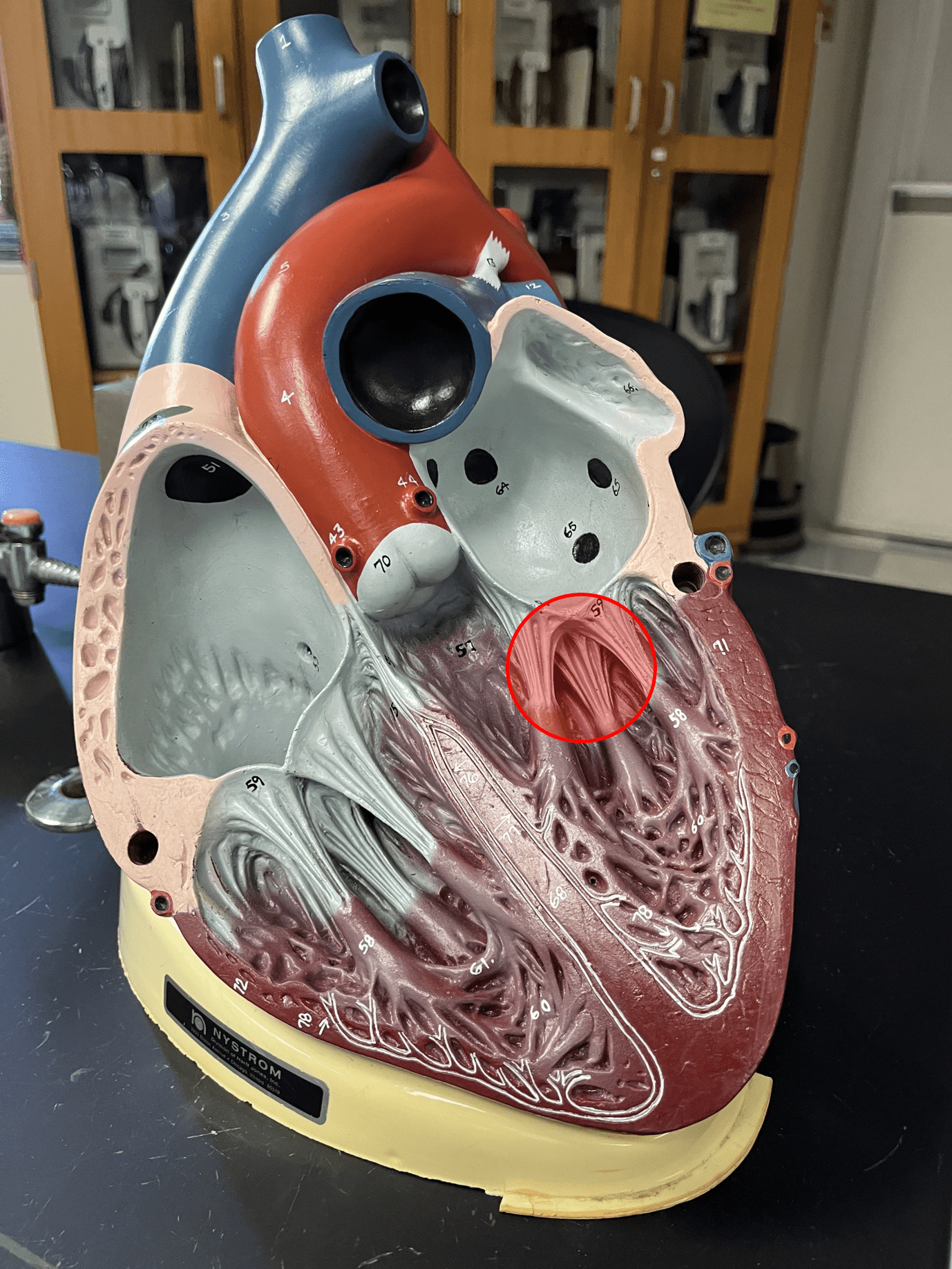
chordae tendineae
• String-like cords of connective tissue that prevents prolapse of the atrioventricular valves.
• Anchored by the papillar muscles.
• Located in the ventricles of the heart.
• Anchored by the papillar muscles.
• Located in the ventricles of the heart.
35
New cards
right coronary artery
• Part of the coronary circulation.
• Passes around the lateral right side in the coronary sulcus of the heart and then posterior to the ventricles.
• Arises from the base of the aorta.
• Branches into the posterior interventricular and marginal arteries.
• Passes around the lateral right side in the coronary sulcus of the heart and then posterior to the ventricles.
• Arises from the base of the aorta.
• Branches into the posterior interventricular and marginal arteries.
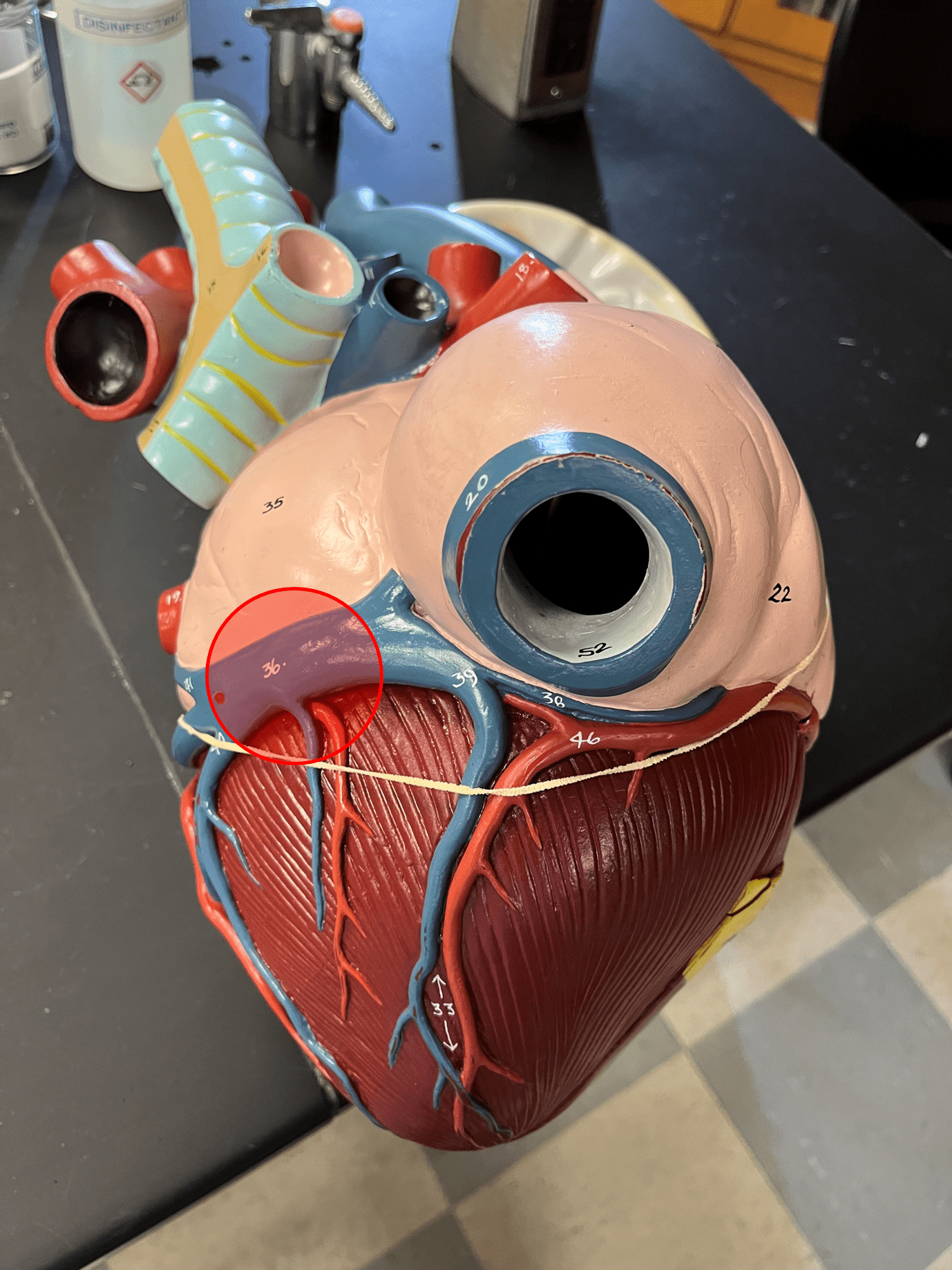
36
New cards
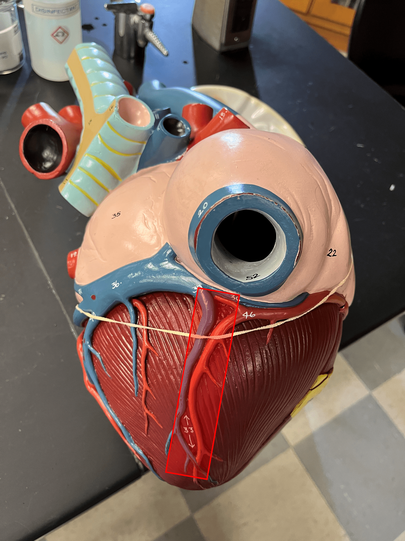
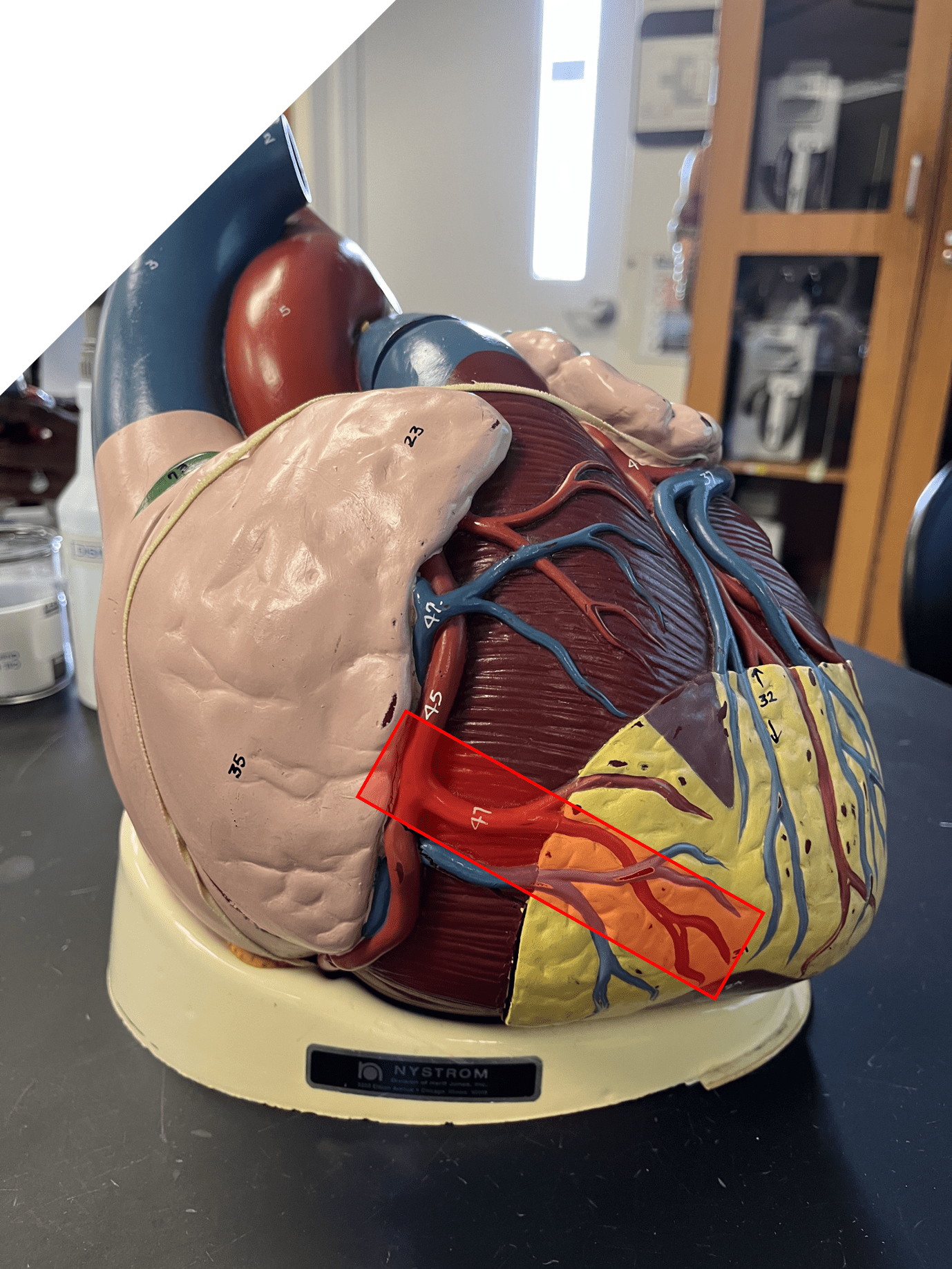
posterior interventricular artery
• Part of the coronary circulation.
• Located in and supplies the tissue around the posterior interventricular sulcus.
• One of the branches of the right coronary arteries.
• Located in and supplies the tissue around the posterior interventricular sulcus.
• One of the branches of the right coronary arteries.

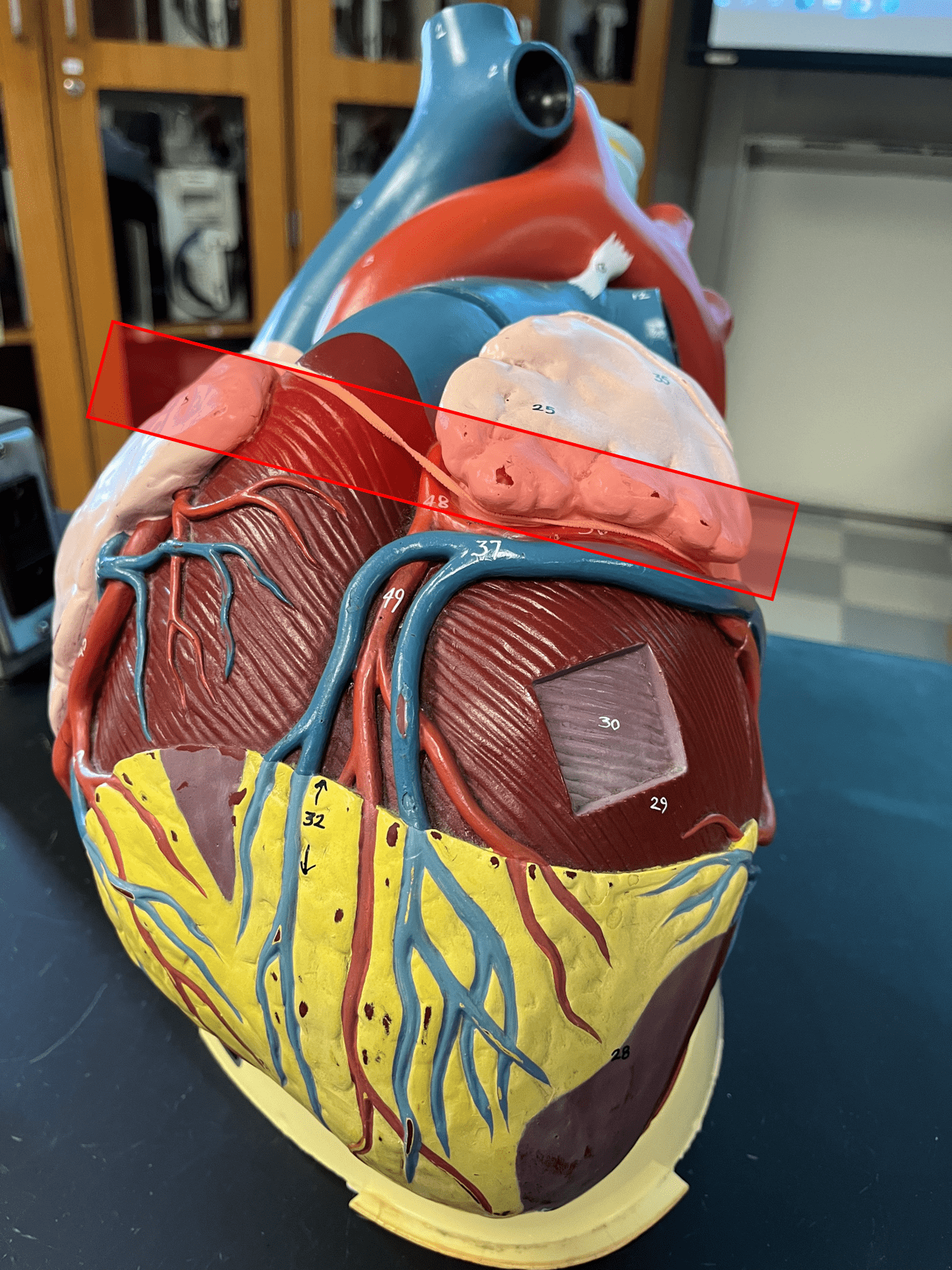
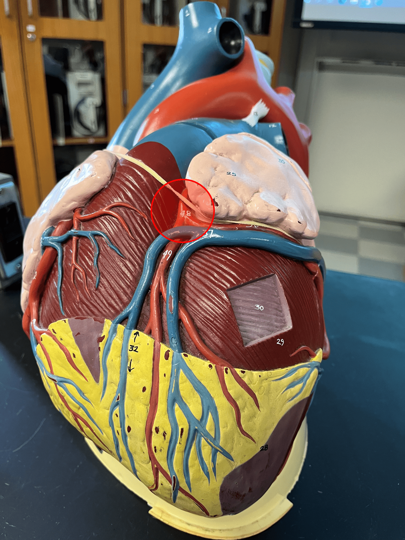
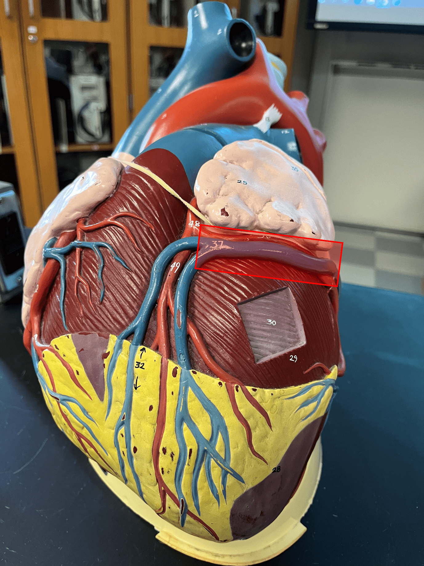
37
New cards
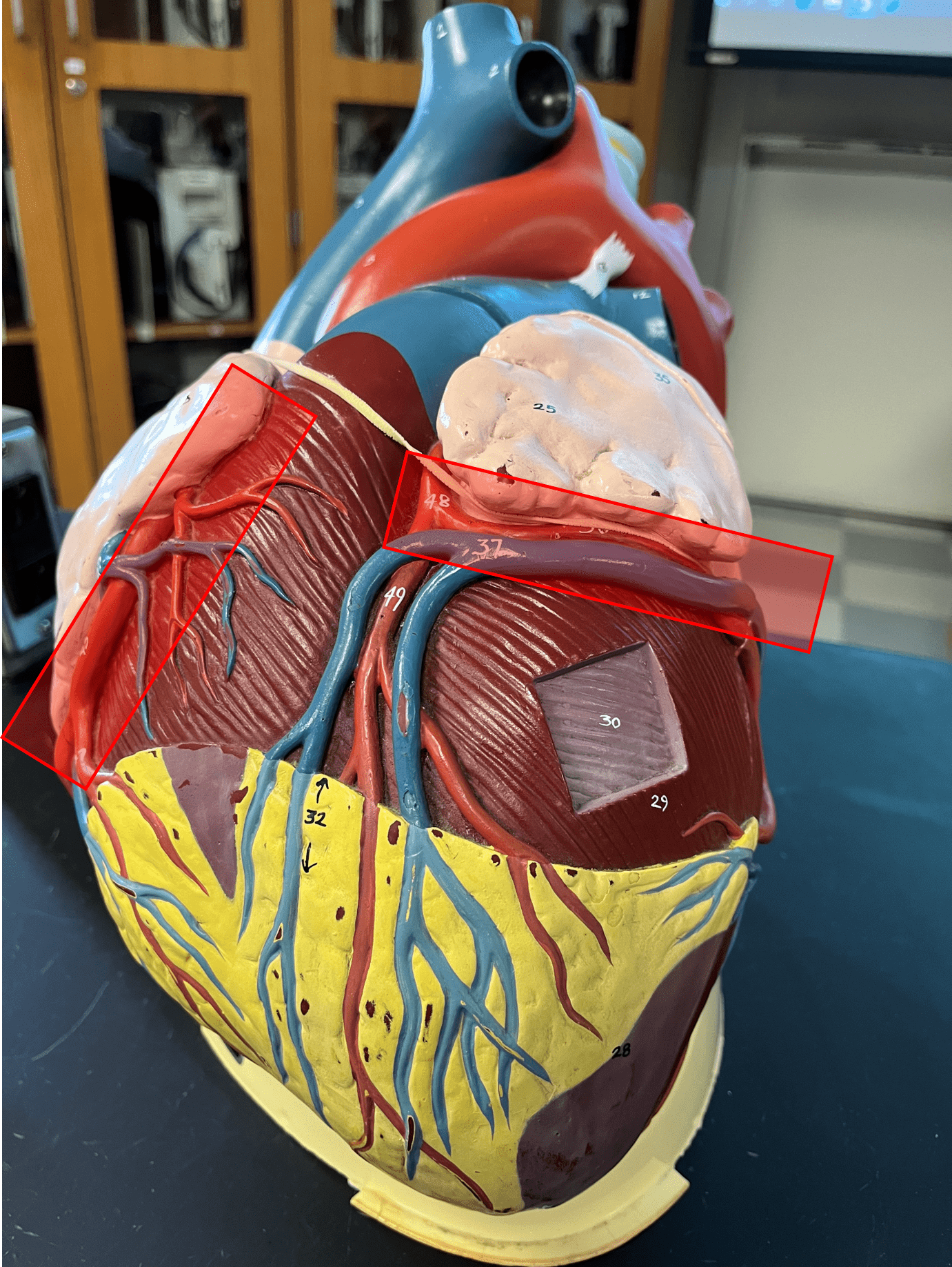
marginal artery
• Part of the coronary circulation.
• Supplies the lateral right side of the heart moving toward the apex.
• One of the branches of the right coronary artery.
• Supplies the lateral right side of the heart moving toward the apex.
• One of the branches of the right coronary artery.
38
New cards
left coronary artery
• Part of the coronary circulation.
• Supplies the anterior side of the ventricles and laterodorsal left side of the heart.
• Arises from the base of the aorta.
• Branches into the anterior interventricular and circumflex arteries.
• Supplies the anterior side of the ventricles and laterodorsal left side of the heart.
• Arises from the base of the aorta.
• Branches into the anterior interventricular and circumflex arteries.
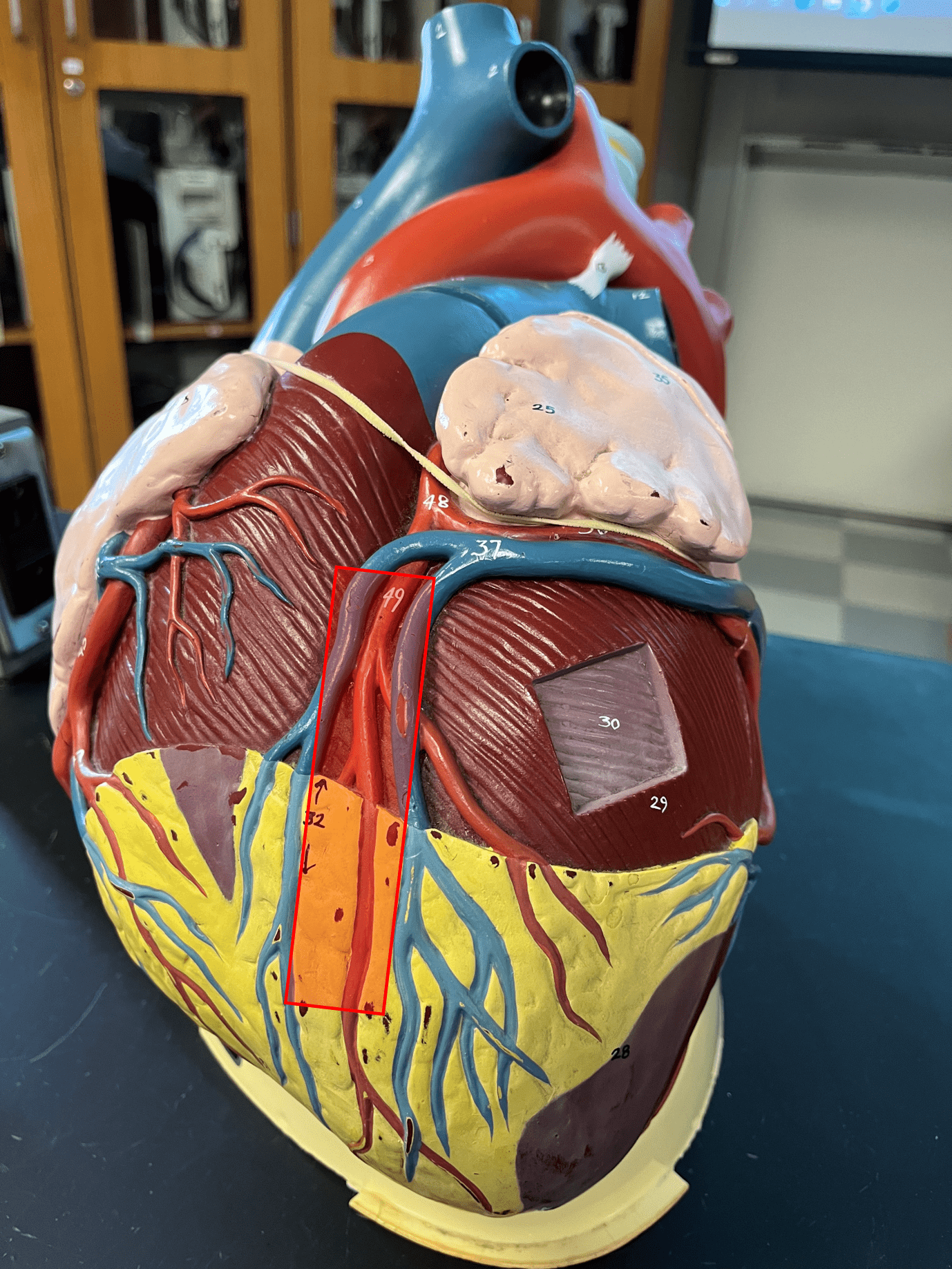
39
New cards
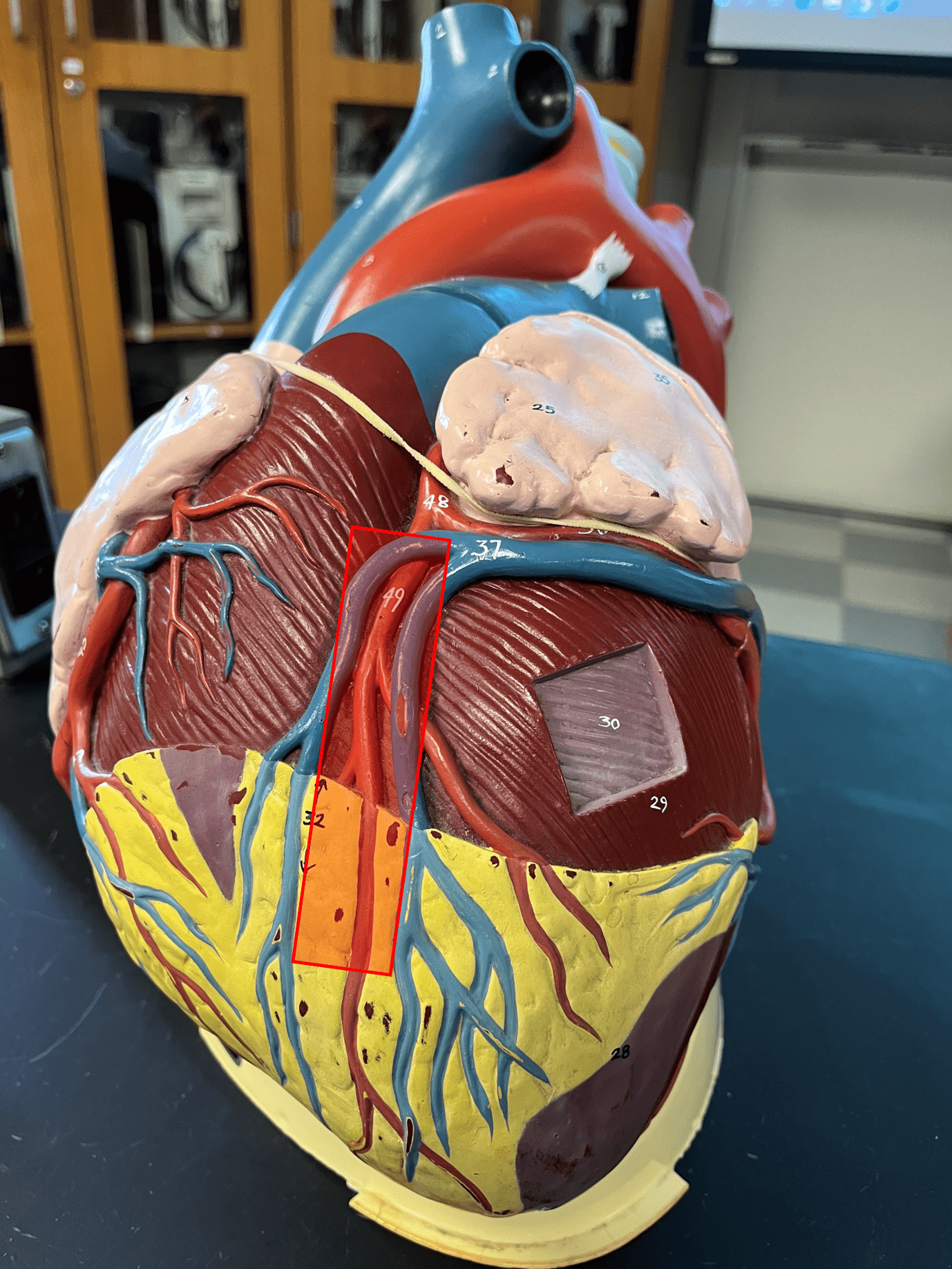
anterior interventricular artery
• Part of the coronary circulation.
• Located in and supplies the tissue around the anterior interventricular sulcus.
• One of the branches of the left coronary artery.
• Located in and supplies the tissue around the anterior interventricular sulcus.
• One of the branches of the left coronary artery.
40
New cards
circumflex artery
• Part of the coronary circulation.
• Wraps around the left side of the heart underneath the left auricle.
• One of the branches of the left coronary artery.
• Wraps around the left side of the heart underneath the left auricle.
• One of the branches of the left coronary artery.

41
New cards
small cardiac vein
• Part of the coronary circulation.
• Drains the myocardium on the lateral right side of the heart underneath the right auricle.
• Leads to the coronary sinus.
• Drains the myocardium on the lateral right side of the heart underneath the right auricle.
• Leads to the coronary sinus.
42
New cards
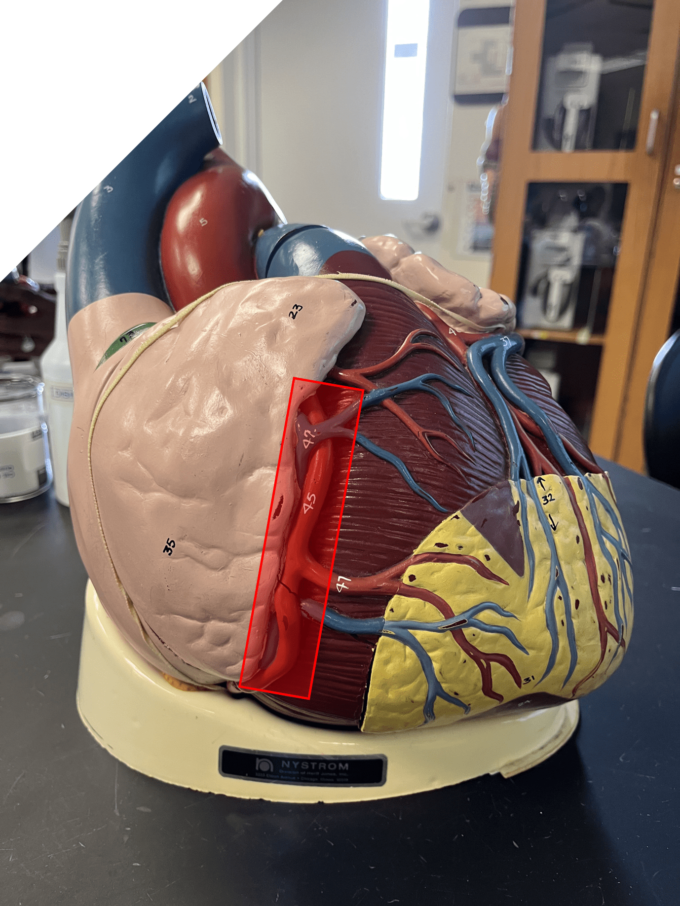
middle cardiac vein
• Part of the coronary circulation.
• Drains the myocardium on the posterior side of the heart, just inferior to the coronary sinus.
• Leads to the coronary sinus.
• Drains the myocardium on the posterior side of the heart, just inferior to the coronary sinus.
• Leads to the coronary sinus.
43
New cards
great cardiac vein
• Part of the coronary circulation.
• Drains the myocardium around the anterior interventricular sulcus.
• Leads to the coronary sinus.
• Drains the myocardium around the anterior interventricular sulcus.
• Leads to the coronary sinus.
44
New cards
coronary sinus
• Part of the coronary circulation.
• Located on the posterior of the heart.
• Receives from the small, middle, and great cardiac veins.
• Empties into the right atrium.
• Located on the posterior of the heart.
• Receives from the small, middle, and great cardiac veins.
• Empties into the right atrium.