MS
 Call Kai
 Learn
Learn Practice Test
 Spaced Repetition
 Match
Match Flashcards
 Knowt Play
1/26

There's no tags or description
Looks like no tags are added yet.
Name | Mastery | Learn | Test | Matching | Spaced | Call with Kai | Chat |
|---|
No analytics yet
Send a link to your students to track their progress
27 Terms
Describe MS
Epi
Age?
Ratio
Lat. Gradient?
Race?
RR %?
Correlations:
What are the two correlations that increases risk of MS
Genetics
Average Risk?
If somebody w/in first degree?
Twins?
Specific Gene Involved?
MS Epi:
Peak Age: 20-30 ((24.5 years))
2:1 ratio women to men
Latitudinal gradient
Greater rates @ farther from equator
Race:
Highest: northern Europeans
Lowest: Asians, African Blacks
85% Initial RR (Relapsing-Remitting)
MS Correlations:
Smoking:
Relative Risk of Smokers: 1.5
Risk of dev. SPMS = 3x higher in smokers
Low Vit D
Mixed results in literature
Genetics
Average Risk: 1/750 (0.1%)
If a first degree relative have MS: < 5%
Twins
Monozygotic Twins: 31%
Dizygotic Twins: 5%
Specific Gene Involved:
HLA-DR2 allele (Chromosome 6)
Increases risk
Describe the Immunopathogenesis of MS
Inflammation → Demyelination → Axonal Loss

Myelin is stained red and axons green. Arrowheads indicate axons undergoing active demyelination. One demyelinated axon ends with an ovoid (arrow).
The irregular and discontinuous distribution of myelin around axons indicates areas of continuous demyelination.
Axonal transection commonly occurs in both chronic and acute plaques; it is more common than formerly appreciated, and varies in degree among patients.

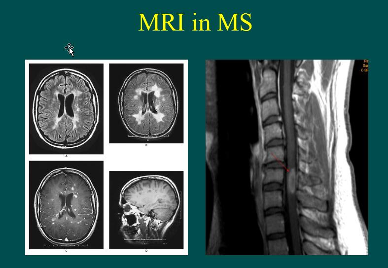
What is the interpretation of these images?
T1-weighted scan w/ Gd contrast
shows areas of inflammation @ sites of Gd penetration
blood vessel -> broken BBB -> CNS.
T2-weighted (hyperintense) scans
visualize heterogeneous lesions
represent cumulative burden of disease.
T1-weighted hypointense images
visualize areas of substantial structural injury (axonal loss and gliosis)
AKA “black holes.”
What are the four different MS Clinical Subtypes

Draw out the progression of MS
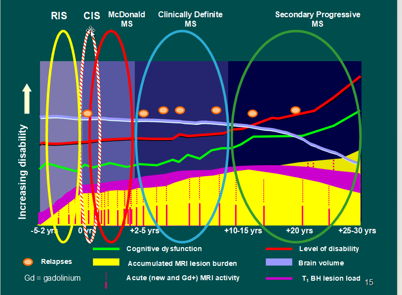
Describe the MS Diagnosis Criteria:
History
PE
Labs
MS Diagnosis Criteria
History:
(2 episodes time and space)
Relapsing-Remitting Course
PE
APD
Afferent Pupillary Defect
INO
Inter-Nuclear Ophthalmoplegia
Labs
CSF;
IgG index,
OCB
Evoked Potentials
(VER, BAER, SSEP)
Blood
Rule out mimickers
MRI Brain/Spine w/ + w/o Gd
MS Misdiagnosis Stats:
20%
Most Common Alt. Disease
Migraine: 16%
Radiologically Isolated Syndrome (RIS): 9%
Spondylopathy: 7%
Neuropathy: 7%
List the Clinical Features Suggestive of MS
Disease Course
Epi
Key Symptoms
List the Clinical Features NOT Suggestive of Multiple Sclerosis
Epi
Disease Course
Symptoms
Clinical Features Suggestive of MS
Disease Course:
Relapses and Remissions
Onset btw 15 - 50 y/o
Epi:
Northern European ancestry
Female
Key symptoms:
Optic neuritis
APD
L’hermitte's sign
Shock-like sensation
Diplopia-INO
Fatigue
Uhthoff's phenomenon
(Worsening of existing symptoms and fatigue)
Clinical Features NOT Suggestive of Multiple Sclerosis
Epi
Onset: <10 + >50
Disease Course
Steady progression
Deficit developing within minutes
Symptoms:
Cortical Defects
aphasia, apraxia, alexia, neglect
Rigidity, sustained dystonia
Convulsions
Early dementia
list the different differentials for white matter lesions

Describe the relationship between age and white matter lesions
66-80% of MRI scans done on > 65 years of age → has periventricular white matter(PVWM) abnormalities
Part of Normal Aging Process


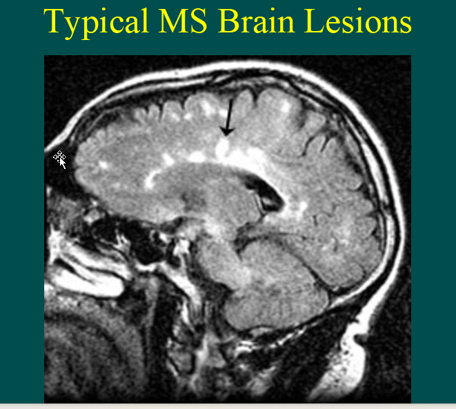
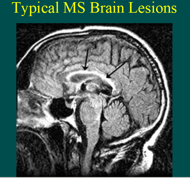
Describe where MS White matter lesions occur
Location:
Corpus callosum
U-Fibers
white matter tracts that connect adjacent gyri (ridges)
Temporal Lobes
Brainstem/Cerebellum
SC
90% involvement in MS
Rare in normal aging/small vessel disease

List the two labs that help in diagnosing MS
Of the two labs, what other differentials can exist besides MS
IgG Index Differential
MS (80-90% sensitive/94% specific)
Other Differentials
Aseptic meningitis
Lymphoma
Neurosyphilis
GBS
Cerebral Lupus
Oligoclonal banding
MS-83-94% specific
Other Differentials
Encephalitis
Meningitis
Polyneuritis
Headache
SSPE
Other CNS Inflammatory Disorders
Vasculitis
Neurosarcoid
CNS Lupus
GBS
Behcet’s
Describe the Evoked potentials in MS
VER-80-85% abnormal in definite MS
Visual evoked potentials
SSEP-65-80%
Somatosensory evoked potentials
BAER-50-65%
Brainstem auditory evoked responses

List the MS Lab Workups
Lab Workups for Atypical Cases
MS Lab Workups
Vasculitis Labs:
[CRP, ESR, C3, C4, ACE]
HIV, HTLV 1-2
Vitamin D
RPR, VDRL
Screening for Syphilis
CADASIL: [Notch 3 mutation]
hereditary small‑vessel disease caused by a mutation in the NOTCH3 gene
Thyroid Panel
Lupus Panel
ANA, Rheum Factor
Autoimmune Screen
Anticardiolipin Ab
Part of antiphospholipid syndrome work‑up.
Lyme
B12/Folate
NMOSD Labs
neuromyelitis optica spectrum disorder
For Atypical cases:
WBC Arasulfatase A
(MLD)-Mayo Lab, yellow top
Very Long Chain Fatty Acids (ALD, AMN)
Mayo Lab, green top
Hexosaminidase A
(Tay Sachs)
Galactocerebrosidase C
(Gal-C)
Fasting blood lactate, Quantitative plasma AA, and Urine organic acid analysis (MELAS)
Palmitoyl Protein Thioesterase 1 (adults), tripeptidyl peptidase 1, CLN3, and CLN5 (NCL) (Neuronal Ceroid Lipofuscinosis)
NPC-1 (Niemann Pick)
Buccal or rectal mucosa biopsy for EM or skin biopsy
fibroblast culture or peripheral blood smear
(look for vacuolated monocytes)
HIV
Vasculitis W/U:
Behcet’s-Pathergy skin test or skin biopsy and/or derm/rheum referral
[CRP, ESR, C3, C4, ACE]
Brain Biopsy
NCL shows fingerprint profiles and curvilinear and rectilinear bodies as well as diffuse white matter gliosis
Helpful in Systemic Histiocytosis or Whipple’s or Lymphoma



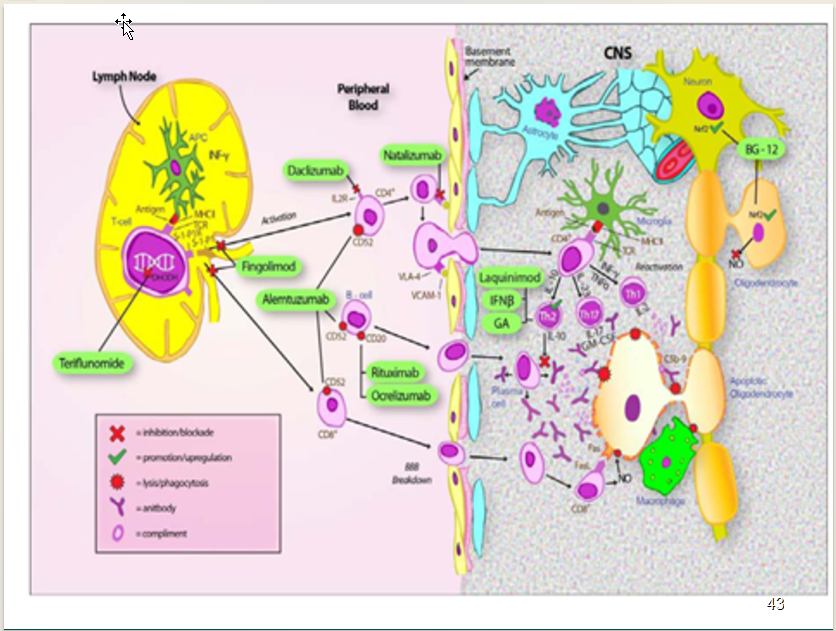
List Olek’s Rationale for using Tysabril
Limiting factor of using Tysabril
68% Relapse Rate Reduction
42% Slowing of Confirmed Disability
92% Reduction in MRI Gd Lesions
83% Reduction in MRI T2 Lesion
76% Reduction in MRI T1 Lesions
Limiting Factor:
PML: Progressive Multifocal Leukoencephalopathy]
List the CIS associated w/ MS
What do you do to determine if its CIS or Possible MS Attack in ER?
Clinically Isolated Syndromes (CIS) Associated w/ MS
Optic Neuritis (ON)
Acute loss of vision
Transverse Myelitis (TM)
Spinal cord lesion
Brainstem syndrome
Diplopia, face numbness or weakness, dysarthria
Cerebellar syndrome
Balance problems
CIS or Possible MS Attack in ER Protocol
Rule out infection
Treat w/ Abx if infection
Fundoscopy -> Spinal Tap + Blood
IgG index
OCB
Tx: Set-up Outpatient IV methylprednisolone
1000mg Daily x 5 days w/ no oral taper
Dose 1 may be given in ER AFTER LP
What is RIS?
Describe the relationship between RIS and MS
RIS:
white matter anomalies characteristic of MS that are found on Brain/SC MRI
BUT Pt does not have characteristics of MS at that time
NO relapsing or progressive symptoms typical of inflammatory demyelination.
RIS and MS
34% w/in 5 yrs to have an MS event
10% fulfilled the criteria for PPMS
Significant Predictors of First Event:
Age + male + lesions @ Cervical/Thoracic Spine
Describe the relationship between MS and ONTT/CIS
Describe Interferon (IFN) Neutralizing Antibodies
What what are they?
Drugs Affected
List the biomarkers that rules out MS
ONTT ((Optic Neuritis Treatment Trial)):
15 years -> 50% developed MS
Baseline MRI
NO Lesions: 25% developed MS
>1 lesion: 72% developed MS
CIS:
% of developing MS
NO Lesions on Initial MRI: 19% MS in 14 years
>1 lesion on Initial MRI: 88% MS in 14 years
% Developing EDSS (Expanded Disability Status Scale)
NO lesions on initial MRI: 6% EDSS (>6) in 20 yrs
>10 lesions on initial MRI: 45% EDSS (>6) in 20 yrs
Interferon (IFN) Neutralizing Antibodies
What are they?
antibodies pt develop against interferon‑β medications -> reduces effectiveness
Drugs Affected:
Avonex-5%
Rebif-25%
Betaseron-38%
Biomarkers that rule out MS
Aquaporin-4
[Neuromyelitis Optica-NMO]
Serum: 91% specific and 73% sensitive
CSF: 60% predictive
Myelin Oligodendrocyte Glycoprotein (MOG)
Marker for MOG‑associated disease (MOGAD)
JC Virus Antibodies
Describe the Ms Medications Decision Tree
current therapy doing well
New diagnosis RRMs
Current therapy not doing well
Ms Medications Decision Tree
current therapy doing well: No Changes
New diagnosis RRMS
NO JC Ab: Tysabri
Jc Ab Pos:
Tecfidera
Tysabri for 12 months -> Tecfidera
Current therapy NOT doing well:
Switch to other DMT
Add Immunosuppressant
Switch to IVIG or IV Cytoxan
List the Potential Indicators of Suboptimal Response
Clinical Indicators
Functional Indicators
Radiologic Indicators
Clinical Indicators
Relapse frequency
≥1 severe relapse or ≥2 relapses of any severity in 1 year
Relapse frequency not decreasing after pt on DMT for ≥6 to 12 months
Incomplete recovery at 6 months from one or more attacks
Involvement of multiple neurologic systems or functional domains
Functional Indicators
Progressive motor or cognitive impairment -> interferes w/ daily activities
(after ruling out contributing factors)
Increasing EDSS score or permanent residual disability
***NOTE: Changes in EDSS scores <3.0 should not be used isolation to define treatment failure***
*** Increase in EDSS of ≥1 from prev score of 3.0–5.5 or 0.5 point increase from prev score of ≥6.0 = RRMS -> SPMS or partial response to treatment in SPMS***
Radiologic Indicators
New or recurrent brain stem or spinal cord lesions
Increasing lesion number/Size
***(eg, ≥3 enhancing lesions, or >3 new T2 lesions in 1 year, or ≥2 new T2 lesions on each repeated scan performed at no less than quarterly intervals)***
***especially Significant if increase in >2 types of lesions or in the presence of EDSS worsening independent of attack***
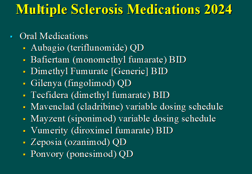
List the MS Medications based on type
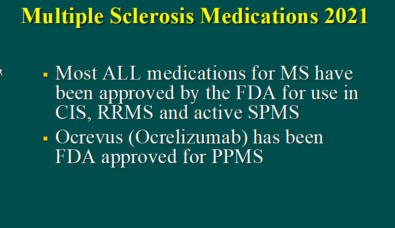
PPMS
SPMS
SPMS w/ attacks
List the Treatments for Acute Attacks
List Tx for NMO
PPMS
Ocrevus
SPMS
No RRMS medications
(Except Active SPMS-see below)
Trial of immunosuppression
MTX, Imuran, Cellcept, CTX, IV steroids
IVIG
PE (Plasma Exchange)
BMT (Bone Marrow Transplantation)
SPMS with Attacks
Tecfidera
Tysabri
Mavenclad
Mayzent
Acute Attacks Treatment
Rule out Infection
IV/Oral steroids or Achtar Gel
NMO
Rituxan
Soliris (eculizumab)
Enspryng (satralizumab)
Uplizna (inebilizumab)
Other immunosuppressants