Chapter 6 - THE DIGESTIVE SYSTEM SUPPLIES NUTRIENTS FOR THE BODY
1/33
There's no tags or description
Looks like no tags are added yet.
Name | Mastery | Learn | Test | Matching | Spaced | Call with Kai |
|---|
No analytics yet
Send a link to your students to track their progress
34 Terms
digestive system
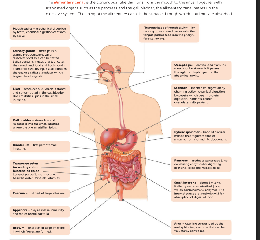
The digestive system extracts nutrients from the food we eat and absorbs them into the body for use by the cells. The organs of the digestive system are structured and arranged so that they can carry out six basic activities:
1 ingestion of food and water
2 mechanical digestion of food
3 chemical digestion of food
4 movement of food along the alimentary canal
5 absorption of digested food and water into the blood and lymph
6 elimination of material that is not absorbed. The structures of the digestive system work together to fulfil these functions.
Key concept Digestion
Digestion is the process of breaking down food into particles small enough to be absorbed into the bloodstream.
Mechanical digestion
Mechanical digestion is the physical breakdown of food particles. It involves the following processes in the mouth, stomach and small intestine:
• The teeth cut, tear and grind the food.
• Churning action in the stomach breaks the food down further. • The gall bladder releases bile into the small intestine. Bile salts act as emulsifying agents, breaking fat down into smaller droplets.
The aim of mechanical digestion is to break the food down into smaller pieces so that the total surface area increases. This allows more effective chemical digestion, as the chemicals can access more of the food.
Mechanical digestion
Mechanical digestion is the physical breakdown of food into smaller pieces to increase the surface area.
Chemical digestion
During chemical digestion, chemicals break down large, complex molecules into smaller, simpler molecules. These smaller molecules are then small enough to be absorbed into the bloodstream.
• Carbohydrates split into monosaccharides such as glucose, fructose and galactose.
• Proteins are split into peptides and amino acids.
• Lipids are split into fatty acids and glycerol.
• Nucleic acids are split into nucleotides.
Chemical digestion is achieved by enzymes. Enzymes are biological catalysts – chemicals that are able to increase the rate of a reaction without being consumed.
Chemical digestion
Chemical digestion uses enzymes to break large, complex molecules into small, simpler molecules.
The mouth
Intake of food, called ingestion, occurs at the mouth. Here the food is chewed in a process called mastication. Both mechanical and chemical digestion commence before the food is swallowed.
Saliva and chemical digestion
As the food is chewed it is mixed with saliva, a fluid that is secreted into the mouth cavity by three pairs of salivary glands. It contains mucus to lubricate the food and a digestive enzyme – salivary amylase – which begins the chemical digestion of starch into the disaccharide maltose.
The teeth and mechanical digestion
The action of the jaws and teeth begins mechanical digestion. There are four types of teeth, each with a different function in mechanical digestion. A full adult set of teeth in the lower jaw consists of:
• four incisors – chisel-shaped teeth used for biting or cutting, as when taking a bite of an apple
• two canines, one on each side of the incisors. These are conical teeth used for tearing. Human canines are the same length as the other teeth
• four premolars, two on each side of the jaw
• six molars, three on each side of the jaw. The premolars and molars have broad crowns with rounded cusps. The cusps of the teeth of one jaw fit into depressions on the surface of teeth on the other jaw, making the premolars and molars ideal for crushing and grinding food. The same number and type of teeth occur in the upper jaw.
The oesophagus
The oesaphagus is a tube about 23–25 cm long that connects the pharynx to the stomach. The wall of the oesophagus, like the rest of the alimentary canal, has a double layer of muscle. Circular muscle has muscle fibres arranged in a circle, and longitudinal muscle has fibres arranged along the length of the canal.
food enters pharynx and oesophagus
As the lump of food enters the pharynx and oesophagus, the circular muscle behind it contracts to narrow the tube. The contraction of successive bands of circular muscle causes the constriction to move in a wave called Peristalsis. This movement pushes the food in front of it, assisted by the secretion of mucus that lubricates the inner lining.
Peristalsis
Peristalsis is the wave-like muscle contractions that move food through a tube such as the oesophagus.
The stomach
The oesophagus passes through the diaphragm, a sheet of muscle that separates the thoracic cavity from the abdominal cavity. Just after passing through the diaphragm it opens into the stomach, a roughly J-shaped, enlarged section of the alimentary canal.
Mechanical digestion
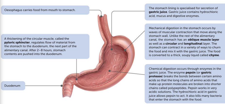
Mechanical digestion in the stomach is achieved by waves of muscular contraction that move along the stomach wall. Unlike the rest of the alimentary canal, the stomach has an oblique muscle layer as well as a circular and longitudinal layer. This enables the stomach to contract in a variety of ways to churn the food and mix it with the stomach juices until the food is converted to a thick, soupy liquid called chyme.
mucosa
The lining of the stomach, the mucosa, is specialised for the secretion of gastric juice by gastric glands located in narrow, tube-like structures called gastric pits.
Gastric juice
Gastric juice is a digestive juice containing hydrochloric acid, mucus and digestive enzymes. Each of these is secreted by a different type of cell in the gastric pits. Gastric juice is responsible for chemical digestion in the stomach, which is mainly the start of protein digestion. The pH in the stomach is approximately 2–3, due to the hydrochloric acid. The cells lining the stomach are protected from the acid by a layer of mucus. The acidic environment allows the enzyme pepsinogen to be converted to pepsin, an active form of the enzyme. Pepsin is able to break proteins down into shorter peptides. It also breaks down the nucleic acids DNA and RNA.
Nutrients
Nutrients are not absorbed into the bloodstream through the stomach, because the internal surface is covered by a thick layer of mucus. However, some alcohol and a few other drugs such as aspirin are absorbed. At the lower end of the stomach there is a thickening of the circular muscle, which results in a constriction called the pyloric sphincter. The constriction is sufficient to prevent the stomach contents moving through unless pushed along by peristalsis. After 2 to 8 hours, the stomach contents are gradually pushed into the next part of the alimentary canal, the small intestine.
The small intestine
The small intestine gets its name from the narrow diameter of its tube. It is the longest part of the alimentary canal, at approximately 6–7 m in length. It receives material pushed through the pyloric sphincter from the stomach. There are three regions of the small intestine:
1 Duodenum: the first part of the small intestine. It is the shortest section at only 25 cm. The duodenum extends from the bottom end of the stomach in a curve around the pancreas. Most of the chemical digestion occurs here before the chyme moves further along the small intestine.
2 Jejunum: the middle section of the small intestine. The lining of the jejunum allows effective absorption of carbohydrates and proteins.
3 Ileum: the final part of the small intestine. It is here that vitamin B12, bile salts, and any remaining products of digestion are absorbed.
Digestion continues in the small intestine under the influence of:
• pancreatic juice – secreted by the pancreas via the pancreatic duct
• bile – produced by the liver, but stored in the gall bladder and secreted into the small intestine via the bile duct
• intestinal juice – secreted by glands in the lining of the small intestine.
Pancreatic juice
Pancreatic juice enters the duodenum through the common bile duct. It helps to neutralise the acid that has come with the material from the stomach and contains many of the enzymes involved in the digestion of food. The enzymes include:
• pancreatic amylase, which breaks down starch into the disaccharide maltose
• trypsin (or pancreatic protease), which splits proteins into peptides
• pancreatic lipases – enzymes that break down fats into fatty acids and glycerol
• ribonuclease and deoxyribonuclease – enzymes that digest RNA and DNA.
Bile
Bile is secreted into the small intestine through the common bile duct. Although it does not contain any digestive enzymes, bile salts are very important in the mechanical digestion of fats. They act like a detergent and emulsify the fat, breaking it into tiny droplets. This is a form of mechanical digestion, as it increases the surface area on which the lipases can act to bring about the chemical digestion of fat.
Intestinal juice
Intestinal juice contains many enzymes that complete the digestion of carbohydrates, proteins and lipids. These include: • peptidase to break down peptides into amino acids
• sucrase, lactase and maltase to break down sucrose, lactose and maltose, respectively, into the monosaccharides glucose, fructose and galactose
• lipases to break down lipids into fatty acids and glycerol
Enzymes in pancreatic juice and intestinal juice
Enzymes in pancreatic juice and intestinal juice facilitate chemical digestion, while bile salts emulsify fat droplets.
Absorption of nutrients
When chemical digestion is complete, the complex carbohydrates will have been broken down into simple sugars, the proteins into amino acids, and the fats into fatty acids and glycerol. These products of digestion, along with substances such as vitamins, minerals and water, are then absorbed through the wall of the small intestine into the blood. Nutrients are absorbed through the internal surface of the small intestine, so efficient absorption requires a large surface area. A large internal surface area is achieved in a number of ways:
• The small intestine is very long – about 6–7 m.
• The inner lining, known as the mucosa, has folds that extend into the interior of the small intestine.
• The mucosa has small, finger-like projections called villi that extend from the folded surface.
• The cells covering the outside of the villi have tiny microscopic projections from their external surfaces. These are the microvilli.
The structure of a villus
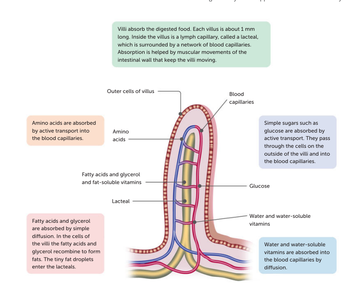
The structure of a villus is ideally suited to its function of nutrient absorption. Each villus is about 1 mm long, although the villi in the jejunum are longer than those in the duodenum and ileum. Each villus is covered by a single layer of cells. Inside the villus is a lymph capillary, called a lacteal, which is surrounded by a network of blood capillaries. Absorption is further enhanced by continual movement of the villi brought about by the muscular movements of the intestinal wall. This constantly brings the villi into contact with different parts of the intestinal contents. These contents are constantly changing as new material is emptied into the small intestine from the stomach. Some absorption occurs through simple diffusion, as there is a higher concentration of nutrient materials in the interior of the small intestine than in the cells lining the villi. Absorption also occurs through active transport, which involves the cells of the villi using energy to take in nutrients against a concentration gradient – that is, taking in molecules from a lower concentration to a higher concentration. From the walls of the villi, simple sugars, amino acids, water and water-soluble vitamins are absorbed into the blood capillaries. Fatty acids and glycerol recombine in the cells of the villi to form fats and, along with the fat-soluble vitamins, enter the lacteals. The substances that are absorbed into the blood capillaries are carried by the hepatic portal vein to the liver. Here they may be removed for further processing, or they may remain in the blood to be carried to other body cells. Substances that are absorbed into the lacteals are transported in the lymph system, which eventually empties into the blood through veins in the upper part of the chest.
The lining of the small intestine
The lining of the small intestine has folds, villi and microvilli to maximise the absorption of nutrients.
The large intestine
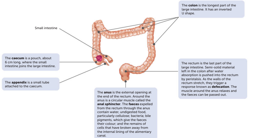
The large intestine is about 1.5 m long, and is so named because it is larger in diameter than the small intestine. It is made up of the caecum, colon, rectum and anus. Additionally, the appendix attaches to the caecum. There are no villi in the large intestine, and no digestive juices are secreted, although the lining does secrete a large amount of mucus. Movement of material through the large intestine is fairly slow, taking 18–24 hours for material to pass through. During this time, most of the remaining water is absorbed so the contents become more solid. Bacteria in the large intestine break down much of the remaining organic compounds. Some bacteria produce vitamins, which are then absorbed through the walls into the blood. Mineral nutrients are also absorbed.The semi-solid material left after water absorption and bacterial action makes up the faeces. Faeces contain water, undigested food material (particularly cellulose), bacteria, bile pigments (which give the faeces their colour) and the remains of cells that have broken away from the internal lining of the alimentary canal. The faeces pass through the rectum and anus to the exterior of the body. Many people refer to defecation as ‘excretion’. Excretion is the removal of metabolic waste – waste that has been produced by chemical activity of the body cells. Except for the bile pigments, the contents of faeces are not metabolic waste, so defecation is better referred to as elimination, rather than excretion.
The importance of soluble fibre in the diet
Both soluble and insoluble fibre are found only in foods derived from plants. Soluble fibre includes pectins, gums and mucilage. Soluble fibre intake has been linked to lower cholesterol levels in the blood, decreased risk of heart disease and cancer, and beneficial effects on blood glucose levels. Fats in the intestines are trapped by soluble fibre, thereby helping to prevent their absorption by the body. This is thought to be the reason that soluble fibre helps to lower blood cholesterol levels. Good sources of soluble fibres are fruits, vegetables, oat bran, barley and soy products.
Bowel cancer
Bowel cancer, or colorectal cancer, is an uncontrolled growth of cells in the wall of the large intestine. Research suggests that bowel cancer may be linked to diet, high alcohol consumption and smoking. A diet high in red and processed meat, and low in fibre (fruit and vegetables), may increase the risk of developing bowel cancer. Being overweight or obese and physical inactivity are also risk factors.