Anatomy II - Mediastinum
 Call Kai
 Learn
Learn Practice Test
 Spaced Repetition
 Match
Match Flashcards
 Knowt Play
1/78

There's no tags or description
Looks like no tags are added yet.
Name | Mastery | Learn | Test | Matching | Spaced | Call with Kai | Chat |
|---|
No analytics yet
Send a link to your students to track their progress
79 Terms
T1-T12 Vertebrae
Posterior boundary of Mediastinum
Lung cavities
Lateral boundary of Mediastinum
Sternum
Anterior boundary of Mediastinum
Diaphragm
Inferior boundary of Mediastinum
Thoracic Inlet
Superior boundary of Mediastinum
Pericardial Sac
Thymus
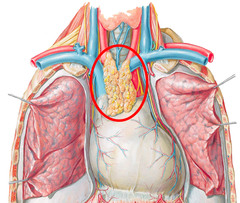
Thymus
T lymphocytes mature here during fetal development.
Fibrous Pericardium
found only outside of Pericardial Sac
tough, prevents rapid overfilling of heart chamber
Parietal Layer of Serous Pericardium
found on the inner surface of Fibrous Pericardium
smooth, protects heart from friction rubs
synthesizes Pericardial Fluid
Visceral Layer of Serous Pericardium
found on the outer surface of Heart
together with underlying Epicardial Fat = Epicardium
smooth, protects heart from friction rubs
synthesizes Pericardial Fluid
Pericardial Effusion
Inflammatory conditions can result in the production of too much Pericardial Fluid
Cardiac Tamponade
Reduces cardiac output when Pericardial Fluid fills pericardial cavity and compresses the heart; Bleeding into the Pericardial Cavity can also cause this
Inferior Vena Cava
returns deoxygenated blood from below diaphragm
Superior Vena Cava
returns deoxygenated blood from above diaphragm
Pectinate Muscles
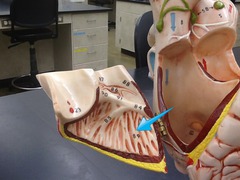
Crista Terminalis
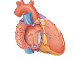
Tricuspid Valve
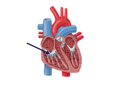
Chordae Tendineae
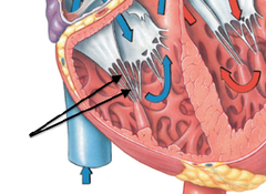
Trabeculae Carniae
J
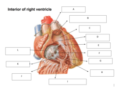
Anterior Papillary Muscle
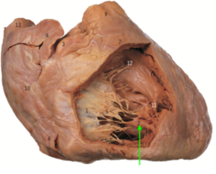
Septomarginal Trabecula
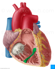
Posterior Papillary Muscle
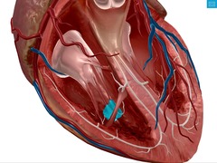
Pulmonary Trunk
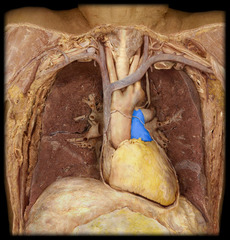
Septomarginal Trabecula
transmits electrical signal triggering contraction of Anterior Papillary Muscle
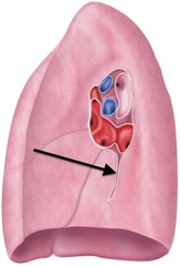
Pulmonary Ligament
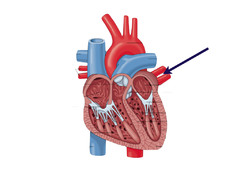
Pulmonary Veins
Bicuspid Valve
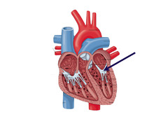
Aortic Valve
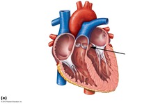
Ascending Aorta
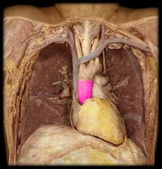
inferior border; diaphragm
Diaphragmatic Surface of heart
Surface of Left Atrium
Base of heart
left border: left lung
Pulmonary Surface of heart
Left Brachiocephalic Vein
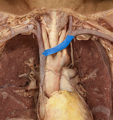
Right Brachiocephalic Vein
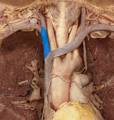
Superior Vena Cava
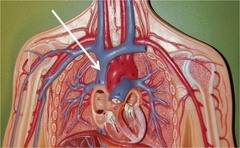
Right Common Carotid Artery
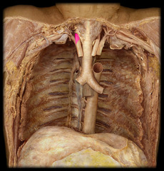
Right Subclavian Artery
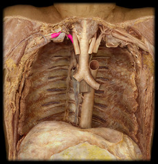
Brachiocephalic Trunk
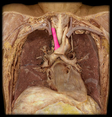
Aortic Arch
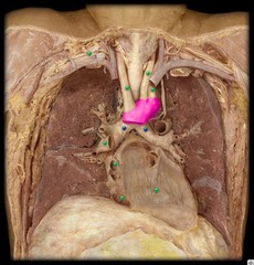
Left Common Carotid Artery
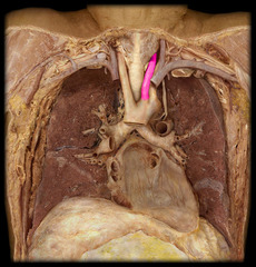
Left Subclavian Artery
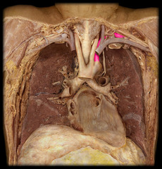
Left Recurrent Laryngeal Nerve
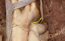
Vagus Nerve
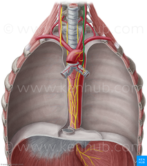
Thoracic Aorta
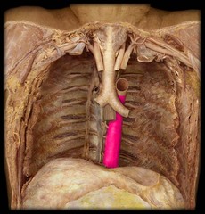
Esophagus
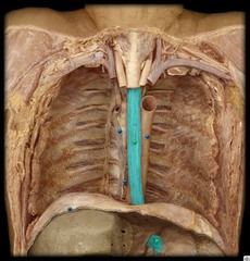
Esophageal Plexus
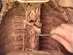
Azygos Vein
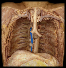
Thoracic Duct
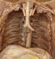
Arch of the Azygos Vein
Phrenic Nerve
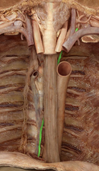
Green arrow
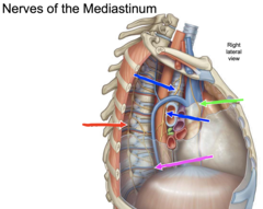
Vagus nerve
Blue arrow
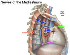
Hemiazygos Vein
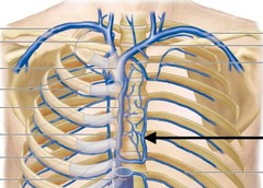
Bronchial Arteries
G
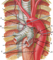
Esophageal Arteries
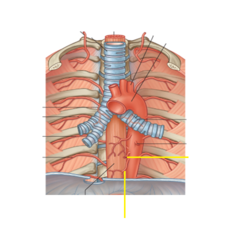
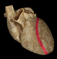
Anterior Interventricular Sulcus
Pulmonary Circuit
Right side of heart
Systemic Circuit
Left side of heart
oxygenated blood
Systemic Circuit has
deoxygenated blood
Pulmonary Circuit has
Anterior Cardiac Veins
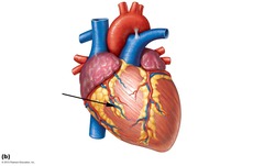
Coronary Sinus
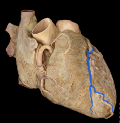
Great Cardiac Vein
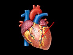
Posterior Interventricular Sulcus
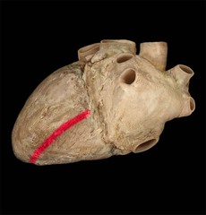
Coronary Sinus
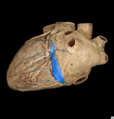
Middle Cardiac Vein
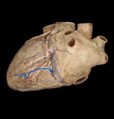
Opening of the Coronary Sinus
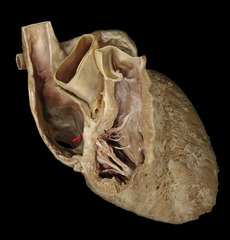
Right Coronary Artery
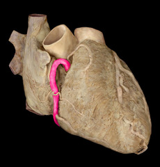
Left Coronary Artery
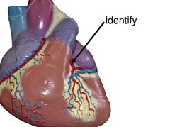
Left Circumflex Artery
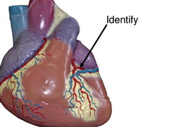
Anterior Interventricular Artery
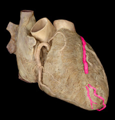
Posterior Interventricular Artery
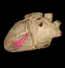
Myocardial infarction
necrosis of an area of the myocardium caused by inadequate blood supply.
75%
Posterior Interventricular Artery arises from the Right Coronary Artery in what % of people (Right dominant)
15%
Posterior Interventricular Artery arises from the Left Coronary Artery in what % of people (Left dominant)
10%
Posterior Interventricular Artery arises from both Right and Left Coronary Artery in what % of people (Co-dominant)
Coronary Artery Bypass Graft
When an obstruction occurs in one or more coronary arteries or their branches, either the great saphenous vein or internal thoracic artery can be grafted to shunt blood to the artery distal to the obstruction.
Oblique Pericardial Sinus
Space within the pericardial cavity, inferior and posterior to the heart. Surrounded by the Inferior Vena Cava and Pulmonary Veins
Transverse Pericardial Sinus
Space within the pericardial cavity, posterior to the Ascending Aorta and Pulmonary Trunk