Exam 2: Chapters 39-41 (Bio2)
1/216
There's no tags or description
Looks like no tags are added yet.
Name | Mastery | Learn | Test | Matching | Spaced | Call with Kai |
|---|
No analytics yet
Send a link to your students to track their progress
217 Terms
Neural signaling
the process by which an animal responds to a stimulus
Neural Signaling Steps:
Reception --> Transmission --> Integration --> Response
Reception (in neural signaling)
performed by neurons and specialized sensory receptors
Transmission (in neural signaling)
sending of a message from one neuron to another; or, from one neuron to a gland or muscle
Integration (in neural signaling)
sorting and interpretation of neural messages & determination of the appropriate response
Response (in neural signaling)
the output or action resulting from the integration of neural messages
Three functional classes of neurons
Afferent neurons
Interneurons
Efferent neurons
Afferent neurons
associate with sensory neurons
transmit stimuli from sensory receptors to interneurons
Interneurons
type of neuron that connect sensory and motor neurons
in the integrating center, process and modify info
Efferent neurons
carry signals indicating a response away from the interneuron to the effectors (muscles or glands) (e.g., motor neuron)
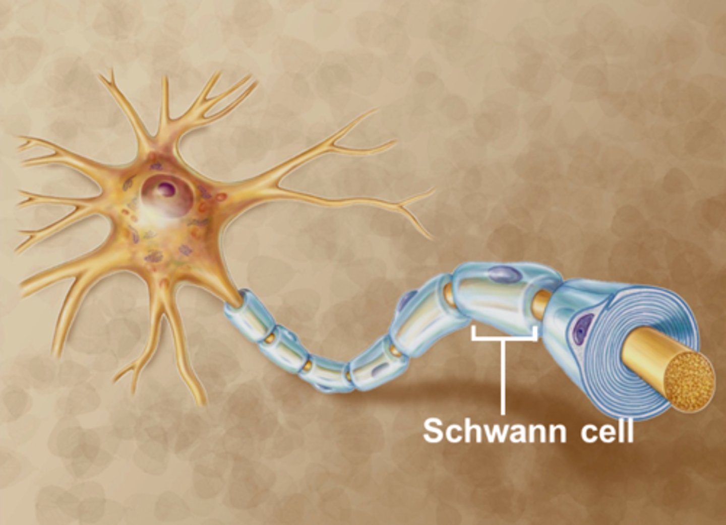
Neuron cell structure
Cell body
Dendrites
Axon
Axon hillock
Axon terminal
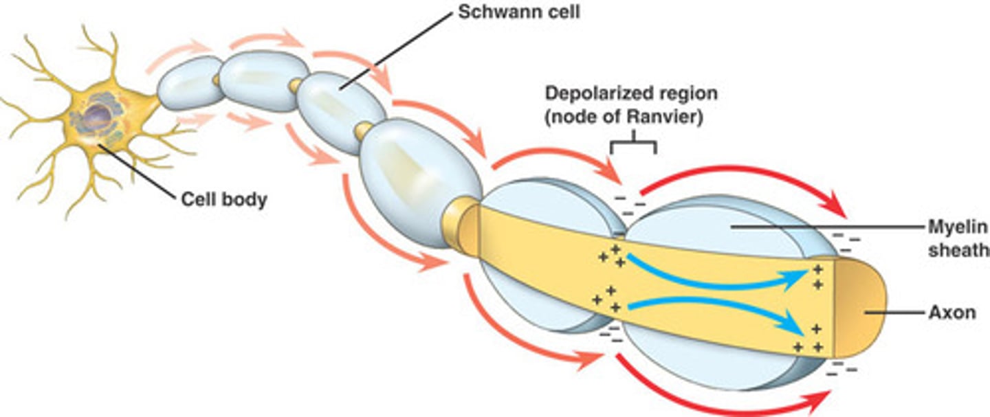
Myelin sheath
Node of ranvier

Cell body
contains the nucleus and most organelles
ganglia
Clusters of cell bodies outside the CNS
(eg. dorsal root ganglia)
nuclei
Clusters of cell bodies within the CNS as gray matter
(eg. basal nuclei of the brain)
Dendrites
highly branched structures which receive signals and transmit them toward the cell body
Axon
conducts signals away from the cell body toward another neuron (or an effector)
Axon hillock
axonal segment between the cell body and the axon
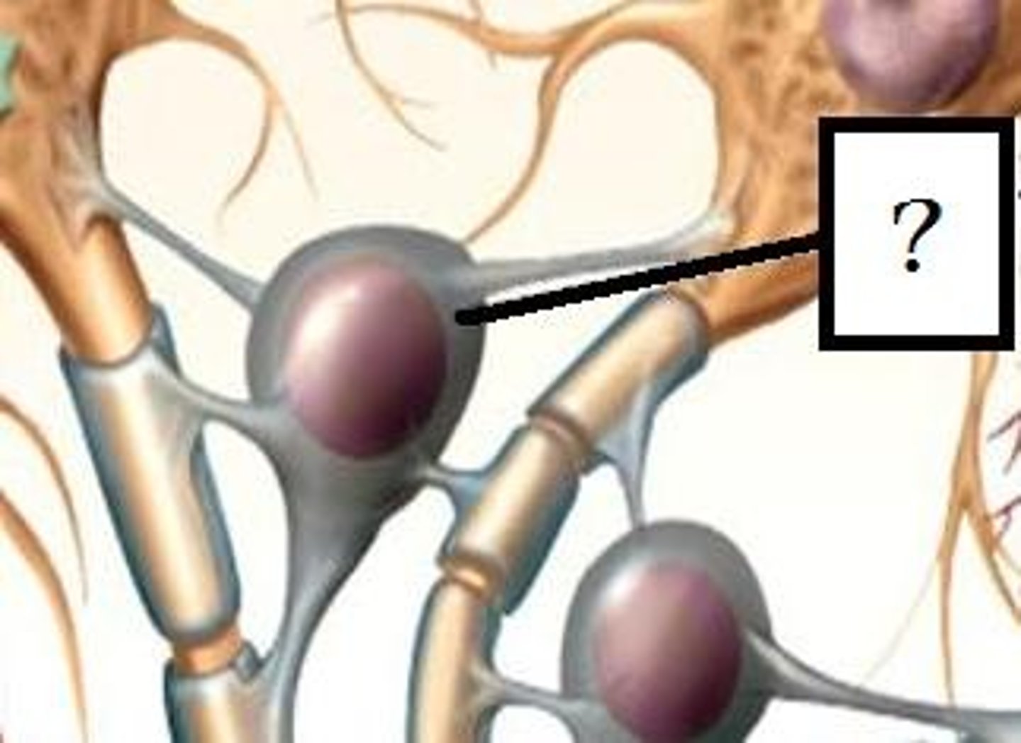
Axon terminals
button-like swellings located at the ends of the axons
Neuronal circuit
a typical neural circuit contains an afferent (sensory) neuron, one or more interneurons, and an efferent neuron
in vertebrates, afferent and efferent neurons form the peripheral nervous system (PNS)
interneurons form the central nervous system (CNS), consisting of the brain and spinal cord
Peripheral nervous system (PNS)
made up of afferent and efferent neurons
Central nervous system (CNS)
includes the brain & spinal cord, consists mostly of interneurons
Neural networks
combined neural circuits that interconnect parts of the nervous system
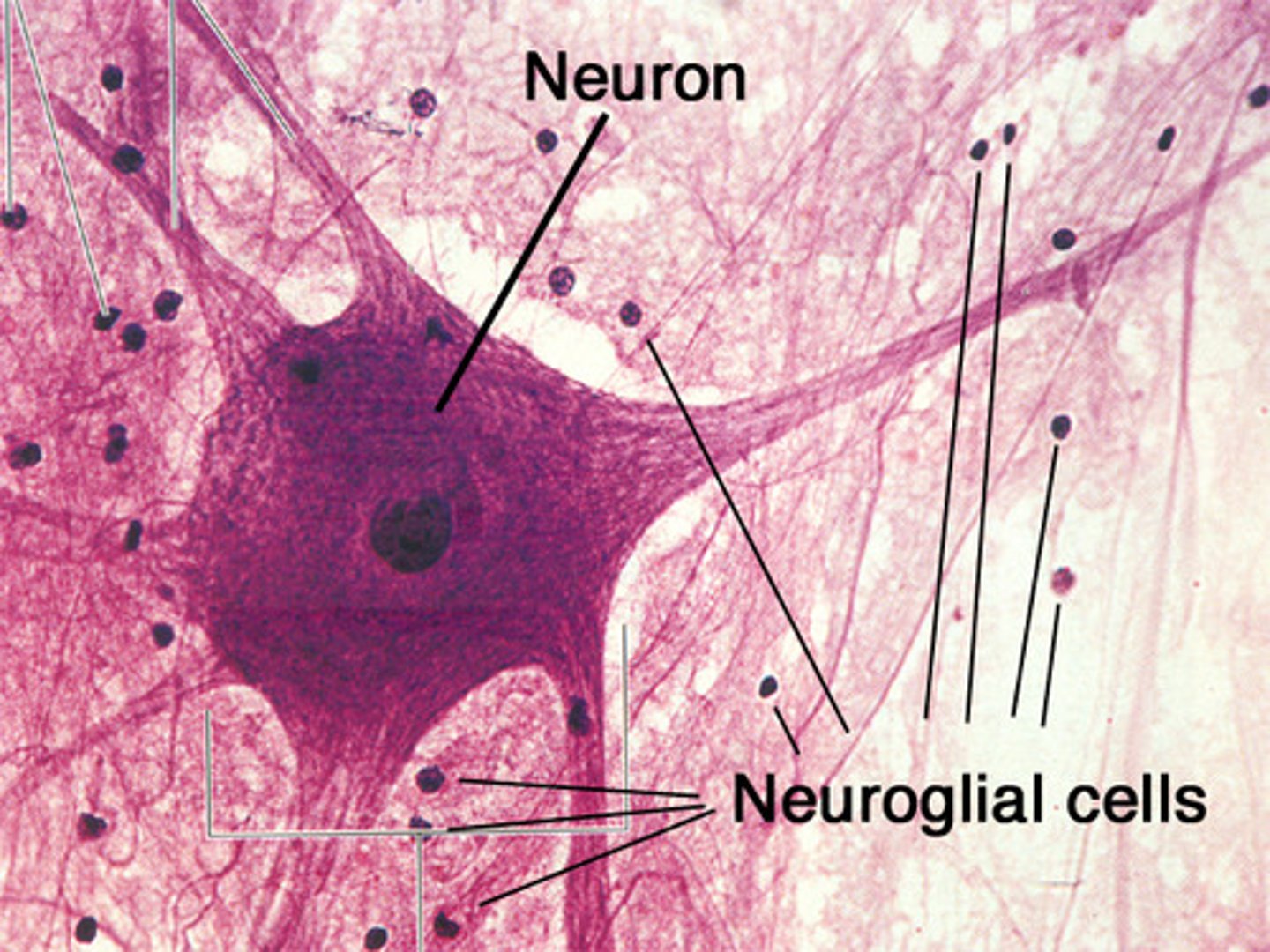
Glial cells (neuroglia)
several types of non-neuronal cells which serve to support the function and survival of neurons (provide nutrition and support)
Astrocytes
located in the vertebrate CNS, covers the surface of blood vessels; physically supports neurons; and, helps maintain ion concentrations of the interstitial fluid
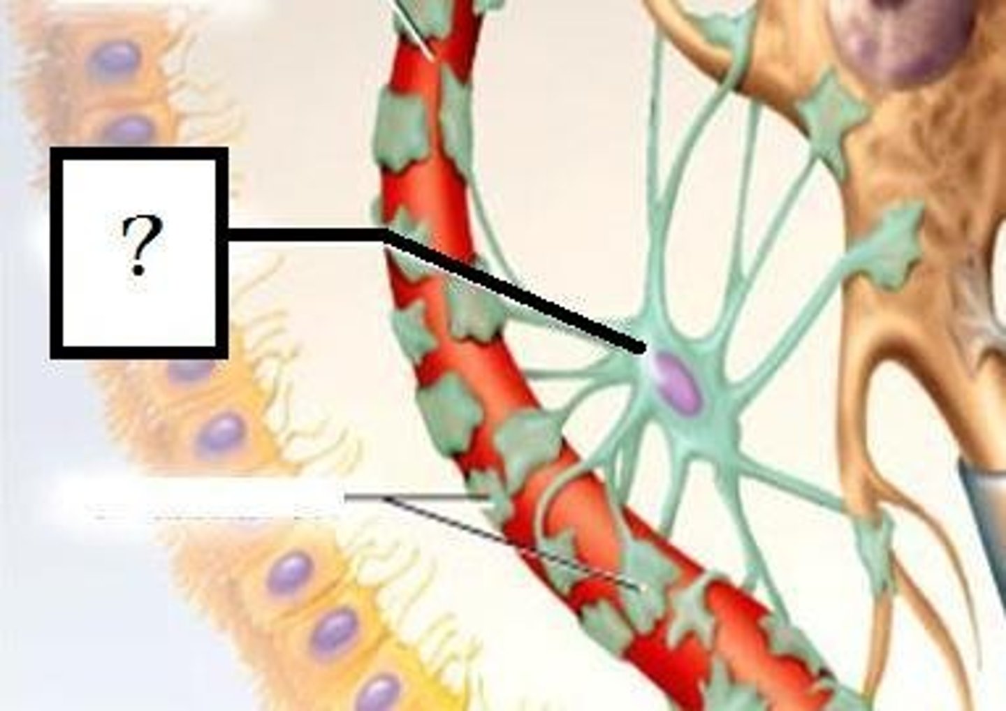
Oligodendrocytes
wraps around CNS axons to form insulating myelin sheaths
Schwann cells
wraps around PNS axons to form insulating myelin sheaths
Synapse
location where a neuron makes a communicating connection with another neuron (or an effector) either via
(a) direct electrical flow; or,
(b) through a chemical neurotransmitter
How many synapses can an individual interneuron form
up to 100,000 synapses
presynaptic cell
The axon terminal which delivers the signal
postsynaptic cell
The dendrite or effector which receives the message
Electrical synapses
the plasma membranes of the pre- and post-synaptic cells are in direct contact
i. Gap junctions allow ions to flow directly between two cells
ii. Allow rapid signal conduction, but difficult to regulate
iii. Egs., cardiac muscle, retina of the eye and pulp of a tooth
Chemical synapse
pre- and post-synaptic cells are separated by a narrow synaptic cleft
i. When an electrical impulse reaches the axon terminal, neurotransmitter is released into the synaptic cleft
ii. Neurotransmitter diffuses across the cleft and binds to a specific receptor in the postsynaptic cell membrane
iii. If enough neurotransmitter binds, the postsynaptic cell generates a new electrical impulse
iiii. Some neurotransmitters are excitatory; while other are inhibitory
Membrane potential
a separation of positive and negative charges across the plasma membrane that produces an electrical potential (voltage)
a. The Na+/K+ pump in the membrane actively removes Na+ from the cytoplasm and brings K+ into the cell
b. Resting membrane potential is produced due to the membrane being more permeable to K+ than to Na+; and, large anionic species being unable to cross the cell membrane
c. Resting membrane potential is -50 to -70mV
Phases of an action potential
threshold
depolarization, repolarization, hyperpolarization
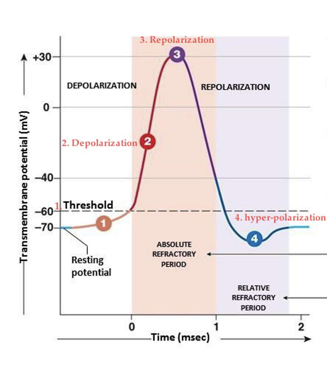
Threshold
for depolarization to occur, the membrane segment must be 10-20mV more positive than resting potential (~55mV)
Depolarization
once threshold is reached, voltage-gated Na+ channels open, and a large amount of Na+ enters causing the cell to depolarize up to +30mV
Repolarization
as Na+ channels are inactivated, K+ channels open allowing K+ to escape the cell and causing the membrane to shift to negative
Hyperpolarization
the K+ channels are slower to fully close which results in more than necessary K+ escaping the cell, and overshooting the resting potential by 10mV to -80mv.
action potential
a series of depolarization events that occur sequentially down the length of an axon.
refractory period
The turning off of Na+ channel activation by inactivation gates creates an absolute refractory period for Na+ channels which keeps the impulse moving forward
(a period when the action potential cant be triggered again)
Nernst-Goldman
equation that can be used to calculate the membrane potential based on the membrane's permeability to specific ions; and, based on the concentration of K+, Na+ and Cl- ions both inside and outside the cell
Tetrodotoxin
Na+ channel blocker which prevents Na+ from entering the cell and keeps the cell more negative than usual (-80mV vs -70mV); the result is typically paralysis
Hyperkalemia
condition in which there is too much K+ in the body which causes the potential to become less negative and makes the neuron more likely to fire (ie., closer to threshold)
The action potential doesn't convey magnitude, instead the:
Strength of a stimulus is based on the frequency of action potentials
(Action potentials are simply on, or off.)
In an unmyelinated axon, the rate of conduction _________ as the diameter of the axon increases
increases
what does myelination do
increases the speed of axon conduction
Saltatory conduction
the mechanism by which myelination permits the “hopping” of action potentials from one Node of Ranvier to another
Voltage-gated Na+ and K+ channels located exclusively at the nodes facilitate saltatory conduction
Process of neurotransmitter release
1. Action potential arrives at an axon terminal of a presynaptic neuron
2. Ca2+ enters the axon terminal
3. Neurotransmitters released by exocytosis
4. Neurotransmitters diffuse across the synapse
5. Neurotransmitters bind to membrane receptors on postsynaptic cell
6. The neurotransmitter stimulates the opening or closing of ligand-gated ion channels
i. Note: ligand is the molecule to which a receptor binds
Neutransmitter
sends information from one neuron to another
a. Some axon terminals release a single type of neurotransmitter; whereas others release several different types
b. Also, excitatory and inhibitory receptors can bind to the same type of neurotransmitter
5 classes of neurotransmitters
1. Acetylcholine
2. monoamines
3. Amino acids
4. Neuropeptides
5. Gaseous neurotransmitters
Acetylcholine
mostly excitatory (eg., neuromuscular junctions)
enables muscle action, learning, and memory
Monoamines (neurotransmitters) include:
dopamine, norepinephrine, serotonin
Norepinephrine
excitatory or inhibitory (eg., CNS interneurons)
helps control alertness and arousal
Dopamine
excitatory (eg., CNS interneurons)
A neurotransmitter associated with movement, attention and learning and the brain's pleasure and reward system.
Serotonin
inhibitory or modulatory (eg., CNS interneurons)
Affects mood, hunger, sleep, and arousal
Selective serotonin reuptake inhibitors
increase serotonin activity in the brain by maintaining levels in the synaptic cleft.
Amino acids (neurotransmitters) include:
Glutamate and GABA
Neuropeptides (neurotransmitters) include:
substance P, endorphins, enkephalins
Gaseous neurotransmitters include:
nitric oxide
Glutamate
A major excitatory neurotransmitter; involved in memory
GABA
inhibitory (many CNS pathways)
Endorphins
inhibitory (modulates pain response)
linked to pain control and to pleasure.
Enkephalins
inhibitory (modulates pain response)
reduce pain
Substance P
excitatory (affects pain perception)
Nitric oxide
modulatory (involved in the PNS)
regulate blood pressure, help immune system
Erectile dysfunction drugs such as Viagra affect this neurotransmitter
Nitric Oxide
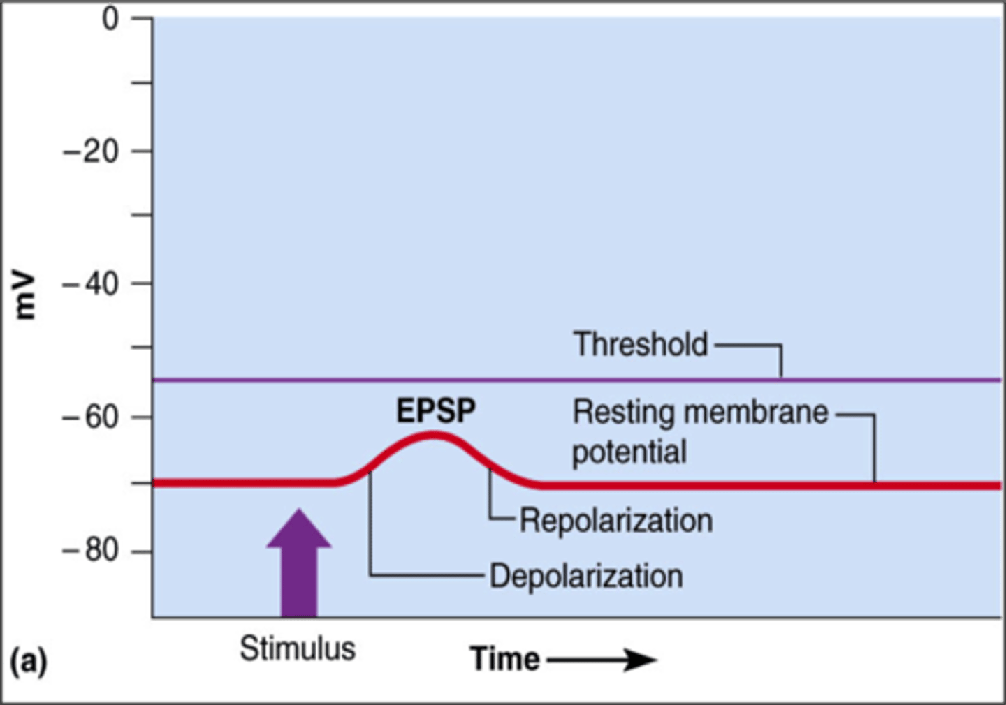
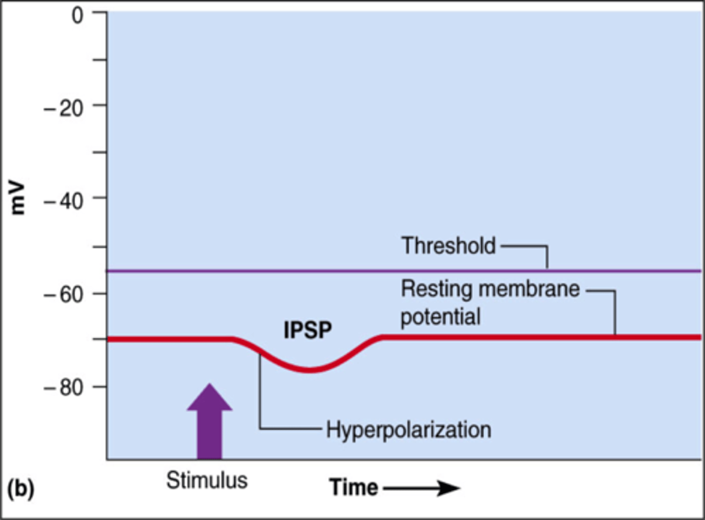
Excitatory and Inhibitory pathways
Excitatory pathways produce graded potentials that potentiate an action potential, while inhibitory pathways produce graded potentials that suppress it
Excitatory postsynaptic potential (EPSP)
a change in membrane potential that pushes the neuron closer to threshold
Inhibitory postsynaptic potential (IPSP)
a change in membrane potential which pushes the neuron farther from threshold (hyperpolarizes it)
Two types of summation
temporal and spatial

temporal summation
The process by which multiple excitatory postsynaptic potentials (EPSPs) from a single neuron combine over time to increase the likelihood of reaching the threshold for action potential generation in a postsynaptic neuron.

spatial summation
The process by which multiple synaptic inputs from different locations on a neuron combine to produce a greater postsynaptic potential, leading to increased likelihood of action potential generation.
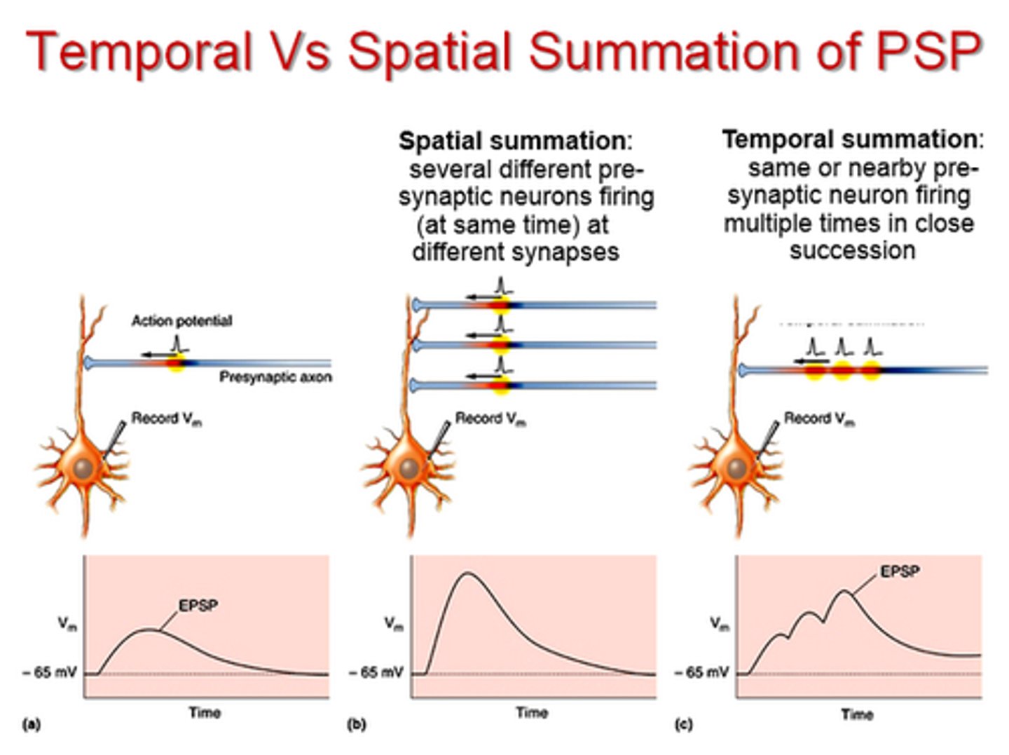
An IPSP and EPSP of equal strength and near proximity can _________
cancel each other
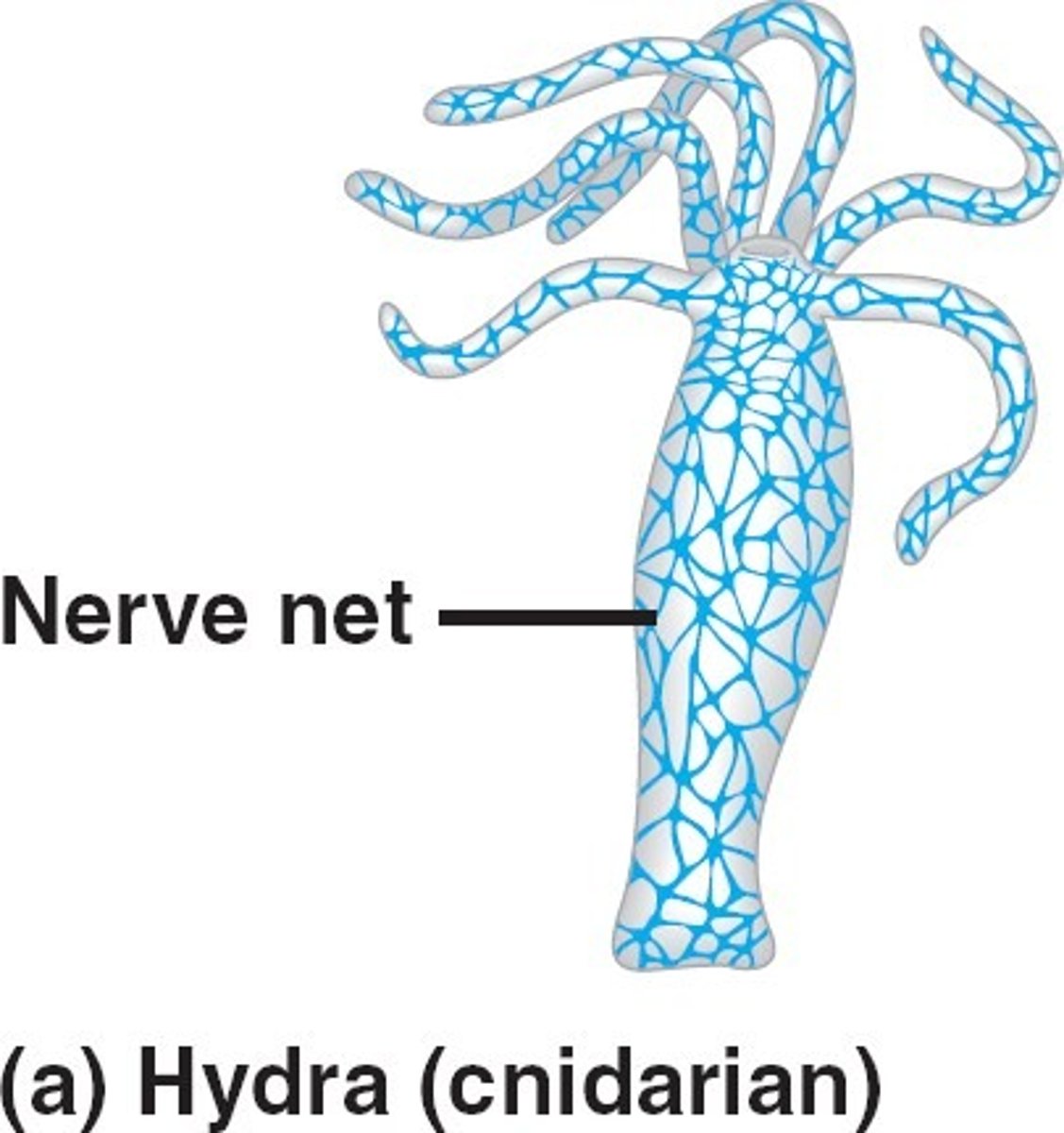
examples of cnidarians
jellyfish/anemone
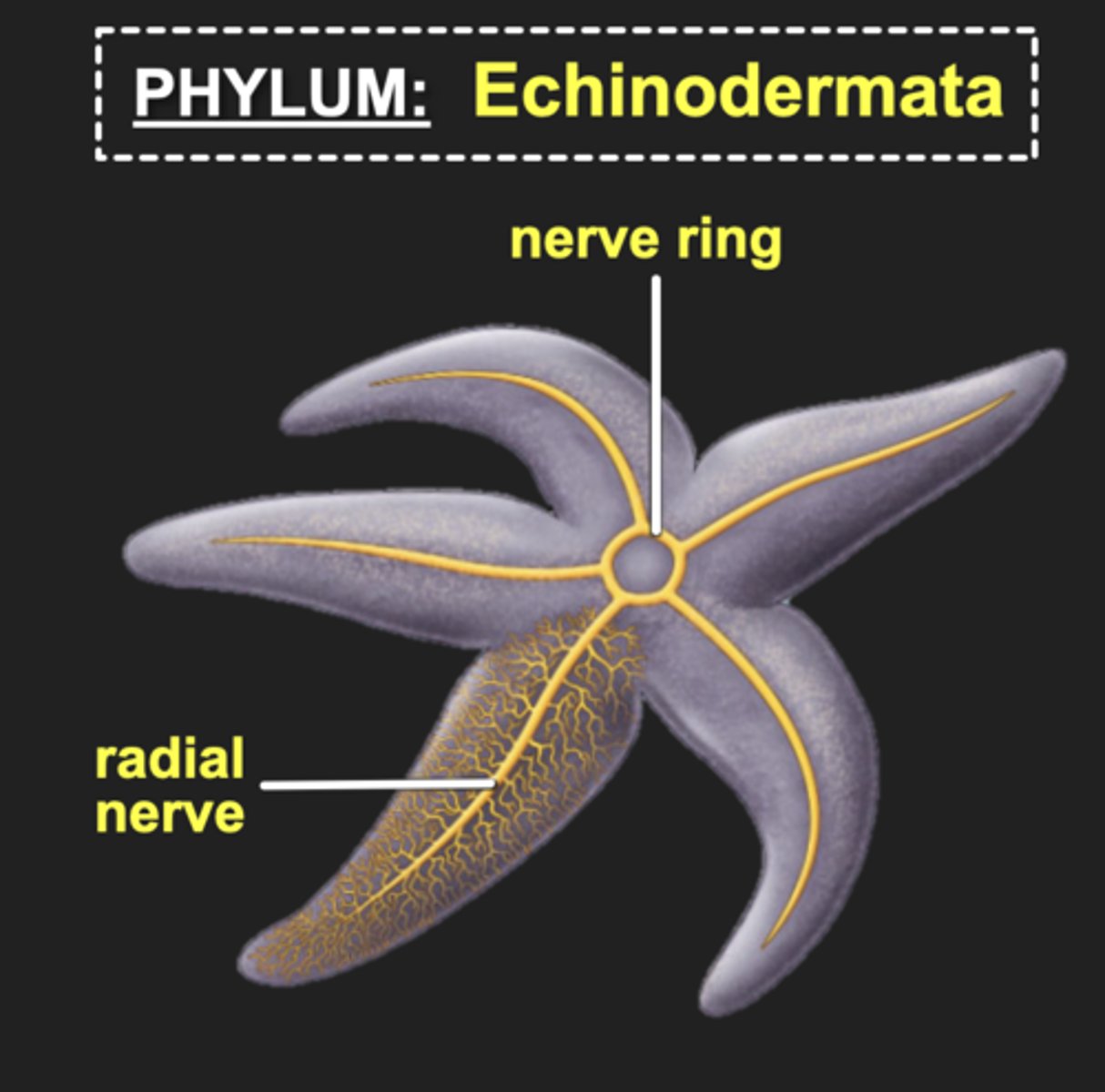
examples of echinoderms
starfish
nervous system of cnidarians
- possess nerve net
- No distinctions between dendrites and axons
- Impulses conducted in all directions from the point of stimulus
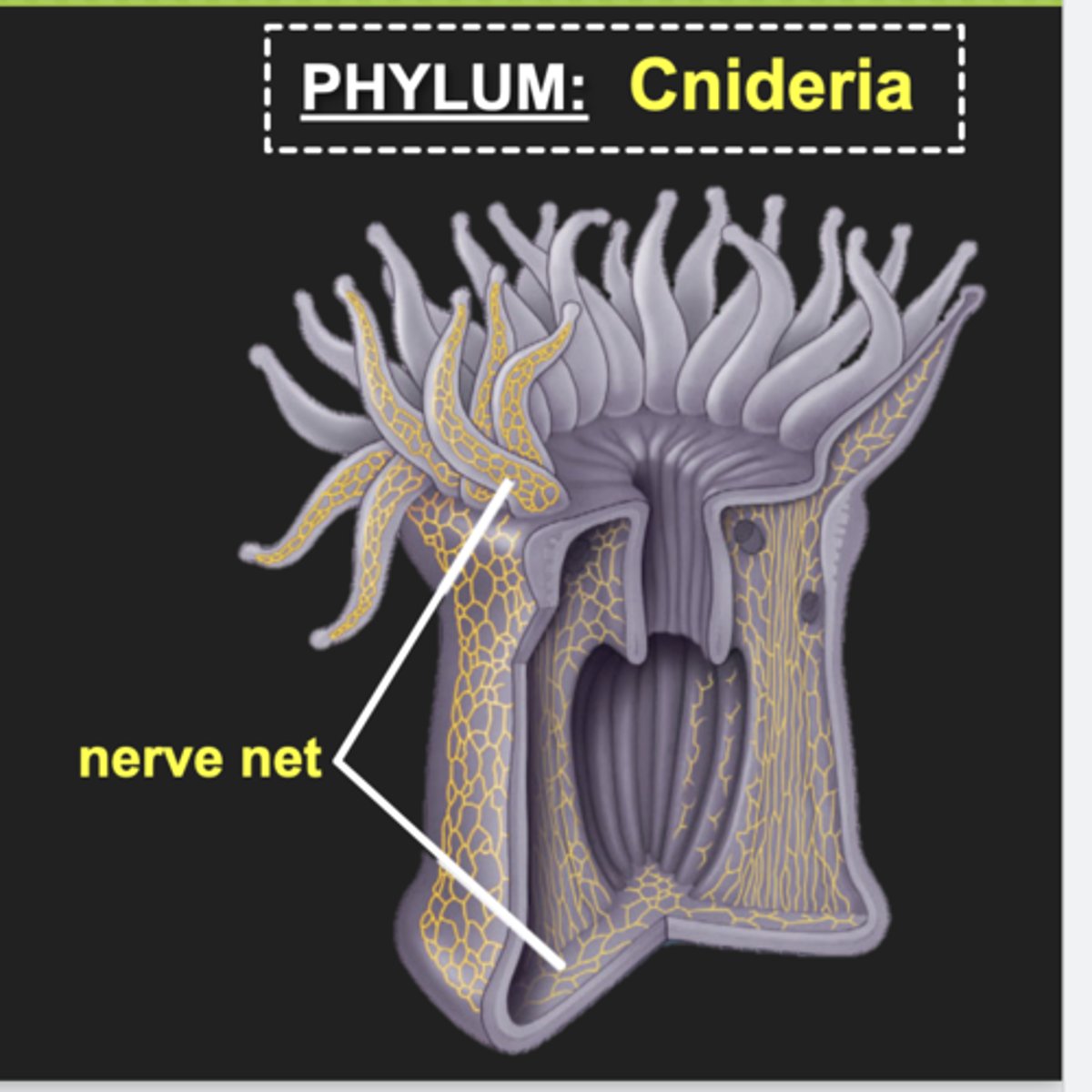
Nerve net
loose mesh of neurons organized within a radial symmetry
found in cnidarians
nervous system of Echinoderms
Radial Nerves: bundles of axons enclosed in connective tissue
Nerve ring: surrounds a centrally located mouth
Nerves
bundles of axons enclosed in connective tissue
Nerve ring
nervous system of echinoderms; surrounds a centrally located mouth
Platyhelminths examples
flatworms
nervous system of Platyhelminths
- A pair of ganglia (clusters of neurons) at its head-end
- Cephalization: formation of a distinct head region containing a control center
- two or more longitudinal nerve cords (CNS) run the length of the body
- nerve nets serve as the PNS
- Eyespots house photoreceptors which detects changes in light
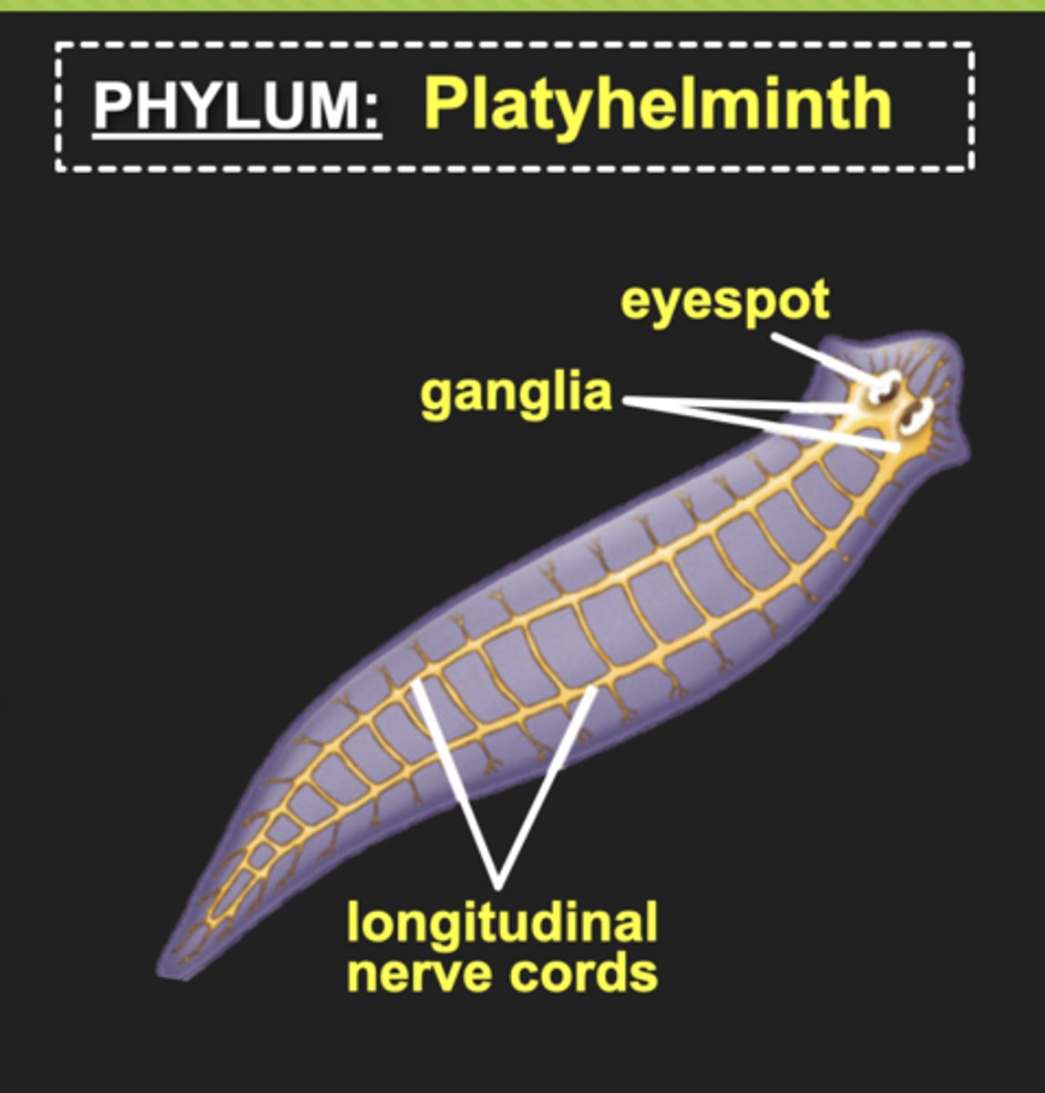
Cephalization
formation of a distinct head region containing a control center
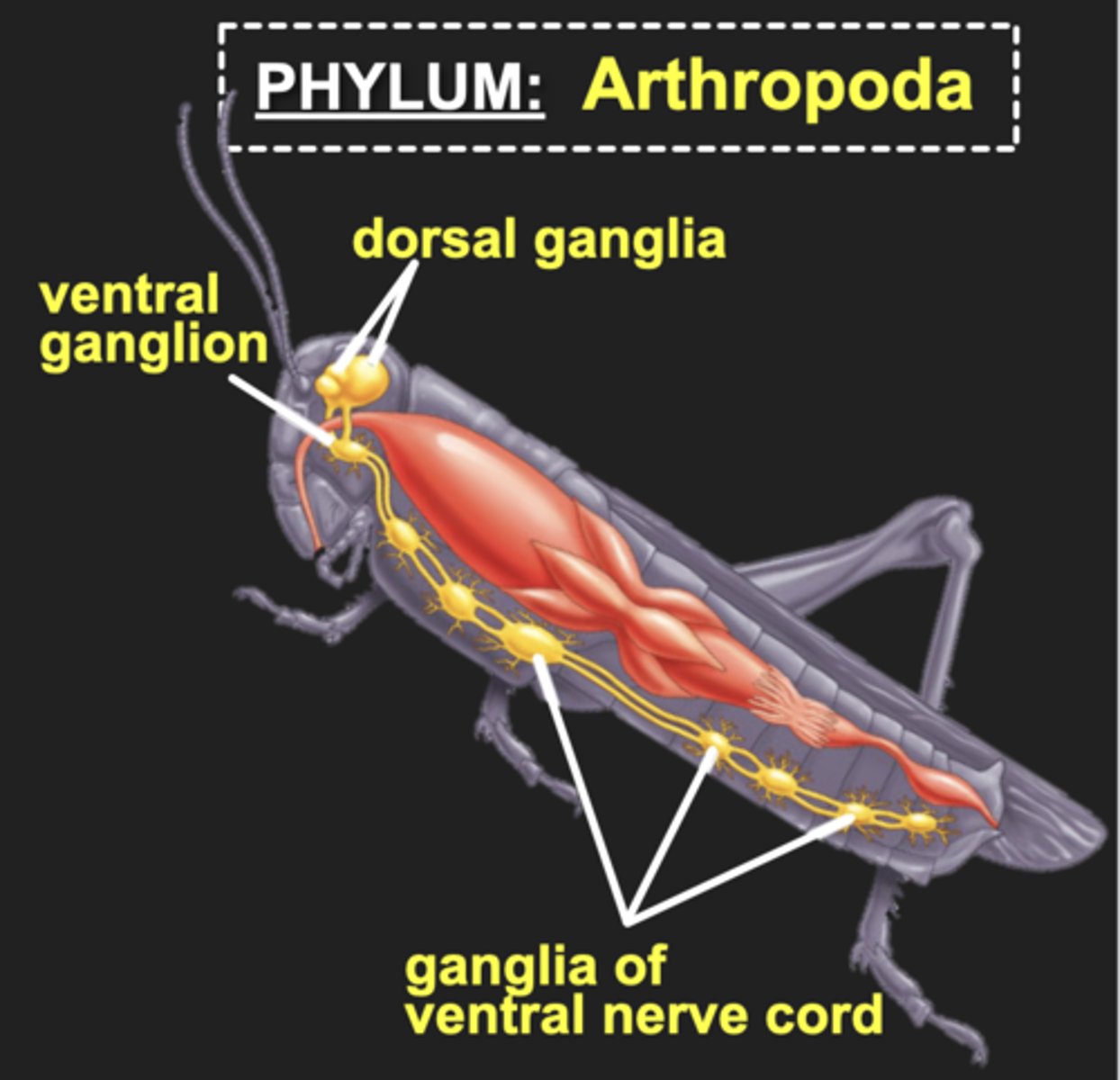
Arthropods examples
insects
Nervous system of arthropods
- Head region with brain (dorsal and ventral pairs of ganglia)
- Sensory structures such as antennae and complex eyes
- Ventral nerve cord enlarges into a pair of ganglia within each body segment
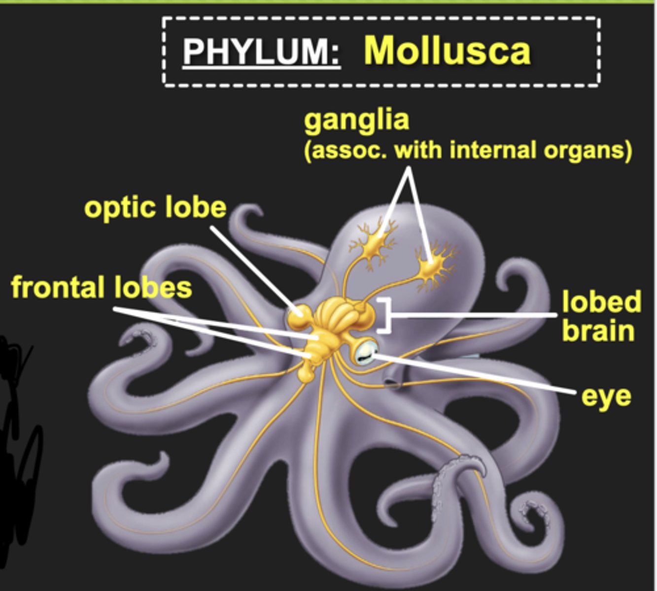
Mollusca examples
includes cephalopods like octopus
nervous system of Mollusca
- Most cephalization of any invertebrate
- Paired ganglia connected by many nerves
- Octopus possess complex, lobed brains with defined sensory and motor regions
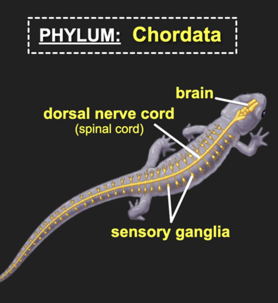
Chordata examples
vertebrates: fish, reptiles, humans, mammals, amphibians
invertebrates: soft bodied marine mammals ect.
(dont need to know all of those, just know what vertebrates are)
Nervous system of chordata
- Vertebrate CNS consists of a brain and spinal cord
- PNS consists of all nerves and ganglia that connect CNS to rest of the body
- Brain and nerve cord are hollow, fluid-filled structures
- Head contains specialized sensory organs connected directly to brain via nerves
In humans, the peripheral nervous system (PNS) is subdivided into:
afferent PNS
efferent PNS
Afferent PNS
transmits sensory information from their receptors
Efferent PNS
carries nerve impulses to effectors
effectors
-produces a response
e.g.muscle contracts to move hand away from stimulus or gland squeezes and releases hormone into blood.
Efferent PNS divided into:
Somatic nervous system (SNS)
Autonomic nervous system (ANS)
Somatic nervous system (SNS)
the part of the peripheral nervous system that controls voluntary movement of skeletal muscles
Autonomic nervous system (ANS)
the part of the peripheral nervous system that controls the glands and the muscles of the internal organs (such as the heart)
Major structures associated with the PNS in vertebrates:
- 31 pairs of spinal nerves
- 12 pairs of cranial nerves
The autonomic nervous system (ANS) controls involuntary processes such as
digestion, sweat gland activation; contraction of smooth muscle
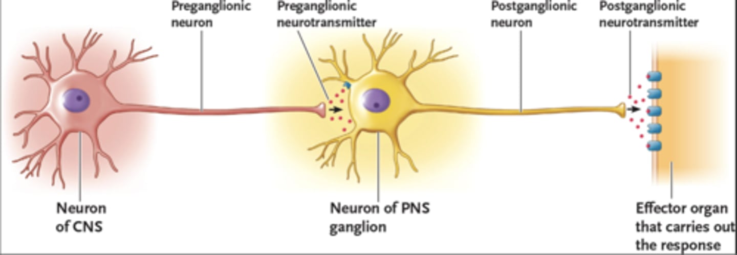
Understand the following diagram
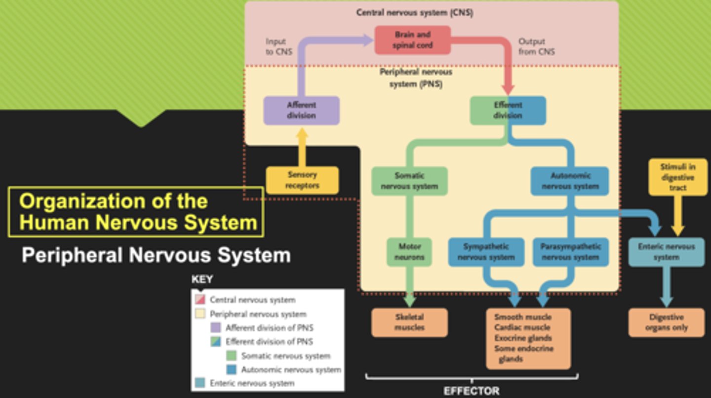
Autonomic neuronal circuits feature
1st preganglionic neuron with cell body located in the CNS
2nd postganglionic neuron which extends from the ganglion to the effector