D2 - Cell and Nuclear Division, Water Potential
1/47
There's no tags or description
Looks like no tags are added yet.
Name | Mastery | Learn | Test | Matching | Spaced | Call with Kai |
|---|
No analytics yet
Send a link to your students to track their progress
48 Terms
Cytokinesis
The division of a cells cytoplasm to form two cells. It occurs after mitosis and happens differently in plant and animal cells.
Cytokinesis in Plant Cells
A new cell wall is made across the equator, with a plasma membrane on both sides. This divides the cell in two.
Cytokinesis in Animal Cells
The cytoplasm is divided by moving the plasma membrane. Movement is due to actin and myosin proteins adjacent to the membrane. Tension is exerted to form the cleavage furrow, with the membrane pulled inward so it eventually splits the cell.
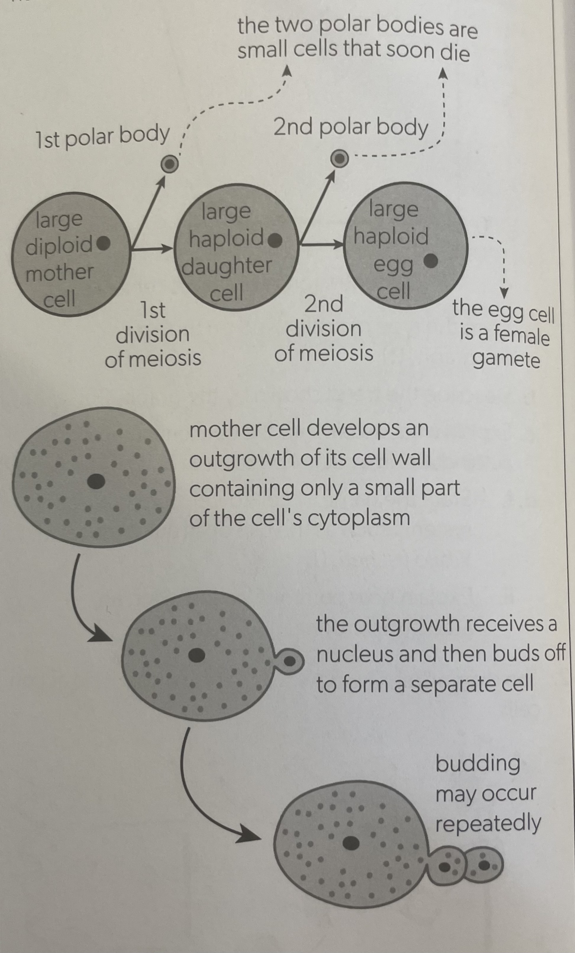
Cytokinesis can be equal or unequal
Cytoplasm of the mother cell is divided into equal halves
Unequal division results in small cells that can grow and further divide with a nucleus
Examples are oogenesis (egg production) in humans, or budding in yeast
Mitosis - For Continuity
Daughter cells receive all the chromosomes and genes of the mother cell. Same chromosome number. Used in asexual reproduction to produce genetically identical offspring or in organisms for body cells
Meiosis - For Change
Diploid nucleus divides into haploid nucleus, halving the chromosome number. Allows for haploid gamete production. Allows for genetic diversity because every haploid cell has different combination of alleles.
Condensation
When DNA is packed up to form shorter and fatter chromosomes. Includes DNA being wrapped twice around 8 histone proteins. These are then linked together and supercoiled
Microtubules
Narrow structures assembled from many molecules of tubulin (a globular protein). More tubulins can be added to extend the microtubules.
Microtubule Motors
Cause movement by removing tubulin subunits from the end of microtubules and shortening them.
Kenetichores
Microtubule motor anchored to chromatid’s centromere to which microtubules attach to during anaphase. They allow chromosomes to be pulled to opposite poles.
Stages of Meiosis
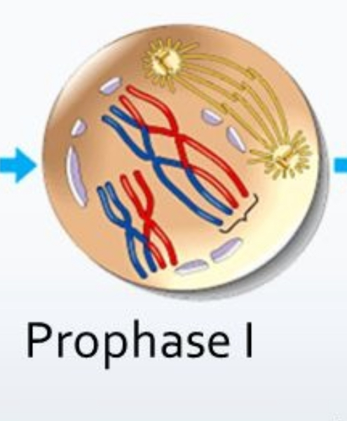
Early prophase
Late prophase
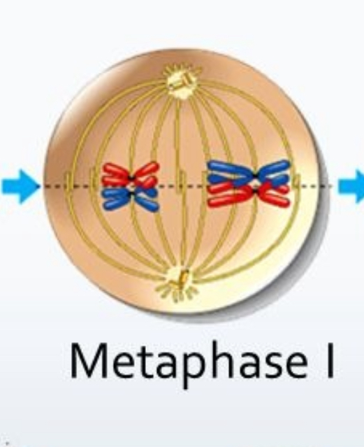
Metaphase
Anaphase
Early telophase
Late telophase
Early Prophase
Microtubules grow from centrioles to form spindles
Chromosomes condensing
Late Prophase
Spindle microtubules extend from each pole to the equator
Each chromosome includes identical sister chromatids
Metaphase
Nuclear membrane broken down
Chromosomes migrate to equator
Microtubules attach to kenetichores
Anaphase
Sister chromatids separate becoming separate chromosomes
Kenetichores shorten spindle microtubules, pulling to opposite poles
Early Telophase
Chromosomes reach the poles and nuclear membranes form around them
Spindle microtubules break down
Late Telophase
Chromosomes uncoil
Cytokinesis divides cells
Viewing Stages of Mitosis
Interphase - no condensation
Prophase - condensation
Metaphase - aligned to equator
Anaphase - V shaped and moving to poles
Telophase - condensing at poles
Meiosis
A reduction division because it halves the number of chromosomes. Mother cell divides a diploid nucleus producing four cells with haploid nuclei
Diploid Nucleus
Has 2 sets of chromosomes
Contains homologous chromosomes, with same genes in same sequence but potentially different alleles
Haploid Nucleus
Has only one set of chromosomes
Represented by the letter n
Includes gametes like sperm and egg to be used during fertilization to produce one diploid cell.
Meiosis Stages
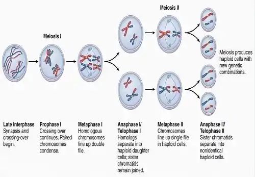
Meiosis Prophase I
Chromosomes pair up, each pair are homologous
Spindle microtubules grow from poles
Meiosis - Metaphase I
Random orientation of homologous chromosomes on the equator
Spindle microtubules attach
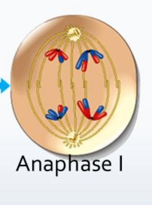
Meiosis - Anaphase I
Homologous chromosomes pulled to opposite poles, halving the chromosome number
Each chromosome still includes 2 chromatids
Homologous chromosomes vs Sister chromatids
Sister chromatids are identical copies of a single chromosome.
Homologous chromosomes are pairs of chromosomes that are similar in size, shape, and gene content. They carry the same genes in the same order, but may have different versions of those genes.
Chromosome vs Chromatid
A chromosome is a long, thread-like structure carrying genetic information in the form of DNA
A chromatid is one of the two identical halves of a replicated chromosome, joined at the centromere
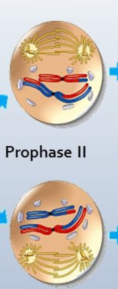
Meiosis - Prophase II
Two haploid cells produced by 1st meiosis division
DNA does not have to replicated because Each chromosome still has 2 chromatids
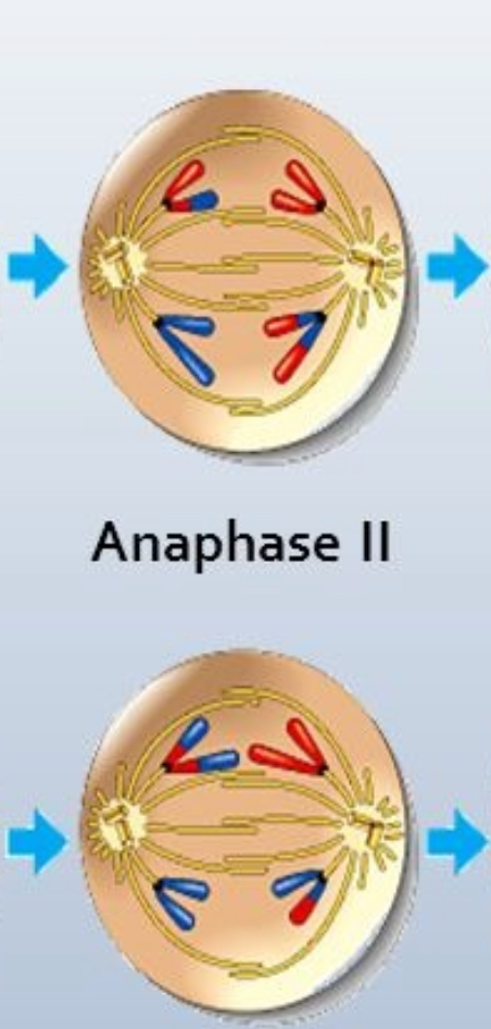
Meiosis - Anaphase II
Kinetochores pull chromatids to the poles
Sister chromatids not identical because of exchange of alleles
Meiosis - Telophase II
Chromosomes decondense inside new nuclear membranes
4 haploid cells produced
Every nucleus produced is genetically different
Trisomy 21 (Down Syndrome)
Caused by an error in meiosis when pair 21 of homologous chromosomes fails to separate in anaphase 1 (non-dysjunction), moving to same pole.
The zygote has 3 copies of chromosome 21.
Trisomy
Having 3 chromosomes instead of 2. Usually results in death of gamete or or early-stage embryo
Meiosis Generating Variation
Creates genetic diversity (different combinations of alleles) in 2 ways:
Random orientation of bivalents
Crossing over
Random Orientation of Bivalents
Bivalent: Pair of homologous chromosomes, one from each parent.
Orientation in metaphase determines to which pole each chromosome moves to.
Crossing Over
Homologous chromosomes pair up in the very early stages of meiosis and non-sister chromatids exchange lengths of DNA at the chiasma.Produces new combinations of alleles. Random places of genetic exchange.
Solvents
Liquids that can dissolve other substances to make solutions
Solutes
Are dissolved substances in solutions
Solvation
The process of dissolving
Types of Solutions
Hypertonic - Higher solute concentration
Hypotonic - Lower solute concentration
Isotonic - Same solute concentration
Net Movement of Water
Up the solute concentration gradient, from the lower to the higher solute concentration.
Dynamic Equilibrium
When the environment of a cell is isotonic the water molecules continue moving but equally so there is no net movement.
Cells in Hypotonic Solution
Water enters cell.
Volume of cytoplasm increases, swelling the cell until bursting.
Cells in Isotonic Solution
Dynamic equilibrium with cell volume not changing
Cells in Hypertonic Solution
Water exits the cell.
Volume is reduced but plasma membrane area stays the same so appears shrivelled.
Regulation of Water Movement
Unicellular organisms in freshwater take on water by osmosis and expel it using contractile vacuoles
Cells surrounded by other cells must be kept in an isotonic environment
Humans use kidneys to regulate solute concentrate of extracellular fluids
Cell Walls in Hypotonic Solution
Water enters cell
Increase in cytoplasm volume creating turgor pressure against wall
Cell Walls in Hypertonic Solution
Water exits cell
Gap develops between cell wall and plasma membrane due to decreased voluume
Called plasmolysed
Isotonic Solutions in Medicine
Intravenous fluids: injected into veins via syringe or bag, must be close to isotonic to avoid osmotic cell damage
Tissues/organs used in transplants must be bathed in isotonic solutions to prevent damage