Lower extremity arterial disease
 Call Kai
 Learn
Learn Practice Test
 Spaced Repetition
 Match
Match Flashcards
 Knowt Play
1/66

There's no tags or description
Looks like no tags are added yet.
Name | Mastery | Learn | Test | Matching | Spaced | Call with Kai | Chat |
|---|
No analytics yet
Send a link to your students to track their progress
67 Terms
Atherosclerosis
Intimal thickening, calcification and loss of wall elasticity
Hardening of the arteries
Most common sites:
#1 SFA in adductor canal
#2 CFA bifurcation
Calf vessels and other vessel origins
Most common cause of arterial occlusive disease
Atherosclerosis obliterans
Atherosclerosis obliterans
Atherosclerotic changes produce progressive obstruction
Accumulation of lipids in intima which cause narrowing
Usually affects extremities in a diffuse manner but increase incidence in popliteal and calf arteries
Focal stenosis and diffuse stenosis can coexist
Symptoms usually are exacerbated with exercise and relived with resting in dependent position
Arteritis
Inflammation of the artery walls causing flow stasis with thrombus formation
Usually affects peroneal arteries, posterior and anterior tibial arteries and smaller distal arteries/capillaries
Thromboangitis
Refers to diffuse inflammation of the intimal wall layer
Kawasaki disease
Affects small to medium arteries
CORONARY ARTERIES most commonly affected; causes dilatation and potential aneurysm formation
Usually seen in pediatric pts age 2-5yrs
More common in Japanese
Buerger disease/Thromboangitis obliterans
Occlusive disease of SMALL to MEDIUM size arteries caused by inflammation of arterial wall and surrounding connective tissue
Digital, plantar, tibial, peroneal and ulnar arteries
Acute onset in plantar or palmar arteries then moves centrally
ALWAYS BILATERAL but can affect one side more than the other
3 of 4 extremities affected
MONOphasic waveforms by Doppler and PPG
Buerger disease/Thromboangitis obliterans is most commonly seen in
Men smokers age 20-30yrs
Buerger disease/Thromboangitis obliterans is associated with:
Collagen disease (lupus), ischemic rest pain and distal ulcerations in the foot/digits/hand
In-step claudication
Corkscrew collateral formation is a sign of:
Buerger disease/Thromboangitis obliterans
Takayasu arteritis
Presents as hypertrophic areas of inflamed tissue usually found in the Aorta and its Branches
90% involve the subclavian arteries
Over half the cases involve the CCA
Can also affect renal arteries
Takayasu arteritis is associated with:
Supraclavicular stenosis and aortic coarctation
Takayasu arteritis Most commons
Most commonly seen in young females (20-40trs)
More common in asia and india than US
Takayasu arteritis symptoms
Absence of peripheral pulse, asymmetric brachial blood pressures, amaurosis fugax, hemiparesis, diplopia, vertigo, upper extremity claudication
Takayasu arteritis USA
Presents similar to stenosis
Clinical exam important for differentiation from atherosclerotic disease
Increased PSV and diastolic flow
2D appearance shows more diffuse involvement and more homogeneous thickening of the vessel walls than seen with atheroma formation
Giant cell arteritis
Presents in the CRANIAL arteries, but can be seen in the extremities
Blood tests are performed to measure the erythrocyte sedimentation rate
Measure C-reactive protein (CPR)
Asymmetric brachial blood pressures seen in pts with arteritis in extremities
Treated with corticosteroids, like prednisone
Symptoms of giant cell arteritis
Pain, and/or stiffness in the neck, jaw claudication, and visual disturbances
Giant cell arteritis is most commonly seen in
Women in their 70-80s
USA Giant cell arteritis
Causes diffuse or focal thickening of the muscle layer
May see increased vascularity in the wall of the inflamed segment
Halo sign - ring of thickened vessel wall tissue surrounding the vessel
Increased resistance to flow
Doppler similar to stenosis
Surgical repair is recommended for aneurysms in the extremity that exceed:
2.5-3cm in diameter
Surgical repair is recommended for aneurysms in the aorta that exceed:
5.5cm in diameter
Most common complication of peripheral artery aneurysms
Embolization
Embolization
Obstruction of a vessel by a foreign substance, such as plaque or thrombus
Most arterial emboli are from a cardiac source
Embolic material ledges in distal artery causing ischemia/necrosis
Air bubble can also be harmful embolism
Most common cardiac abnormality associated with embolization
Atrial fibrillation
Other causes of embolization
Thrombus in aneurysm, arteritis, ulcerated atherosclerotic lesions, some angioplasty procedures
Blue tow syndrome
Discoloration of the digits caused by acute ischemia
Caused by small pieces of plaque or thrombus breaking loose from a more proximal site
Commonly seen in pts with AAA or after a recent angiography procedure
If blue toes are noted during a LEA Duplex exam, an aortic evaluation should also be performed
Pseudoaneurysms are typically caused by
Trauma or invasive procedures
Pseudoaneurysms
Can be iatrogenic
The groin is the most common location for a pseudo
Incidence is INCREASING due to increased numbers of invasive procedures
Blood escapes all 3 layers of the arterial wall into the surrounding tissues and is encapsulated within the tissues
A connection is made through a neck or stalk
Treated by compression or thrombin injection
Anticoagulant therapy should be discontinued
Neck/stalk should be measured
Smaller pseudos can clot spontaneously
Primary symptoms of a Pseudoaneurysms
Palpable, pulsatile mass and a bruit
Contraindications for US guided compression therapy for a pseudo
Pt unable to tolerate procedure due to pain
Infection at the puncture site and surrounding tissues
Diameter >4cm
Stalk diameter >5mm
Present for more than 1 mo
Pts on anticoagulant therapy (must be stopped before treatment)
Contraindications for thrombin injection for pseudo
Small diameter
Indefinable neck/stalk
Associated AVF
Current anticoagulant therapy
USA Pseudoaneurysms
Rounded anechoic structure adjacent to main artery
Measure size of body in 2 planes
Measure diameter of neck at widest point
Color demonstrates turbulent flow → Ying-yang sign
High resistance to and fro flow in stalk
Use PPG to monitor changes with compression
Larger stalk Pseudoaneurysms can take longer than an hour of compression before treatment works
Compression performed in __________ intervals with a re-evaluation of flow after each compression interval
10 one-minute
AVM
Congenital connection between the arterioles and venules (lack of capillary bed)
Has multiple connections and surrounding venous collaterals and dilated veins
AVMS are seen anywhere but COMMONLY in brain, kidney, uterus, pelvis
Vein of Galen is seen in fetal evals
Causes increased volume of flow in the venous system with pulsatility and turbulence
Reduces resistance in the arterial inflow and increases diastolic flow
Large or chronic AV connections can lead to heart failure
AVF is a DIRECT connection between the arterial and venous systems caused by
Trauma or vascular procedure; usually a single connection and NO surrounding collaterals due to acute cause
Traumatic AVF
Can be a complication of an invasive procedure, MOST COMMONLY right heart catheterization
Groin puncture can cause an AF fistula between femoral vessels
Results in high velocity turbulent flow through the tract
Flow decreases in velocity and pulsatility as it enters the venous puncture site into vein
Venous flow proximal to an AV fistula becomes PULSATILE and turbulent due to the inflow of the arterial flow distally
Arterial flow proximal to AV fistula will be low resistance; pressure will decrease in the artery DISTAL to the fistula
If the AV fistula is close to the heart…
Cardiac failure can occur due to increase in venous return with less O2 blood flow reaching distal locations
Larger diameter fistulas and shorter fistulas offer ___________ to the arterial flow
Less resistance
Small diameter fistulas and fistulas with long tract offer more _____________ to the arterial flow
Resistance
Popliteal entrapment
Compression of the artery by the medial head of the gastrocnemius muscle
Commonly found in runners and athletes
Can cause numbness and paresthesia of the foot
About 30% of cases are bilateral
PPG eval can be used to assess with Extension/Flexion of foot
Most common cause of unilateral claudication in young pts
Popliteal entrapment
PW/CW Doppler eval of popliteal entrapment
Place pt in decubitus position with affected side up
obtain resting 2D images of the pop artery diameter in the trans view
Obtain Doppler waveforms
Ask pt to hyperextend leg and flex and hold foot, repeat diameter measurements
The vessel diameter will be reduced and the waveform will demonstrate reduced or absent flow
PPG eval of popliteal entrapment
Place pt in decubitus position with the affected side up
Place sensor on the great toe
Record at baseline tracing at rest
Ask pt to hyperextend leg and flex and hold the foot
Pulse diminishes when calf contracts
Compartment syndrome
Increased pressure in an osteofascial compartment that prevents blood flow in and out of tissues
Can lead to muscle and nerve damage and problems with blood flow
Can be considered a critical finding to prevent muscle and nerve damage
Compartment syndrome is most commonly an acute onset and associated with
Broken leg or arm
Anterior compartment syndrome
Treated with fasciotomy which is a procedure where the fascia is cut to relieve tension or pressure
Acute compartment syndrome
Medical emergency; it is usually caused by a severe injury; without intervention, it can lead to permanent muscle damage
Chronic compartment syndrome
Known as exertional compartment syndrome
NOT a medical emergency; usually related to athletic exertion; stopping the exertion usually clears up the problem
Risk factors for acute compartment syndrome
Broken bone
Large bruise
Re-established blood flow after surgical intervention
Crushing injuries
Anabolic steroid use
Constricting bandages and casts
Symptoms of compartment syndrome
Acute onset of persistent deep ache in an arm or leg
Pain that seems greater than expected for the severity of the injury
Numbness, pins and needles, or electricity-like pain in the limb
Swelling and edema
Skin muscle tightening
USA compartment syndrome
Similar to hematoma but within space between muscles/structures
Adventitial cysts
Rare condition
Collection of mucinous material within the adventitial wall layer of the affected vessel
Predominately affects PERIPHERAL arteries
No systemic vascular disease
Can cause lower extremity pain
Usually presents as multiple anechoic or hypoechoic areas within the wall
No blood flow is detected in the areas
Can cause compression and narrowing of vessel lumen
Adventitial cysts are most commonly in
The pop artery in young to middle aged men
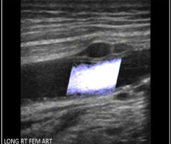
Adventitial cysts
What is the most common location for atherosclerosis formation in the lower extremities?
Distal femoral artery
What correctly describes buergers disease
Associated with heavy smoking
Inflammation of the smaller vessels of the calves, forearms, hands, feet
Always BILATERAL
Which of the following describes a difference between Takayasu arteritis and Buerger disease?
Takayasu arteritis affects the aortic arch, CCA and subclavian arteries, while buerger disease affects small vessels
If the pts chart demonstrates recent lab testing for the erythrocyte sedimentation rate and C-reactive protein, what does the physician suspect is the cause of the pt’s symptoms?
Giant cell arteritis
A popliteal aneurysm is diagnosed with focal dilation when the diameter exceeds:
1cm
You are performing a lower extremity arterial duplex and identify 3 toes on a pt’s foot that are blue and discolored. What other ultrasound exam should be performed for this Pt?
Abdominal aortic duplex
Pseudoaneurysm involves rupture of ____________ of the arterial wall
The intima, media and externa layers
Why is it important to measure the stalk diameter of a pseudo aneurysm
To determine if compression or thrombin injection should be used for treatment
Difference between and AVF and AVM
AVM is a congenital defect and has multiple AV connections found in the brain, kidneys, or uterus. An AVF is a acquired defect that has a SINGLE connection found in the groin or upper extremity
What causes popliteal entrapment syndrome?
Compression of the popliteal artery by the head of the gastrocnemius muscle
What procedure is performed to treat anterior compartment syndrome
Fasciotomy
What are critical findings?
Pseudo, anterior compartment syndrome, and dissection
What sonographic sign is associated with adventitial cyst formation
Scimitar