Module 2- Development of Chondroid Bone, Secondary Cartilage, Mandible, TMJ
1/19
There's no tags or description
Looks like no tags are added yet.
Name | Mastery | Learn | Test | Matching | Spaced |
|---|
No study sessions yet.
20 Terms
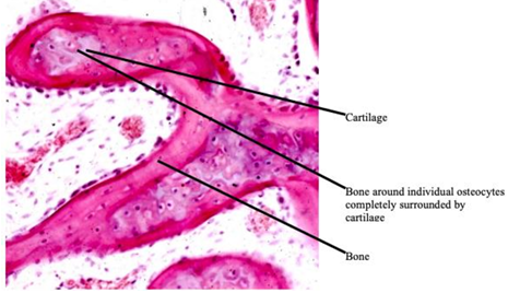
chondroid bone
-trans-differentiation of cartilage into bone tissue in human fetal mandible
development of secondary cartilage
-only found in condylar process
-primitive joint found between first two pharyngeal arches that allowed movements between first arch and rest of the skull
-TMJ formed external (lateral) to primitive joint, then functionally replaces it
secondary cartilage
-normally encountered in development of any diarthrodial joint
-gives rise to articular cartilage
-appear later in development (10-20 weeks as opposed to 6-7 weeks for primary cartilage)
-develops in areas of relative motion and is maintained by motion
-forms from mesenchymal condensations
-somewhat more cellular with larger sized lacunae than primary cartilage
-has ECM with type I collagen in addition to typical cartilage collagens (II/IX/XI)
-retains osteogenic potential
development of diarthrodial joint
-joint cavity and articular cartilages
-initially develop in area where there is no joint space
-area of loose mesenchyme between the 2 cartilage models that is going to give rise to cartilaginous condensation that forms on top of cartilage models
-cells migrate out to the surface where they form a secondary outer layer which is the secondary cartilage; articular cartilage formed as a secondary cartilage
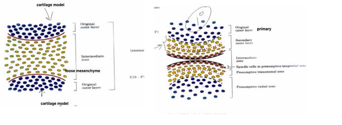
-X = newly formed joint space
development of mandible and TMJ
-TMJ ONLY present in mammals
-remarkable parallelism in ontogenetic development of the mammalian lower jaw and middle ear region
-phylogenetic transition from mammal-like reptile to mammals occurred over a period of about 120 million years
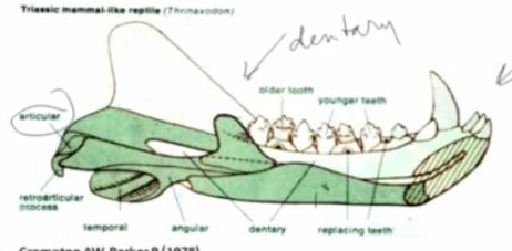
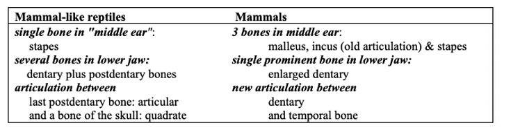
mammal-like reptile
-lower jaw comprises dentary and several postdentary bones
-joint between last postdentary bone: articular and a bone of the skull known as the quadrate
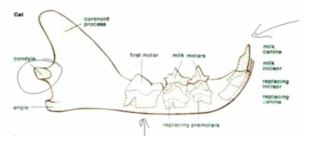
mammal
-single prominent bone in lower jaw: the dentary
-new joint between dentary and temporal bone: TMJ
middle ear ossicles
-1 ossicle → 3 ossicles
-quadrate and articular added as incus and malleus respectively
summary of lower jaw development transition
-during transition from mammal-like reptiles to mammals, following changes have occurred in the first PA region:
-quadrate became incus
-articular (one of postdentary bones) became malleus
-incus and malleus still articulate but now as part of the ossicular chain in the middle ear
-angular (another postdentary bone) became tympanic ring
-dentary became mandible
-new joint formed between dentary and temporal bone (TMJ)
primitive joint of lower jaw
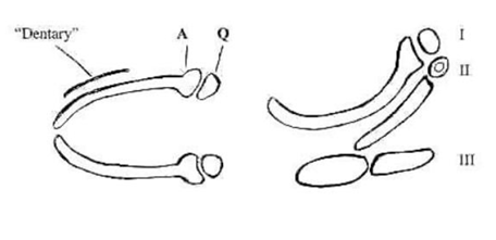
-primitive skeletal element of lower jaw: first PA cartilage (Meckel’s cartilage)
-anterior portion in first PA cartilage is an elongated bar (Meckel’s cartilage) that is somewhat enlarged posteriorly- known as “articular”, articulates with posterior portion of first PA cartilage “quadrate”
-primitive joint between the “articular” and the “quadrate” located immediately outside the otic capsule, on its lateral surface
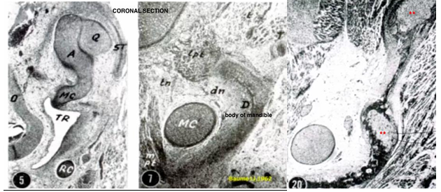
mandible at 6-7 weeks
-after Meckel’s cartilage forms, a separate mesenchymal condensation forms buccal/external to it
-condensation will give rise to first intramembranously formed bone of mandible known as “dentary”
-forms at future mental foramen
mandible development at 8 weeks
-single bone plate transformed into a double-plated bone “trough”
-trough houses inferior alveolar neurovascular bundle as well as developing teeth
-body of mandible appear V shaped
mandible development at 9 weeks
-mandible extends forward as far as the mandibular symphysis and surrounds the anterior portion of Meckel’s cartilage (anterior to mental foramen) almost completely
-posteriorly, mandibular bone extends into ramus area
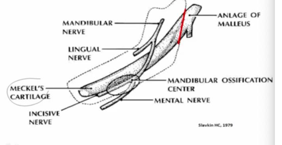
-temporalis and masseter muscle suggest you’re in area of developing coronoid process
-dentary (body of mandible) formed by intramembranous ossification
mandible development at 10 weeks
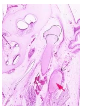
-mandibular bone/dentary so far back extended that it begins to approach the temporal bone and there is motion in lower jaw
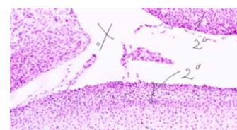
-as a result, secondary cartilage forms
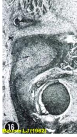
-no joint space, just intermediate zone (red arrow on right)
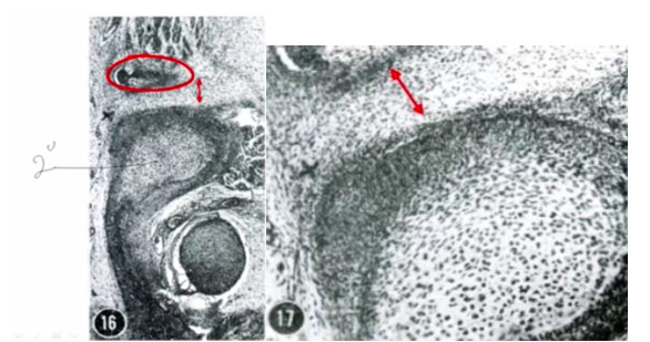
mandible development at 11 weeks
-secondary cartilage (indicated by stars) that are not maintained by motion
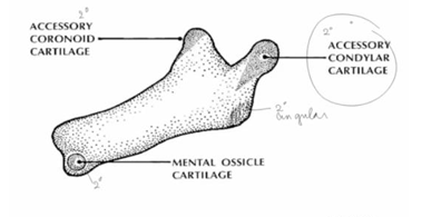
secondary cartilages develop in
-coronoid process, angular process, condylar process, and symphyseal region
-only cartilage that stays is the cartilage of condylar process because this becomes part of a joint (articulates with temporal bone) and motion maintains the cartilage
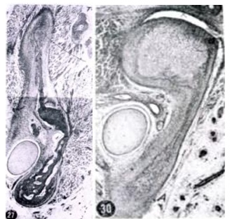
mandible development at 12 weeks
-secondary cartilage disappears (left image), cartilage in condyle still there (right image)
-NO connection between Meckel’s cartilage and body of mandible
-TMJ formation takes place
-TMJ located anterior and slightly lateral to primitive joint between malleus and incus (articular and quadrate)
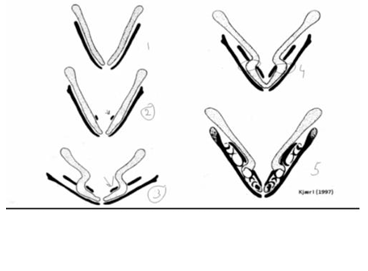
expansion of mandible (black) buccal to Meckel’s cartilage (grey)
-in stage 2: see beginning appearance of additional piece of bone- forms in a V shape, where teeth are going to develop
-between stages 2 and 3: shape of lower jaw changes A LOT, secondary palate forms and face gets wider and shorter in AP direction
-stages 3 and 4: developing bone of mandible (Meckel’s cartilage) gets incorporated in a zig-zag fashion; area of Meckel’s cartilage that will contribute to the mandible in endochondral ossification
-stage 5: developing teeth