lecture 7- Hoof
1/57
Earn XP
Description and Tags
Dr. anderson lecture
Name | Mastery | Learn | Test | Matching | Spaced | Call with Kai |
|---|
No analytics yet
Send a link to your students to track their progress
58 Terms
protective layer to prevent desiccation analogous to a cuticle
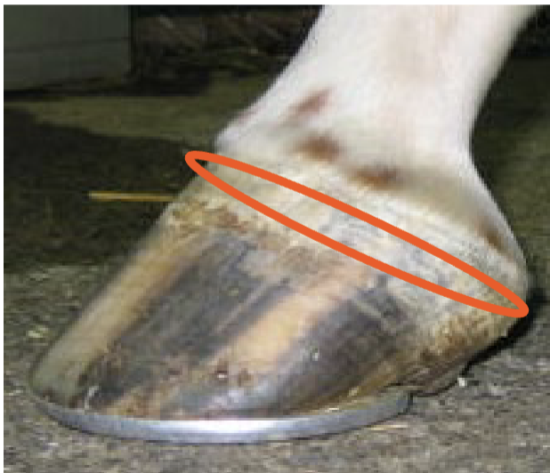
periople
primary weight bearing structure protexting, force dissipation, traction keratinized, tubular epidermis
wall
extension of wall
provides additional weight bearing surface
prevents excessive expansion —> buttress
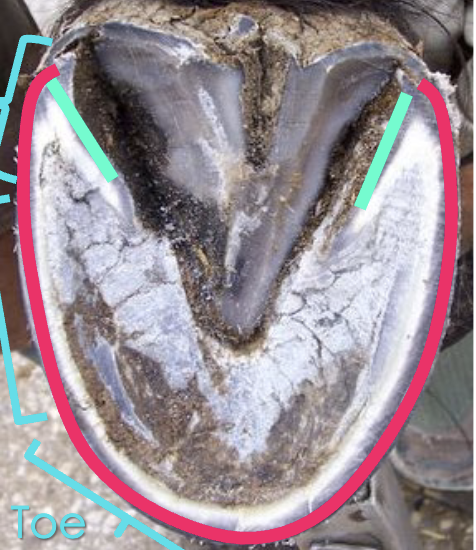
bars
what are the two teal lines
bars
shock absorption —> distributes of forces
heels
where are the heels located on the hoof
towards the back
junction between solar and laminar epidermis
area where nails are driven in on a shod horse
white line
T/F the white line is white
false it actually is really dark
what is this
white line
protects bottom of P3
bears less weight compared to wall (bottom of hoof is concave)
provides traction
sole

what is the black arrow
angle of sole
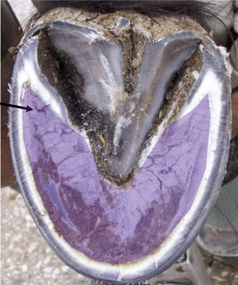
what is the purple
sole
lies over the digtal cushion —> peripheral pump
provides traction
frog

what is the blue
frog
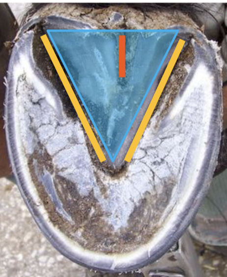
what is the orange
central sulcus

what is the yellow
collateral sulci
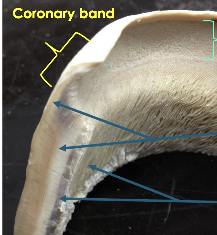
hoof wall (coronary epidermis)
overlying coronary dermis
coronary band
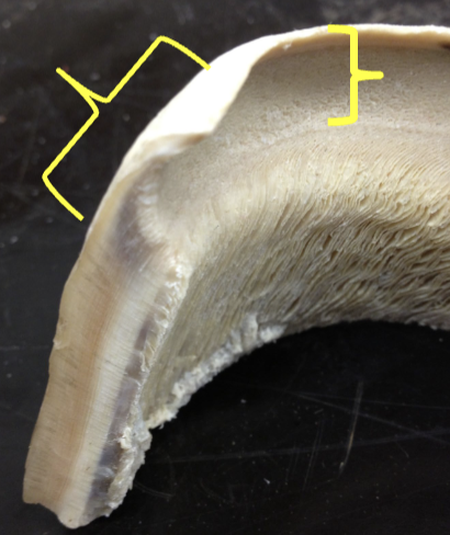
what is the yellow
coronary band
junction between hoof wall (coronary epidermis) and skin (epidermis)
coronet
what is the blue
coronet
dermis hoof epidermis is keratinized
corium
supplies keratinized coronary epidermis that forms tubule and intertubule horn
coronary dermis
supplies laminar epidermis
laminar dermis

what is the top arrow
coronary dermis
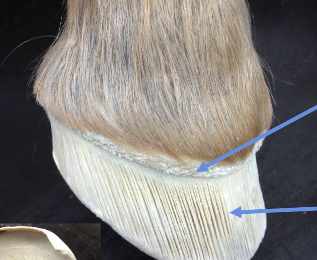
what is the bottom arrow
laminar dermis
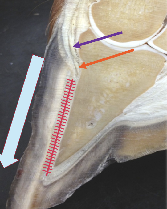
what is known as the sulcus
coronary groove
what is known as the stratum medium
coronary epidermis
what is known as the stratum internum
laminar epidermis

what is the top teal area
coronary groove

what is the middle area
coronary epidermis
what is the bottom area
laminar epidermis
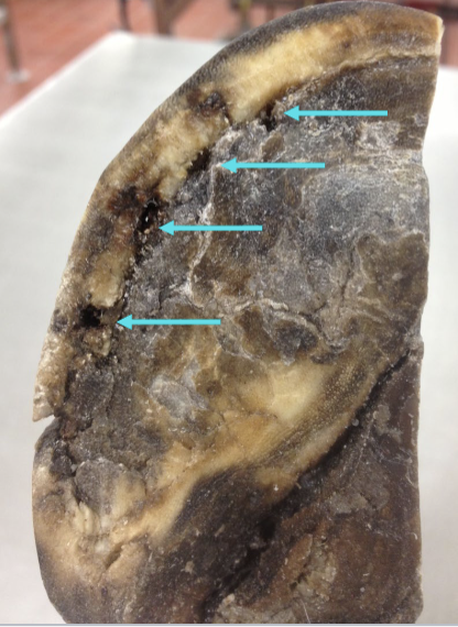
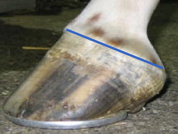
hoof growth rate
6-9 mm per month
when do hooves grow slower
in winter around ~3 mm per month
explain the purple
keratinized coronary epidermis formed by basal epidermal cells at coronary bands **tubular

explain the orange
continuous with keratinized laminar epidermis

how does the laminar epidermis “ratchet” or move down
desmosomes detach between epidermal and dermal lamina to let coronary and laminar epidermis “ratchet” or move down
explain sole growth
horn tubules orientated vertically
growth limited by curling of tubules at ground
slower than wall ground
what does the suspensroy apparatus of P3
laminar dermis interdigitates and supplies laminar epidermis
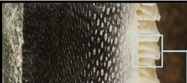
what is the white flaky area
laminar epidermis

what is the dark area
coronary epidermis
why does the suspensory apparatus of P3 fail
• Still under investigation
Insulin dysregulation
Endothelial cell and vascular dysfunction
• Conditions
Metabolic syndrome and insulin resistance
Pars Pituitary Intermedia Dysfunction (PPID or Cushing's)
Endotoxemia
Colic
Retained placenta
Colitis
Physical demand
what is the main blood and nerve supply to the hoof
medial/ lateral palmar/ plantar digital
how do vessels enter the hoof
on palmar/plantar aspect of P3
where is palmar venous plexus
in sole corium, around toe between sole and digital cushion
where is dorsal venous plexus
in laminar corium
where is the coronary venous plexus
at coronet- in coronary and perioplic corium
drains into digital veins cuts at coronet bleed
where does the collateral cartilages attach
to P3
what is known as the structural support for heel bulbs
collateral cartilages w
what is the job of collateral cartilages
shock absorption
dissipates force through their expansion
involved in blood pumping action of foot
explain the hoof as a pump
• Muscle normally required to achieve effective venous drainage in periphery
No muscle in digit
• Digital cushion functions as “pump”
When horse bears weight digital cushion expands and veins compressed → blood pools in venous plexus and digital cushion
Diastolic phase
• When horse lifts hoof digital cushion contracts and veins
open → blood forced out
Systolic phase
explain the purpose of the navicular bone
• Same function as any other sesamoidean bone
Acts as pulley
↓ strain on DDFT as it changes direction to cross coffin joint
↓ work of DDFT to act on P3 (↑ its mechanical effect)

what is the top pink line
flexor tendon sheath
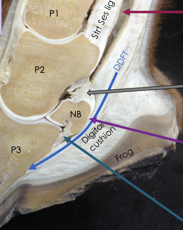
what is the grey arrow
collateral sesamoidean (suspensory) ligament of the navicular bone

what is the purple line
navicular bursa

what is the blue line
impar ligament
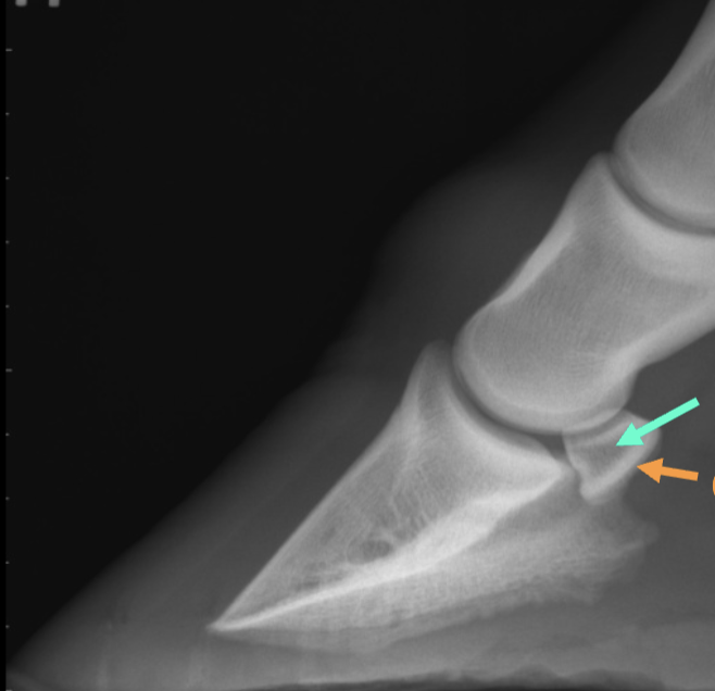
what is the teal arrow
medullary cavity

what is the orange arrow
cortex
what are the hoof functions
Traction
Frog, shape of hoof (concave), wall
Shock absorption
Digital cushion, ability to expand at heels
Support of skeletal column
Weight of horse transmitted via lamina to hoof wall
Suspends P3
Protection of inner structures
Periople protects from evaporation
Hoof wall protects lamina
Venous return (peripheral pump)
Frog, digital cushion, collateral cartilages