Nervous System, Spinal Nerves, and Vertebral Anatomy: Key Concepts for Students
 Call Kai
 Learn
Learn Practice Test
 Spaced Repetition
 Match
Match Flashcards
 Knowt Play
1/81

There's no tags or description
Looks like no tags are added yet.
Name | Mastery | Learn | Test | Matching | Spaced | Call with Kai | Chat |
|---|
No analytics yet
Send a link to your students to track their progress
82 Terms
What are the two main divisions of the nervous system?
CNS (brain and spinal cord) and PNS (spinal nerves and cranial nerves).
What does the somatic nervous system serve vs the autonomic (visceral) nervous system?
Somatic: skin, bones, voluntary (skeletal) muscle.
Autonomic: organs, glands, involuntary (smooth/cardiac) muscle — at an unconscious level.
What do efferent and afferent mean?
Efferent = motor, carrying signals out of the CNS
Afferent = sensory, carrying information into the CNS.
What are the two nicknames and roles of the autonomic divisions?
Sympathetic (""fight or flight"")
Parasympathetic (""rest & digest"")
Which two roots form a spinal nerve, and what does each carry?
The dorsal (posterior) root carries sensory fibers in
The ventral (anterior) root carries motor fibers out.
Where are the cell bodies of somatic sensory vs somatic motor neurons?
Sensory: dorsal root ganglion (DRG), in the PNS, no synapse.
.
Motor: ventral (anterior) horn of the spinal cord gray matter.
After the two roots join, what is the functional nature of the spinal nerve?
Mixed — it carries both motor and sensory fibers.
Clinically, how does a whole-nerve lesion differ from a single-root lesion?
Whole nerve → both sensory and motor deficit
Vs
Single root → one or the other.
Into which two rami does a spinal nerve split, and what does each supply?
Dorsal ramus (small): intrinsic/deep back muscles and overlying skin only.
.
Ventral ramus (large): everything else — limbs, body wall, abdominal wall, extrinsic back muscles.
Which rami form plexuses such as the brachial plexus?
The ventral rami.
Name the four intrinsic (deep) back muscle groups.
All DORSAL Rami
1) Splenius
2) Erector spinae
3) Transversospinal
4) Suboccipital groups
Name the extrinsic (superficial) back muscles and their innervation.
Supplied by VENTRAL rami (they act on the upper limb).
Trapezius
Latissimus Dorsi
Rhomboids
Levator scapulae
What is the tested exception to the extrinsic-back-muscle rule?
The trapezius is supplied by the spinal accessory nerve (CN XI), not a ventral ramus.
How many pairs of spinal nerves are there, and by region?
31 pairs:
8 cervical,
12 thoracic
5 lumbar
5 (fused) sacral
1 (fused) coccygeal.
Why are there 8 cervical nerves but only 7 cervical vertebrae?
C1-C7 emerge ABOVE their same-numbered vertebra C8 emerges below C7.
From T1 down, every nerve emerges BELOW its same-numbered vertebra.
List the circulatory loop in order from the heart (7)
Heart (left ventricle) → arteries → arterioles → capillaries → venules → veins → back to the heart.
What defines an artery — content or direction?
Direction: arteries carry blood away from the heart (the pulmonary artery carries deoxygenated blood).
Where does exchange occur, and what is the role of arterioles?
Exchange happens only at the capillary
.
Arterioles are the resistance vessels.
What is an anastomosis, and what is an end (terminal) artery?
An anastomosis is a communication between two vessels (a detour);
an end artery has no anastomotic partner, so occluding it kills the tissue it feeds.
Why does the speed of an arterial occlusion matter?
A slow, gradual occlusion lets collaterals enlarge; a sudden occlusion does not.
Name the five vessel-naming patterns with an example.
Position/direction (superior/inferior epigastric);
depth (""profunda"" = deep)
adjacent bone (vertebral, radial, ulnar)
region supplied (posterior intercostal)
branching order (common → external → internal).
Trace the main arterial supply of the upper limb from the subclavian (5)
Subclavian (Neck) a. →
axillary a. (armpit) →
brachial a. (Arm)→
radial a.
ulnar a.
Which artery ascends through the cervical transverse foramina?
The vertebral artery (a branch of the subclavian).
What are the three main functions of the vertebral column?
Protect the spinal cord
Transfer load from head/trunk to the pelvis
Give attachment to postural muscles.
In which plane are the four vertebral curvatures, and how does the newborn spine differ?
Sagittal plane
.
The newborn spine is a single curve concave anteriorly, whereas the adult has four alternating curves.
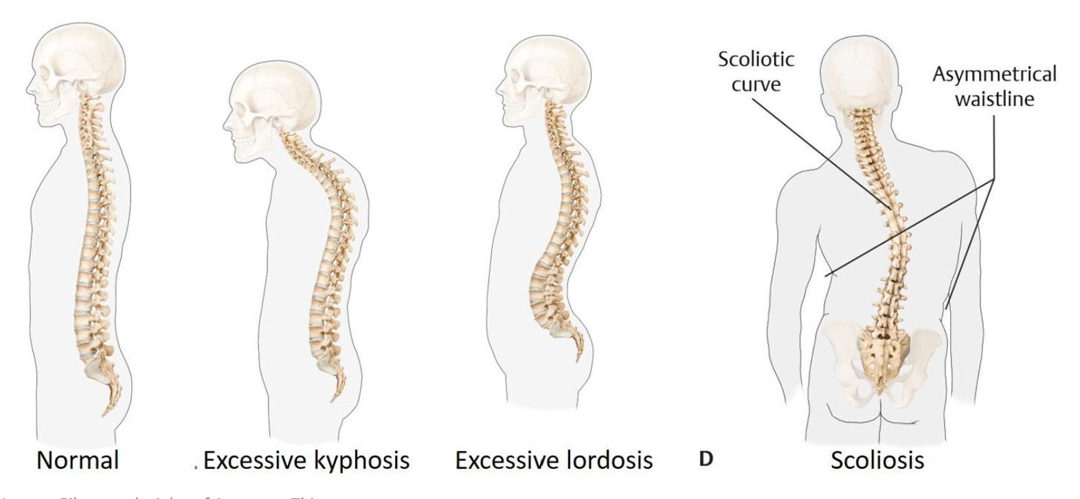
What is a primary curve (kyphosis) — shape, timing, regions?
Concave anteriorly, present from fetal life, never lost
Thoracic
Sacral.
What is a secondary curve (lordosis) — shape, timing, regions?
Concave posteriorly, acquired (cervical as the infant lifts the head, lumbar as the toddler stands/walks)
Cervical
Lumbar
Why do alternating curves help the column mechanically?
They let it act like a spring rather than a rigid post, distributing load along its length.
"What does ""neutral"" mean mechanically, and what happens when you lean forward?"
Neutral: the line of gravity passes near the inflection points between curves, so muscular effort is minimal.
.
Leaning forward shifts the line of gravity anteriorly, creating an external moment that the posterior muscles must resist continuously (sustained static loading).
Define excessive kyphosis, excessive lordosis, and scoliosis.
Excessive kyphosis: thoracic curve increased (head/shoulders forward).
.
Excessive lordosis: lumbar curve increased (often with anterior pelvic tilt).
.
Scoliosis: lateral deviation with rotation — a curve in a plane the spine doesn't normally curve.
What are the three building blocks of a typical vertebra?
The Vertebral body
The Vertebral arch (pedicles + laminae)
The Processes.
How many processes does a typical vertebra have?
Seven:
One spinous (posterior)
Two transverse (lateral)
Four articular (two superior, two inferior).
What forms the vertebral foramen, and what do stacked foramina create?
Body + arch = vertebral foramen
Stacked, they form the vertebral canal that houses the spinal cord.
Summarize the job of each vertebral part (3)
Body bears weight
Arch protects the cord
Processes are levers for muscles and stops for movement.
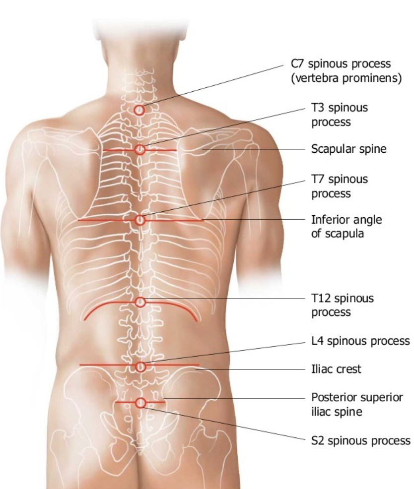
Give the six palpable spinous-process landmarks and their levels.
C7 vertebra prominens
T3 spine of scapula
T7 inferior angle of scapula
T12 12th rib
L4 highest point of iliac crest
S2 posterior superior iliac spine (PSIS).
.
(1 - C, 3 - T, 1 - L, 1 - S)
What Main feature that proves a vertebra is cervical?
The transverse foramen — no other region has one; it transmits the vertebral artery.
Give the key features of a cervical vertebra (4)
Small wide body; large triangular vertebral foramen;
Bifid spinous process (C3-C6; C7 is long and single);
Uncinate processes
Transverse foramen.
What are the uncovertebral joints (of Luschka), and why do they matter clinically?
Joints formed where the uncinate processes (C3-C7) meet the body above;
they sit at the anteromedial wall of the intervertebral foramen
So anything enlarging them encroaches on the exiting nerve root.
Outline the four-step cascade of cervical spondylosis.
1) Disc height falls
2) load shifts onto uncovertebral and facet joints
3) bone responds (hypertrophy, osteophytes)
4) the intervertebral foramen narrows, crowding the nerve root.
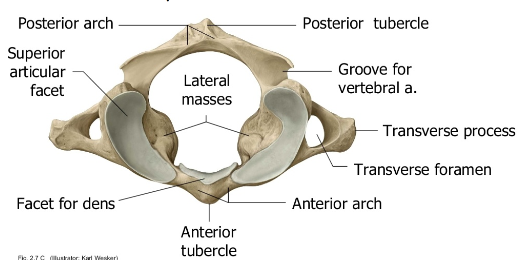
What are the defining features of the atlas (C1)? (2)
No body and no spinous process
ring of anterior and posterior arches with lateral masses that bear the load facet for the dens on the anterior arch groove for the vertebral artery on the posterior arch.
"Which is the ""yes"" joint and what movement does it give? "
The atlanto-occipital joint (occiput on C1)
concave superior facets receive the occipital condyles → nodding (flexion/extension).
What are the defining features of the axis (C2)?
The dens (odontoid process) projecting up, with anterior facet (meets C1's anterior arch) and posterior facet (meets the transverse ligament).
"Which is the ""no"" joint and how much rotation occurs there? "
The atlanto-axial joint (C1 on C2)
the atlas and skull rotate around the dens;
roughly half of all cervical rotation happens here.
What single feature proves a vertebra is thoracic?
Costal facets (they articulate with ribs) — only thoracic vertebrae carry ribs.
Give the key features of a thoracic vertebra.
Heart-shaped body;
small circular vertebral foramen
long inferiorly-sloping spinous process
costal facets/demifacets
Each one has 2 demifacets on the verterbral body and a facet on the transverse process
What is a demifacet, and how does a rib head articulate with the column?
A demifacet is a half costal facet
A rib head spans two bodies, meeting the inferior demifacet of the vertebra above and the superior demifacet of the vertebra below (with the disc between).
Classify ribs 1-7, 8-10, and 11-12 by anterior attachment.
Ribs 1-7: true (cartilage reaches the sternum directly).
.
Ribs 8-10: false (cartilage joins the rib above).
.
Ribs 11-12: floating (no anterior attachment).
How are T1, T11, and T12 atypical thoracic vertebrae?
T1: a complete facet for rib 1 plus a demifacet for rib 2
T11/T12: complete facets for their own ribs and NO costal facet on the transverse process (so ribs 11-12 have only one attachment — why they float).
What single feature identifies a lumbar vertebra?
A large kidney-shaped body with no costal facet and no transverse foramen.
Give the key features of a lumbar vertebra.
Large kidney-shaped body;
Triangular vertebral foramen
Short blunt spinous process
Mammillary and accessory processes for muscle attachment.
What is the pars interarticularis?
The short bridge of the vertebral arch between the superior and inferior articular processes
The narrowest part of the arch and the part that fails.
Distinguish spondylolysis from spondylolisthesis.
Spondylolysis: a pars defect/stress fracture without vertebral body displacement.
.
Spondylolisthesis: a pars defect PLUS anterior slippage of the body on the one below (most often L4-L5 or L5-S1).
Why is the lower lumbar pars the predictable failure point?
Sagittal lumbar facets resist rotation but transmit shear forward
Combined with the largest column loads and the narrowest part of the arch.
What is the sacrum, structurally and functionally?
Five fused vertebrae (S1-S5) forming a load-transferring wedge that shifts weight from the lumbar spine into the pelvis.
Which rami pass through the anterior vs posterior sacral foramina?
Anterior sacral foramina → ventral rami;
Posterior sacral foramina → dorsal rami.
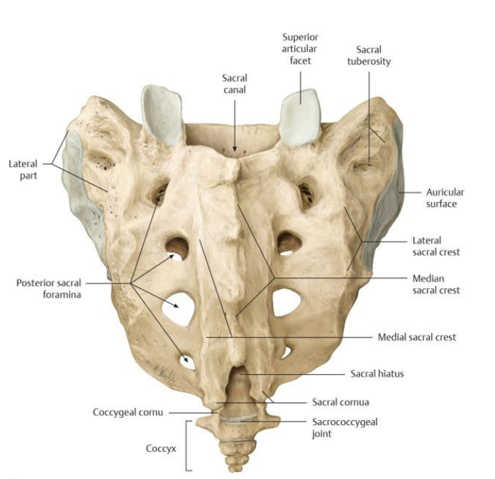
What are the major structural features of the sacrum? (7)
Base and promontory (S1 body)
Ala (wing)
Anterior sacral foramina
Posterior sacral foramina
Median Sacral Crest
Sacral canal and hiatus
Auricular surface (articulated with ilium)
Describe the coccyx.
4 fused rudimentary vertebrae (Co1-Co4) with no vertebral arch and no canal
a muscle attachment site.
How is facet orientation and favoured motion related, by region?
Cervical ≈45° from horizontal → most mobile, all movements.
Thoracic near coronal → rotation/lateral flexion (flexion/extension limited by ribs).
Lumbar near sagittal → flexion/extension (rotation restricted; facets interlock).
How many joints are between two adjacent vertebrae, and of what types?
Three
1) One secondary cartilaginous joint (symphysis — the intervertebral disc) between the bodies,
2 & 3) Two synovial zygapophysial (facet) joints between the arches.

Which vertebral joints have NO intervertebral disc?
The craniovertebral joints — none between the occiput and C1, or between C1 and C2.
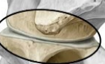
Describe the structure of a facet (zygapophysial) joint.
Synovial: articular surfaces of hyaline cartilage,
enclosed by a capsule with synovial fluid;
a single level glides little,
but ~30 levels together give the column its range.
List the five factors that determine range of motion at a vertebral level.
1) Intervertebral disc (composition/height);
2) Facet joint morphology;
3) Muscle and ligament resistance;
4) Whether the vertebra carries a rib;
5) Surrounding soft-tissue bulk.
What are the two parts of the intervertebral disc, and where does the nucleus sit?
1) Anulus fibrosus (outer fibrocartilaginous rings)
2) Nucleus pulposus (gelatinous, high water content),
Which sits slightly posterior to center.
Over what extent are Intervertebral discs present, and how are they numbered?
C2 to the lumbosacral junction;
Each disc is numbered for the vertebral body immediately above it.
State the ligament continuity rule (three that change name from the occiput).
Anterior atlanto-occipital membrane → anterior longitudinal ligament;
Tectorial membrane → posterior longitudinal ligament;
Ligamentum nuchae → supraspinous ligament.
What does the transverse ligament (of the cruciform ligament) do?
It holds the dens against the anterior arch of C1, keeping it from moving back into the cord.
What do the alar ligaments connect and limit?
Dens to the margins of the foramen magnum; they limit rotation of the head.
Where does the anterior longitudinal ligament run and what does it limit?
Down the anterior/lateral surfaces of the bodies (occiput/C2 → sacrum)
It is broad and strong and limits EXTENSION — the only ligament that does.
Where does the posterior longitudinal ligament run, and why is its shape significant?
Down the posterior surface of the bodies inside the vertebral canal (C2 → sacrum);
it narrows over each body (scalloped), leaving the posterolateral disc least supported; limits flexion weakly.
What is the ligamentum flavum made of, what does it limit, and why must it be elastic?
High elastin (yellow);
connects lamina to lamina forming the posterior wall of the canal;
limits flexion; elastic recoil keeps it taut rather than buckling inward toward the cord.
What do the interspinous, supraspinous, and intertransverse ligaments limit and where are they located?
interspinous → flexion (between adjacent spinous processes in the sagittal plane)
.
supraspinous → flexion (Along the posterior tips of the spinous processes; continuation of the ligamentum nuchae)
.
intertransverse - Between adjacent transverse processes, in the frontal plane Limits: Lateral flexion
processes; continuation of the ligamentum
intertransverse → lateral flexion (Between adjacent transverse processes, in the frontal plane)"
Which ligament limits extension, and what do all the others limit?
Anterior longitudinal ligament limits extension; every other ligament limits flexion or lateral flexion.
What are the two rib-to-vertebra joints?
Costovertebral joint (rib head to the demifacets on the bodies, with the disc between)
Costotransverse joint (rib tubercle to the transverse process of the same-numbered vertebra).
Only for Thoracic RIbs
Name the ligaments connecting the spine/sacrum to the pelvis, and why the group matters.
Iliolumbar — Transverse processes of L4 and L5 to the iliac crest
Sacroiliac (ant., post., interosseous) — Sacrum to ilium across the sacroiliac joint
Sacrospinous — Sacrum to the ischial spine
Sacrotuberous — Sacrum to the ischial tuberosity
Sacrococcygeal — Sacrum to the coccyx
the sacroiliac joint is where the whole column's load leaves the spine and enters the pelvis (loaded all day when seated)
Give the three anatomical reasons a disc herniates posterolaterally.
1) The nucleus already sits posteriorly
2) the anulus is thinnest behind
3) the PLL reinforces the midline but narrows laterally — so the posterolateral corner (where the nerve root sits) is least supported.
Summarize favoured vs restricted motion and its limiter by region.
Cervical: all movements, least restricted (limited by ligaments/muscles; alar ligs at C1-C2)
Thoracic: rotation/lateral flexion; flexion/extension restricted by rib joints.
Lumbar: flexion/extension; rotation restricted by interlocking facets.
Sacral: fused — motion only at the sacroiliac joint.
What one sentence links facets, ligaments, and ribs to movement?
Facets set the direction; ligaments and ribs set the limit.
Name the four abdominal wall muscle groups.
external oblique,
internal oblique
transversus abdominis
rectus abdominis (all ventral rami).
What forms the exit for the spinal nerve in the lumbar vertebrae?
The superior and inferior vertebral notches together form the intervertebral foramen.
What does the apical ligament connect?
The tip of the dens to the anterior margin of the foramen magnum