nervous coordination and muscles
 Call Kai
 Learn
Learn Practice Test
 Spaced Repetition
 Match
Match Flashcards
 Knowt Play
1/86

There's no tags or description
Looks like no tags are added yet.
Name | Mastery | Learn | Test | Matching | Spaced | Call with Kai | Chat |
|---|
No analytics yet
Send a link to your students to track their progress
87 Terms
what happened as species have evolved?
their cells have become adapted to perform specialist functions
what do cells do by specialising in one function?
they lose the ability to perform other functions, with different groups of cells carrying out their own functions. this makes cells dependant on others to carry out the functions they no longer specialise in
eg. cells specialising in reproduction:
they depend on other cells to obtain oxygen for their respiration, to provide glucose or to remove their waste products. these different functional systems must be coordinated if they are to perform efficiently. no body systems work in isolation
what are the 2 main forms of coordination in animals as a whole?
the nervous system
the hormonal system
the nervous system:
use nerve cells to pass electrical impulses along their length
they stimulate their target cells by secreting chemicals, known as neurotransmitters, directly onto them
this results in rapid communication between specific parts of an organism
the responses are often short lived and restricted to a localised region of the body
example of nervous coordination:
a reflex action, like the withdrawal of the hand from an unpleasant stimulus - short lived, rapid and restricted to one region of the body
the hormonal system:
produces chemicals (hormones) that are transported in the blood plasma to their target cells
the target cells have specific receptors on their cell surface membranes and the change in the concentration of hormones stimulates them
this results in a slower, less specific form of communication between parts of an organism
the responses are often long lasting and widespread
example of the hormonal system:
the control of blood glucose concentration, which produces a slower response but has a more long term and widespread effect
what do both systems do?
work together and interact with one another
diagram to show comparison of hormonal and nervous systems:

what are neurones (nerve cells)?
specialised cells adapted to rapidly carrying electrochemical changes called nerve impulses from one part of the body to another
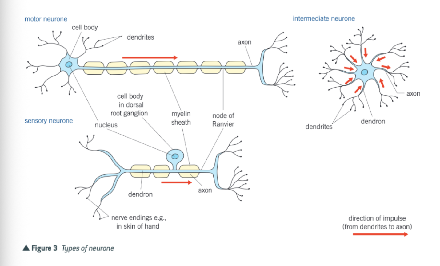
what is a mammalian motor neurone made up of?
a cell body
dendrons
axon
Schwann cells
myelin sheath
nodes of ranvier
cell body:
contains all the usual cell organelles, including a nucleus and large amounts of rough endoplasmic reticulum. this is associated with the production of proteins and neurotransmitters
dendrons:
extensions of the cell body which subdivide into smaller branched fibres called dendrites that carry nerve impulses towards the cell body
axon:
a single long fibre that carries nerve impulses away from the cell body
Schwann cells:
surround the axon, protecting it and providing electrical insulation. they also carry out phagocytosis (the removal of cell debris) and play a part in nerve regeneration. Schwann cells wrap themselves around the axon many times, so that layers of their membranes build up around it
myelin sheath:
forms a covering to the axon and is made up of the membranes of the Schwann cells. these membranes are rich in a lipid known as myelin. neurones with a myelin sheath are called myelinated neurones
nodes of ranvier:
constrictions between adjacent Schwann cells where there is no myelin sheath. these constrictions are 2-3 micrometres long and occur every 1-3mm in humans
diagram to show a myelinated motor neurone:

sensory neurone:
transmit nerve impulses from a receptor to an intermediate or motor neurone. they have one dendron that is often very long. it carries the impulse towards the cell body and one axon that carries it away from the cell body
motor neurones:
transmit nerve impulses from an intermediate or relay neurone to an effector, such as a gland or a muscle. motor neurones have a long axon and many short dendrites
intermediate or relay neurones:
transmit impulses between neurones, for example, from sensory to motor neurones. they have numerous short processes
diagram to show types of neurone:
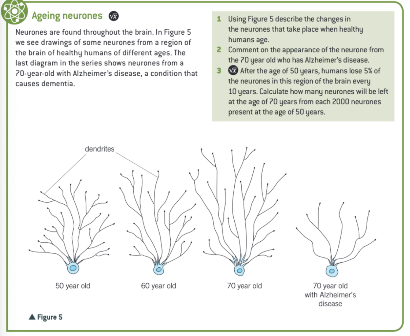
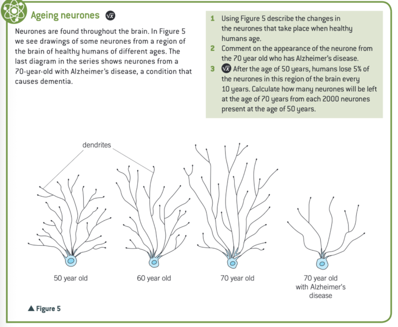
extra info: ageing neurones:
nerve impulse:
a self propagating wave of electrical activity that travels along the axon membrane. it is a temporary reversal of the electrical potential difference across the axon membrane. this reversal is between 2 states - resting potential and action potential
how is the movement of ions, like Na+ ions and K+ ions, across the axon membrane controlled?
phospholipid bilayer of the axon plasma membrane prevents sodium and potassium ions diffusing across it
proteins (channel), span the phospholipid bilayer. they have ion channels which pass through them. some channels have gates, which can be opened or closed so that Na+ or K+ ions can move through them by facilitated diffusion at any one time, but not on other occasions. there are different gated channels for Na+ or K+ ions. Some channels, however, remain open all the time, so the ions move unhindered through them by facilitated diffusion
some carrier proteins actively transport K+ ions into the axon and Na+ ions out of the axon - sodium potassium pump
what happens as a result of the various membrane controls?
the inside of an axon is negatively charged relative to the outside - resting potential
what does the resting potential range between in humans?
50-90 mV (often 65 in humans)
what is the axon said to be in this condition?
polarised
what is the establishment of this potential difference (difference between inside and outside of axon) due to?
sodium ions are actively transported out of the axon by the sodium potassium pumps
potassium ions are actively transported into the axon by the sodium potassium pumps
the active transport of sodium ions is greater than that of potassium ions, in a 3:2 ratio
although both sodium and potassium are +, the active transport movement is different, so there are more sodium ions in the tissue fluid surrounding the axon than in the cytoplasm, and more potassium ions in the cytoplasm than in the tissue fluid, creating an electrochemical gradient
the sodium ions begin to diffuse back naturally into the axon while the potassium ions begin to diffuse back out of the axon
however, most of the gates in the channels that allow the potassium ions to move through are open, while most of the gates in the channels that allow the sodium ions to move through are closed
diagram to show the distribution of ions at resting potential
what happens when a stimulus of sufficient size is detected by a receptor in the nervous system?
its energy causes a temporary reversal of the changes either side of this part of the axon membrane
what happens if the stimulus is great enough?
the negative charge of -65mV inside the membrane becomes a positive charge of 40mV - the action potential
what is this part of the axon membrane said to be in this condition?
depolarised
why does this depolarisation occur?
because the channels in the axon membrane change shape, hence opening or closing depending on the voltage across the membrane. they are therefore called voltage gated channels
what is important to stress about the action potential?
the events described relate to a particular point on the axon membrane and not the whole of the membrane
diagram to show the action potential:

process of the action potential:
at resting potential some K+ voltage gated channels are open (those that are permanently open) but the Na+ voltage gated channels are shut
the energy of the stimulus causes some Na+ voltage gated channels in the axon membrane to open and therefore sodium ions diffuse into the axon through these channels along their electrochemical gradient. Being + charged, they trigger a reversal in the potential difference across the membrane
as the Na+ ions diffuse in the axon, more Na+ channels open, causing a greater influx of Na+ ions by diffusion
once the action potential of 40mV has been established, the voltage gates on the sodium ion channel shut and the voltage gates on the K+ ion channels begin to open
with some of these channels open, the electrical gradient that was preventing further outward movement of K+ ions is now reversed, causing more K+ ion channels to open. this means more K+ ions diffuse out, starting repolarisation of the axon
the outward diffusion of these K+ ions causes a temporary overshoot of the electrical gradient, with the inside of the axon being more negative relative to the outside than usual (hyperpolarisation)
the closable gates on the K+ ion channels now close and the activities of the sodium potassium pumps works again
the resting potential of -65mV is reestablished and the axon is said to be repolarised.
what is the movement of Na+ ions inwards during the action potential due to?
diffusion
what is the movement of Na+ ions during the resting potential due to?
active transport
what does the term action potential simply mean?
the axon membrane is transmitting a nerve impulse
extra info: measuring action potentials

what happens once an action potential has been created?
it moves rapidly along an axon
does the size of an action potential always remain the same from one end of the axon to the other?
yes
strictly speaking, does anything physically move from place to place along the axon of the neurone?
no
what happens as one region of the axon produces an action potential and becomes depolarised?
it acts as a stimulus for the depolarisation of the next region of the axon
what happens in this manner?
action potentials are generated along each small region of the axon membrane, therefore being a travelling wave of depolarisation
what happens in the meantime to the previous part of the membrane?
it returns to its resting potential, and undergoes repolarisation
idea of this movement of an action potential in an example:

how a nerve impulse is propagated in an unmyelinated axon:
step 1:
at resting potential the conc of Na+ ions outside the axon membrane is high relative to the inside, whereas that of the potassium ions is high inside the membrane relative to the outside
the overall concentration of + ions is, however, greater on the outside, making this positive compared with the inside
the axon membrane is polarised

step 2:
a stimulus causes a sudden influx of sodium ions and hence a reversal of charge on the axon membrane
this is the axon potential and the membrane is depolarised
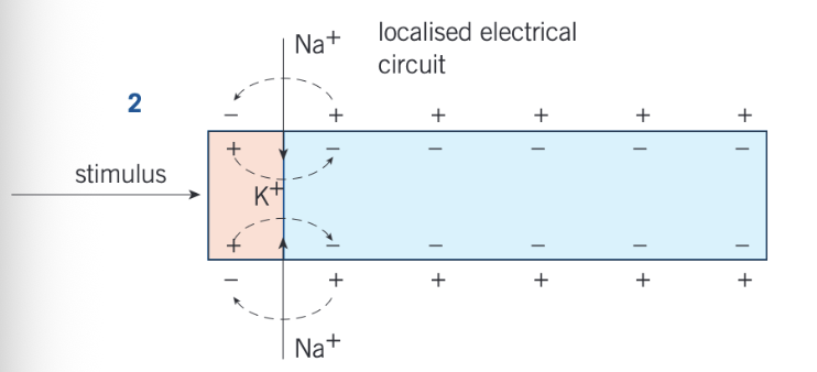
step 3:
the localised electrical currents established by the influx of sodium ions cause the opening of sodium voltage gated channels a little further along the axon
the resulting influx of sodium ions in this region causes depolarisation
behind this new region of depolarisation, the sodium voltage gated channels close and the potassium ones open
potassium ions begin to leave the axon along their electrochemical gradient, so, once initiated, the depolarisation moves along the membrane

step 4:
the action potential (depolarisation) is propagated in the same way further along the axon
the outward movement of the potassium ions has continued to the extent that the axon membrane behind the action potential has returned to its original charged state (+ outside, - inside), that it, it has been repolarised

step 5:
repolarisation of the axon allows sodium ions to be actively transported out, once again returning the axon to its resting potential in readiness for a new stimulus if it comes

what is the case in myelinated axons?
the fatty sheath of myelin around the axon acts as an electrical insulator, preventing action potentials from forming
what is there at intervals of 1-3mm?
there are breaks in this myelin insulation, called nodes of ranvier - action potentials can occur at these points
where do the localised circuits therefore arise between?
adjacent nodes of ranvier and the action potentials in effect jump from node to node in a process known as saltatory conduction
what can then happen as a result of saltatory conduction?
the action potential passes along a myelinated neurone faster than along the axon of an unmyelinated one of the same diameter
why does it move faster than an unmyelinated axon?
in an unmyelinted neurone, the events of depolarisation have to take place all the way along an axon and this takes more time
diagram to show passage of an action potential along a myelinated axon:

what happens once an action potential has been set up?
it moves rapidly from one end of the axon to the other without any decrease in size
what is the transmission of an action potential along the axon of a neurone known as?
the nerve impulse
how can the speed of an action potential be affected?
from as little as 0.5ms-1 or as much as 120ms-1 depending on factors
what are these factors?
the myelin sheath
the diameter of the axon
temperature
the myelin sheath:
passage of an action potential, that the myelin sheath acts as an electrical insulator, preventing an action potential forming in the part of the axon covered in myelin
it does, however, jump from one node of the ranvier to another (saltatory conduction). this increases the speed of conductance from 30ms-1 in an unmyelinated neurone to 90ms-1 in a similar myelinated one
the diameter of the axon:
the greater the diameter of an axon, the faster the speed of conductance. this is due to less leakage of ions from a large axon (leakage makes membrane potentials harder to maintain
temperature:
this affects the rate of diffusion of ions and therefore the higher the temperature the faster the nerve impulse
the energy for active transport comes from respiration
respiration, like the Na+K+ pump, is controlled by enzymes. enzymes function more rapidly at higher temps up to a point
above a certain temp, enzymes and the plasma membrane proteins are denatured and impulses fail to be conducted at all
temp is clearly an important factor in response times in cold blooded (ectothermic) animals, whose body temp varies in accordance with the environment
temp also affects the speed and strength of muscle contractions
what are nerve impulses described as?
all or nothing responses
what is the threshold value?
a certain level of stimulus which triggers an action potential. below the threshold value, no action potential, and therefore no impulse, is generated
what is the’ nothing ‘part?
any stimulus of whatever strength that is below the threshold value will fail to generate an action potential
what is the ‘all’ part?
any stimulus above the threshold value will succeed in generating an action potential and so a nerve impulse will travel
why can’t the strength of a stimulus be detected by the size of the action potentials?
all action potentials are the same size
how then can an organism perceive the size of a stimulus?
by the number of impulses passing in a given time - the larger the stimulus, the more impulses that are generated in a given time
by having different neurones with different threshold values. the brain interprets the number and type of neurones that pass impulses as a result of a given stimulus and thereby determines its size
diagram to illustrate neurone excitability before and after a nerve impulse:
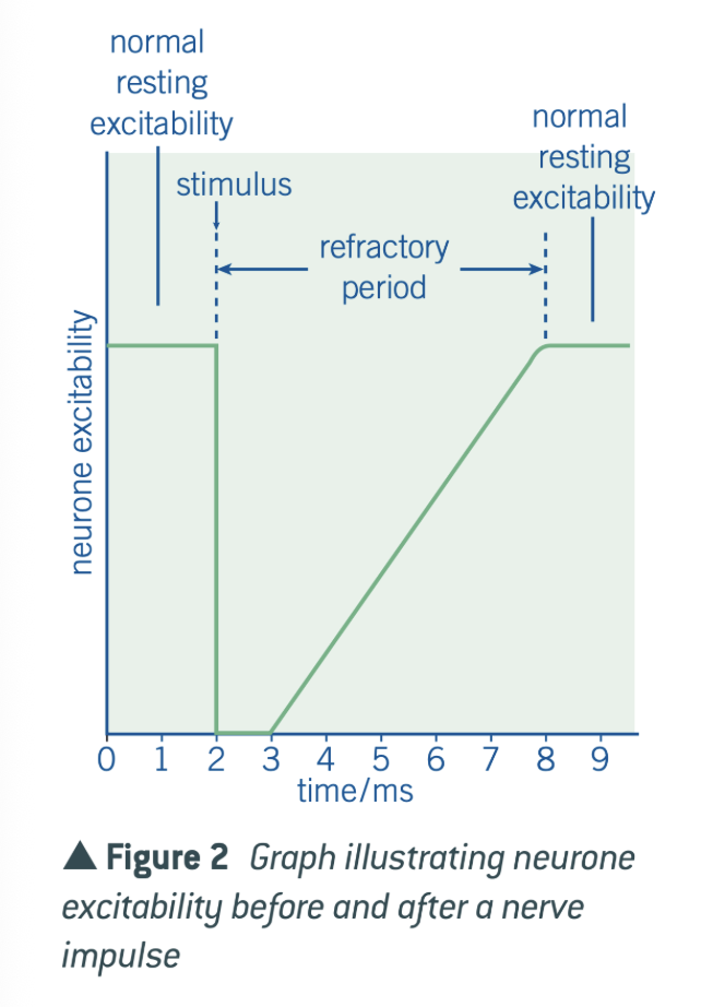
what happens once an action potential has been created in any region of an axon?
there is a period afterwards when inward movement of sodium ions is prevented as the sodium voltage gated channels are closed - during this time it is impossible for a further action potential to be generated
what is this period known as?
the refractory period
diagram to show the effect of stimulus intensity on impulse frequency:

what 3 purposes does the refractory period serve?
ensures that action potentials are propagated in one direction only
produces discrete impulses
limits the number of action potentials
ensures that action potentials are propagated in one direction only:
action potentials can only pass from an active region to a resting region
this is because action potentials can be propagated in a region that is refractory, which means that they can only move in a forward direction
this prevents action potentials from spreading out in both directions, which they would otherwise do
produces discrete impulses:
due to the refractory period, a new action potential can’t be formed immediately behind the first one. this ensures that action potentials are separated form one another
limits the number of action potentials:
as action potentials are separated from one another this limits the number of action potentials that can pass along an axon in a given time, and thus limits the strength of stimulus that can be detected
extra info: different axons, different speeds:
