FoME Anatomy Final
 Call Kai
 Learn
Learn Practice Test
 Spaced Repetition
 Match
Match Flashcards
 Knowt Play
1/95

There's no tags or description
Looks like no tags are added yet.
Name | Mastery | Learn | Test | Matching | Spaced | Call with Kai | Chat |
|---|
No analytics yet
Send a link to your students to track their progress
96 Terms
hypertrophy
A cross section of hearts common seen in our dissections. Compare to the left normal heart, name the pathologic process results in the thickening of the left ventricle wall.
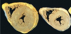
Increase in cell number and size
Smooth muscle in the main component of the uterine wall. Uterus on the left is during pregnancy and normal is on the right. Which is the best description of the enlargement of the left one?
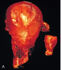
oxygen poor, nutrition rich
Which of the following best describes components of blood carried in the pulmonary arteries
oxygen poor, nutrition rich
Which of the following best describes components of the blood carried in the hepatic portal vein?
Limbs
Valves in the vein can prevent back flow of blood. Which of the following is the best location to visualize?
Deep vein thrombosis (DVT)
Pulmonary embolism, especially main pulmonary arteries, can result in sudden death. What is the most common source of venous emboli?
arteriovenous shunts
In skin, blood flow can be varied according to the external temperature by
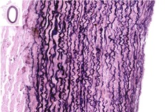
Convey blood from the heart to system
In this image, the elastic fibers are stained black. which of the following best describes this type of vessel?
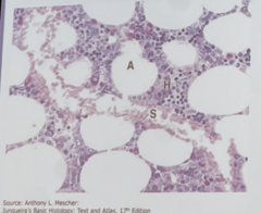
easy movement of blood cells across the endothelium
The sinusoidal capillary shown is specialized for?
cancer cells
fluid
macromolecules
macrophages
Which of the following can be moved from the interstitial space to lymph nodes through lymphoid vessels?
abdominal aorta
What is the most frequent site to see severe atherosclerosis?
congestive heart failure
lymphatic obstruction
venous obstruction
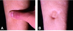
What could be the most likely cause(s) of the problem seen in this leg?
gray matter
Caudate nucleus is a
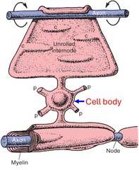
oligodendrocytes
Myelin sheath in the CNS is made by
loss of Schwann cells
Which of the following is true regarding Guillain-Barre Syndrome
oligodendrocyte
Name the type of cell indicated by the blue arrow
Carry motor information from the CNS to muscle
What is the function of the efferent nerve fibers?
left hemisphere
Language is usually controlled by
inability to speak
Which of the following is false, when Wernicke area is affected by stroke?
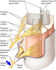
both motor and sensory
Anterior ramus, as shown in the image, consist of
medulla
Mechanosensory information from the right side of the spinal cord to the cortex cross at
medulla
Movement commands from the left side of the cortex to the spinal cord cross at
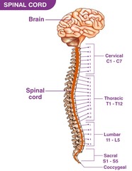
Sacral cord
Which of the following involve parasympathetic regulation?
occipital lobe
Primary visual cortex is in
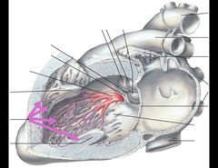
Purkinje fiber
Motor function of the heart is innervated by the autonomic nervous system. Name the structure labeled in purple.
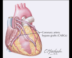
Great saphenous vein
What vessel is most commonly used for coronary triple bypass grafts?
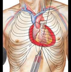
Normal venous pressure
Which of the following is false? Rupture of a severe myocardial infarction could result in
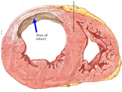
Left ventricle
This old infarct is located in
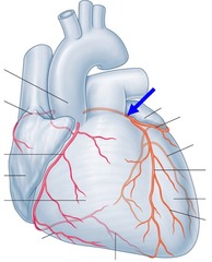
Left coronary artery
Name the coronary artery indicated by the blue arrow
Hilar nodes
Which of the following lymph nodes are likely to find tumor cells when a malignant lung cancer metastasized?
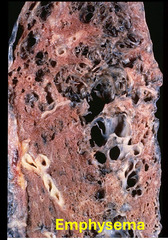
Septa destroyed through inflammation
Emphysema is highly associated to smoking. The enlarged openings in the lung are mainly due to
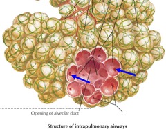
Pore of Kohn
Name the structure indicated by the blue arrows
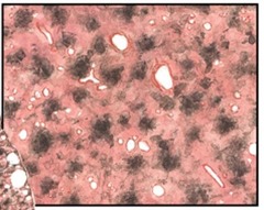
Macrophage
This is a lung section from a patient lived in a heavily polluted area. Which of the following cell types accumulated the black pigments?
Artery
Pulmonary bronchiole is always located close to
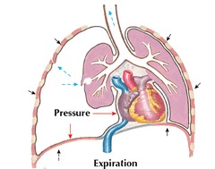
Between parietal and visceral layers of pleura
This image shows a patient with tension pneumothorax. The air is trapped in
Subendocardium
Which of the following area is most vulnerable to ischemia during hypertension?
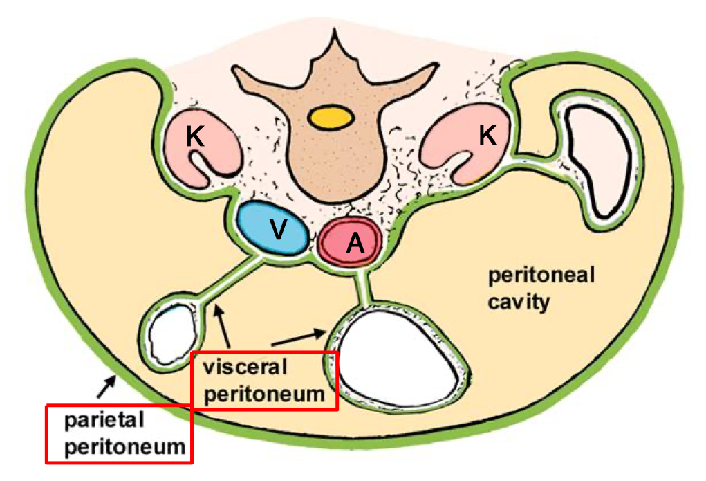
Which peritoneal layer is somatic and produces sharp, well-localized pain?
Parietal peritoneum
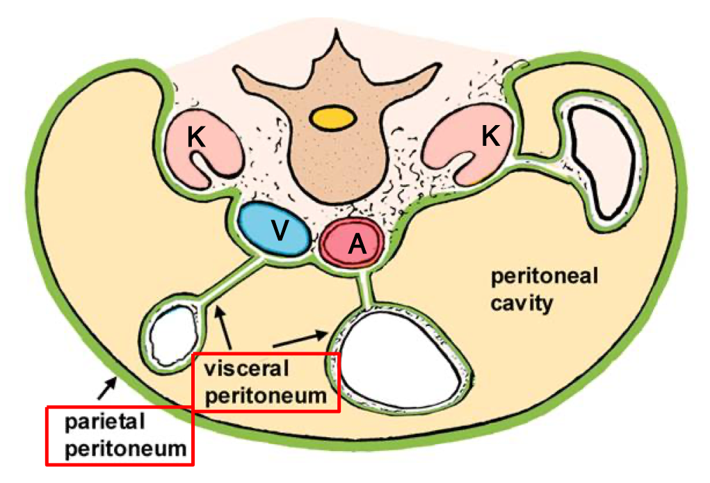
Which peritoneal layer wraps around the organs and is innervated by autonomic afferents?
Visceral peritoneum
Name the anatomical landmark used to divide the abdomen into four quadrants.
The umbilicus (intersection of the sagittal and transumbilical planes)
In the nine-region system, what two sets of planes are used to divide the abdomen?
Midclavicular lines and the subcostal/intertubercular planes
Name the opening in the diaphragm through which the esophagus passes.
Esophageal hiatus
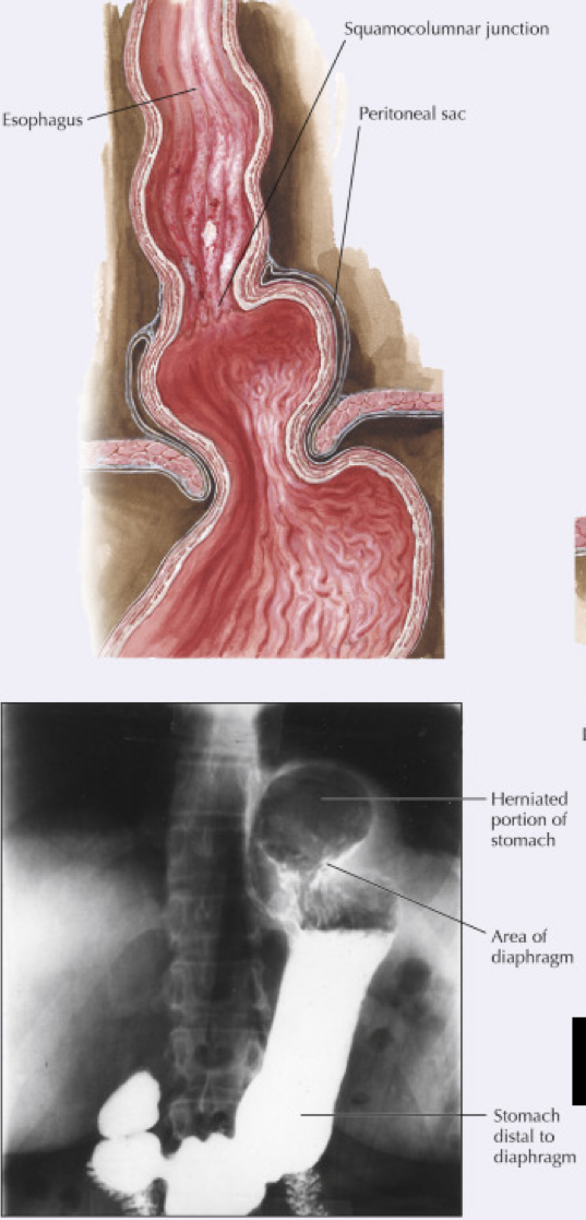
This type of hiatal hernia accounts for >95% of cases and appears like a bell on imaging.
Sliding (axial) hiatal hernia
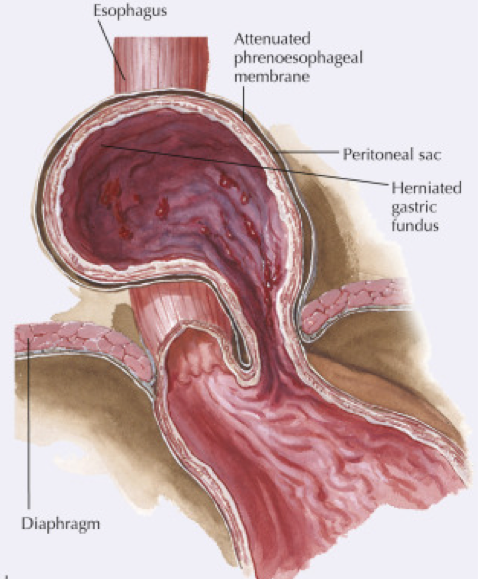
This rarer type of hiatal hernia involves the gastric fundus herniating alongside — not in line with — the esophagus.
Paraesophageal (nonaxial) hernia
Name the epithelium type lining the esophagus.
Non-keratinized stratified squamous epithelium
Name the epithelium type lining the stomach.
Simple columnar (glandular) epithelium
What is the histologic name for the sharp transition between esophageal and gastric epithelium?
Z-line (squamocolumnar junction)
Chronic GERD causes inflammation of the esophageal mucosa and epithelial metaplasia of the lower esophagus — what is this clinical finding called?
Barrett's esophagus
Name the four parts of the duodenum in order.
Superior (1st), descending (2nd), horizontal/inferior (3rd), ascending (4th)
Where do the common bile duct and main pancreatic duct join and empty into the duodenum?
the major duodenal papilla (of Vater)
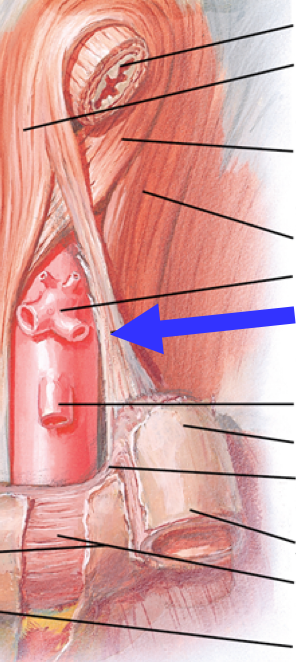
What structure suspends the duodenojejunal flexure?
suspensory muscle of duodenum (ligament of Treitz)
What is the single most common site of peptic ulcers?
First part of the duodenum (duodenal bulb/cap)
Name the two most serious complications of peptic ulcer disease.
Perforation and hemorrhage
Erosions differ from true ulcers in that they only affect this layer.
The superficial epithelium (they do not cross the muscularis mucosae)
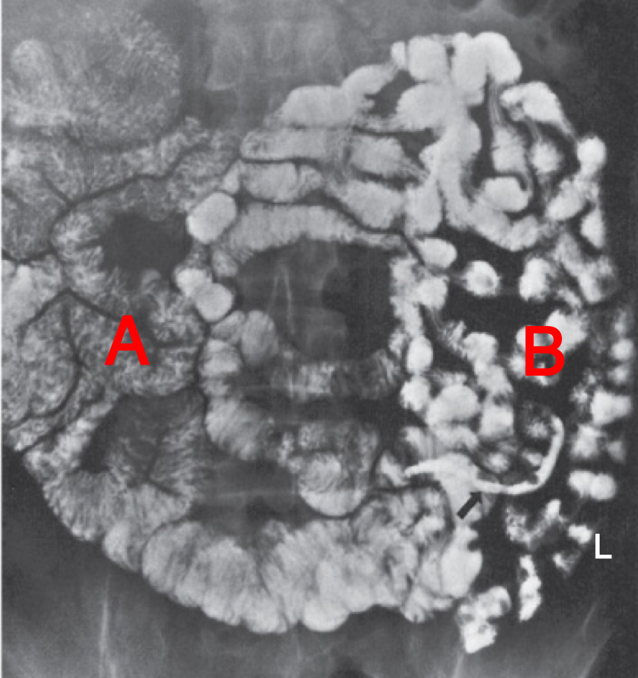
On barium imaging, segment A of small bowel shows a “feathery” pattern from extensive mucosal folds
Jejunum
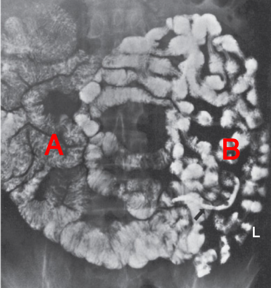
On barium imaging, segment B of small bowel appears fully filled due to fewer mucosal folds.
Ileum
Name the aggregated lymphoid nodules found in the ileum.
Peyer's patches
Name the three structures found only in the large intestine (not the small intestine) that help distinguish it grossly.
Teniae coli, haustra, and omental appendices
What are the three teniae coli bands called?
Free tenia, omental tenia, and mesocolic tenia
Name the flexure between the ascending and transverse colon.
Right colic (hepatic) flexure
Name the flexure between the transverse and descending colon.
Left colic (splenic) flexure
What is the most common positional variant of the appendix?
Retrocecal (~65%)
What anatomical junction is the most constant landmark for locating the appendix, regardless of its positional variant?
The cecum–appendix junction
Where is McBurney's point located?
1/3 of the way from the anterior superior iliac spine to the umbilicus (or 2/3 from the umbilicus)
Name the three unpaired arterial trunks supplying the abdominal GI tract, from proximal to distal.
Celiac trunk, superior mesenteric artery (SMA), inferior mesenteric artery (IMA)
Which artery supplies the embryologic midgut (distal duodenum through the proximal 2/3 of the transverse colon)?
Superior mesenteric artery (SMA)
Nearly all venous blood from the GI tract drains first into this vessel before reaching the systemic circulation.
Hepatic portal vein
Name the two veins that combine to form the portal vein.
Superior mesenteric vein and splenic vein
This part of the colon is the most common site of diverticulosis.
Sigmoid colon
Diverticulosis is a herniation of which two layers through the muscular wall?
Mucosa and submucosa
Colorectal cancer most commonly metastasizes to the liver via which route?
Portal venous tributaries
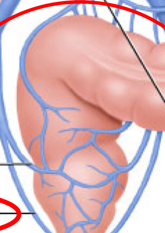
Which two rectal veins are the exception to portal drainage, bypassing the liver directly into the systemic circulation?
Inferior and middle rectal veins
Name the four lobes of the liver.
Right, left, caudate, and quadrate
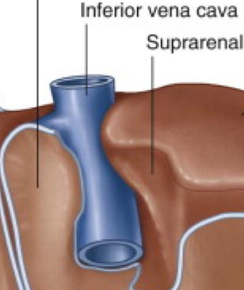
Which liver lobe sits directly against the IVC?
Caudate lobe
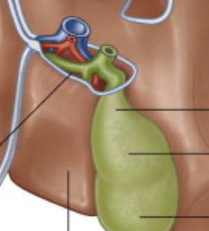
Which liver lobe sits next to the gallbladder?
Quadrate lobe
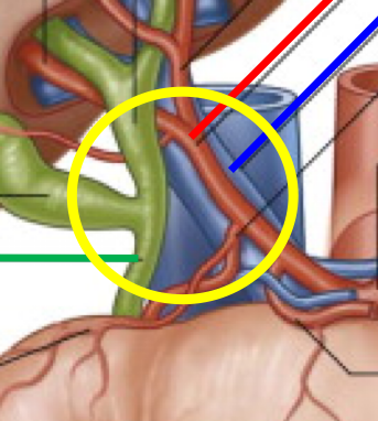
Name the three structures that make up the portal triad.
Hepatic artery proper, hepatic portal vein, and the hepatic duct
What percentage of blood supply to the liver comes from the portal vein vs. the hepatic artery?
75–80% from the portal vein, 20–25% from the hepatic artery
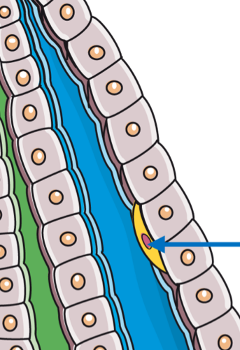
Name the resident macrophages that line the liver sinusoids.
Kupffer cells
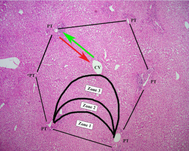
In the liver acinus, which zone is closest to the portal triad and most oxygenated?
Zone 1
In the liver acinus, which zone is closest to the central vein and most vulnerable to ischemia/toxins?
Zone 3
Absorbed dietary lipids bypass the liver and travel via which route to reach systemic circulation?
Intestinal lymphatics → thoracic duct
This early stage of liver damage involves fat accumulation in hepatocytes and is reversible.
Steatosis (fatty liver)
This late stage of liver damage involves diffuse fibrosis and regenerative nodules and is largely irreversible.
Cirrhosis
Portal vein pressure, normally around 10 mmHg, rises to what range in portal hypertension?
20–30+ mmHg
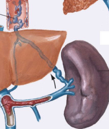
Name three major consequences of portal hypertension.
Esophageal varices, splenomegaly, and portal vein thrombosis
Classify this cause of splenomegaly: portal vein thrombosis.
Pre-hepatic cause
Classify this cause of splenomegaly: cirrhosis or schistosomiasis
Intra-hepatic cause
Classify this cause of splenomegaly: Budd-Chiari syndrome (thrombosis of hepatic vv.) or constrictive pericarditis.
Post-hepatic cause
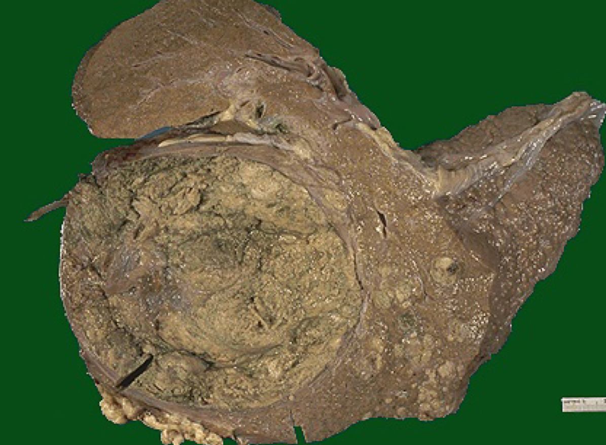
Name the primary liver cancer arising from hepatocytes, often in the setting of cirrhosis.
Hepatocellular carcinoma
Metastatic liver tumors classically appear on gross exam with this feature
Multiple, well-circumscribed nodules of varying sizes
The majority of gallstones are composed of what substance?
bile & cholesterol
Name the 2 narrowest points of the biliary tract where gallstones most commonly become lodged.
(1) neck of the gallbladder & (2) ampulla of Vater
Bile duct obstruction leading to bilirubin accumulation in the skin and sclera causes what clinical sign?
Jaundice
Name the two functional roles of the pancreas.
Exocrine (digestive enzymes) and endocrine (insulin)
What percentage of pancreatic cancers occur in the head of the pancreas?
About 60%
Why does a pancreatic head tumor commonly cause jaundice?
It obstructs the common bile duct as it passes through the pancreatic head
Although not a digestive organ, the spleen is vascularly connected to the GI tract via which vessels?
Splenic artery & vein