Cat Pelvic Limb ID - Dissection video
1/94
There's no tags or description
Looks like no tags are added yet.
Name | Mastery | Learn | Test | Matching | Spaced |
|---|
No study sessions yet.
95 Terms

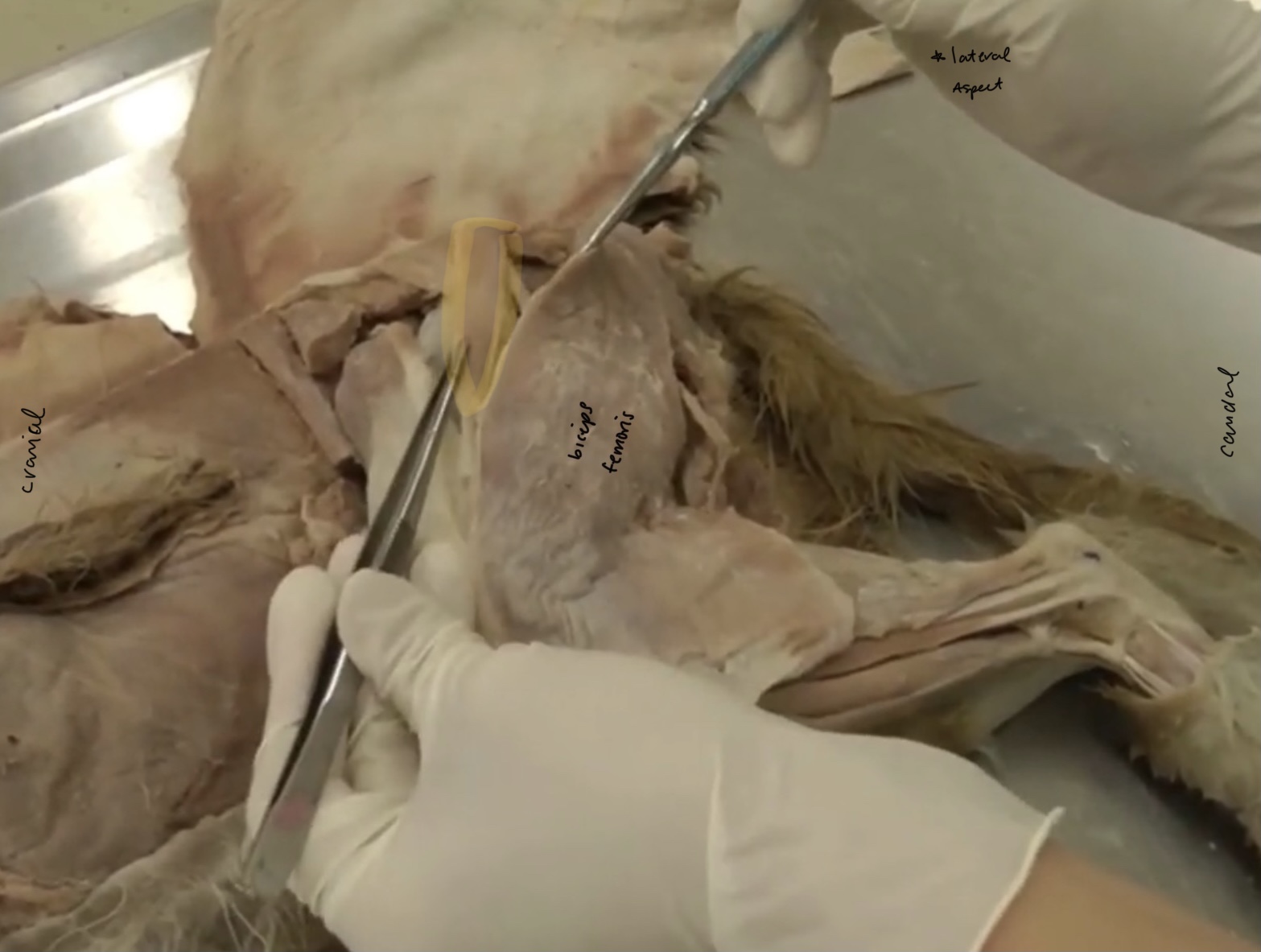
What is the yellow highlighted muscle?
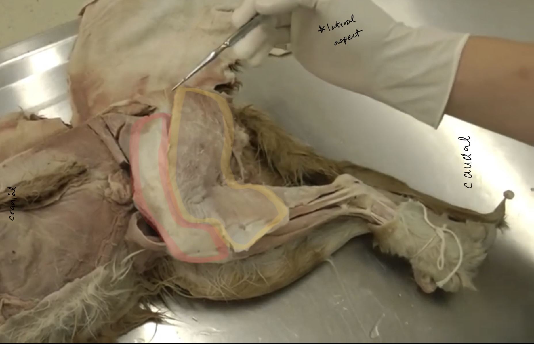
biceps femoris m.
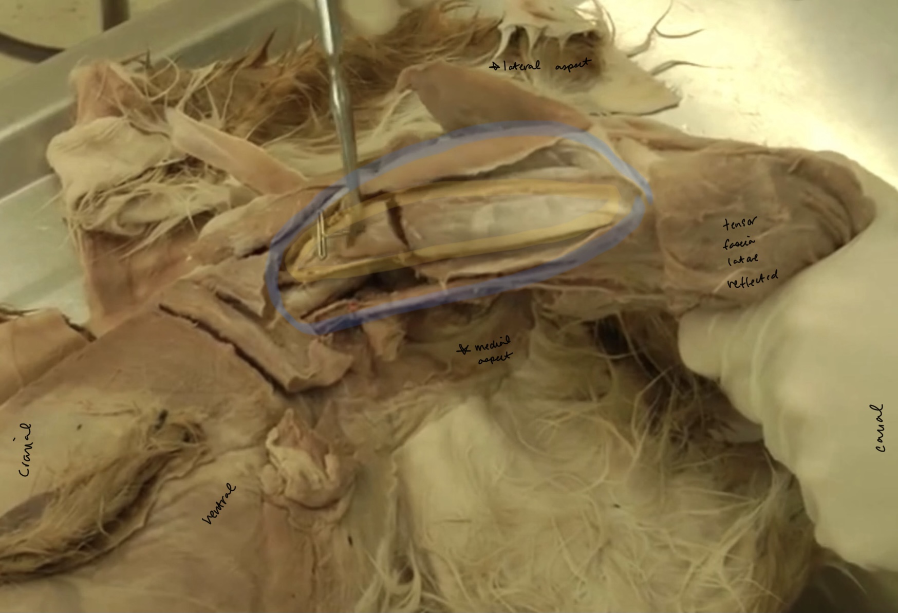
What is the red highlighted part?
tensor fasciae latae m.
What is the yellow highlighted structure?
semitendinosus m.
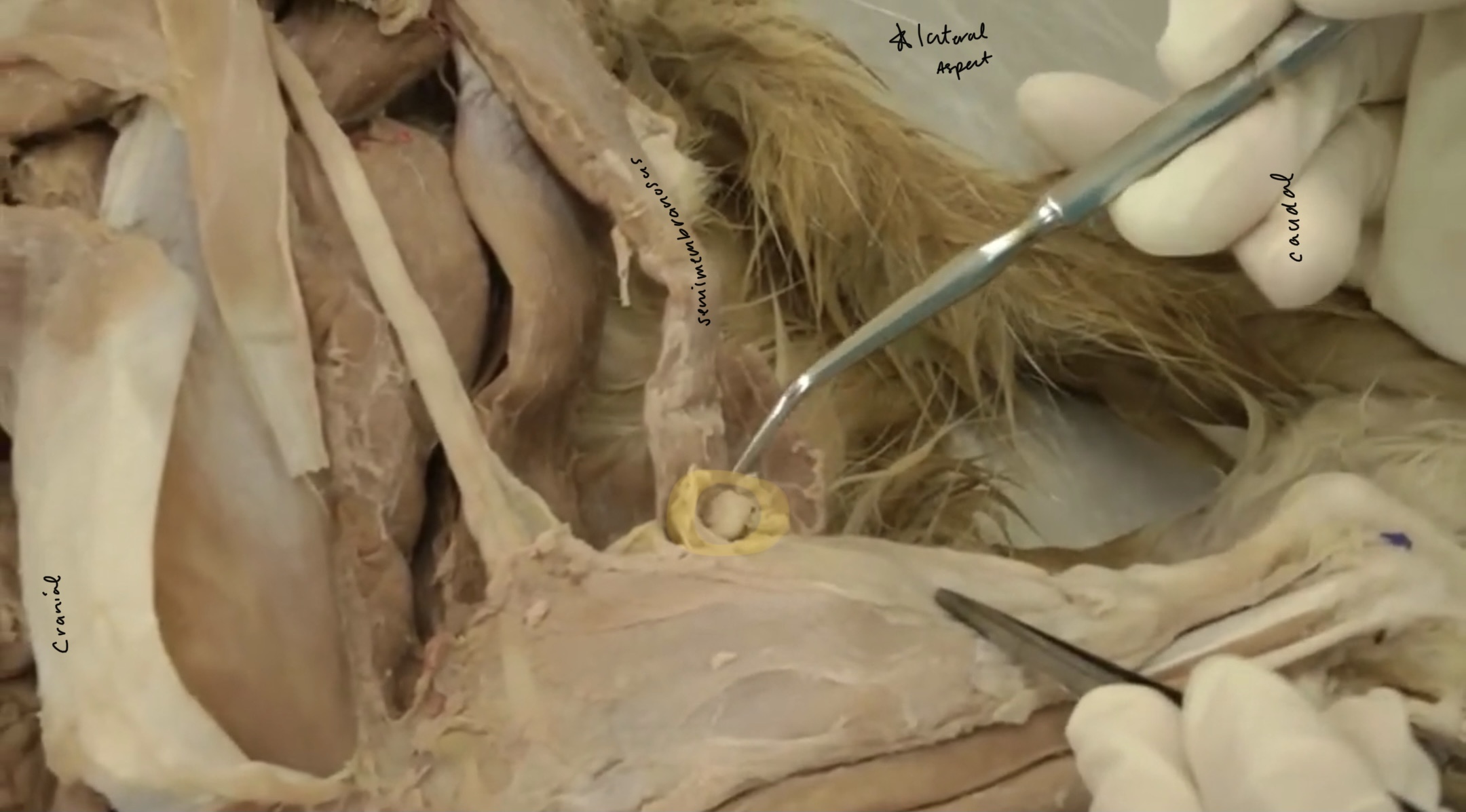
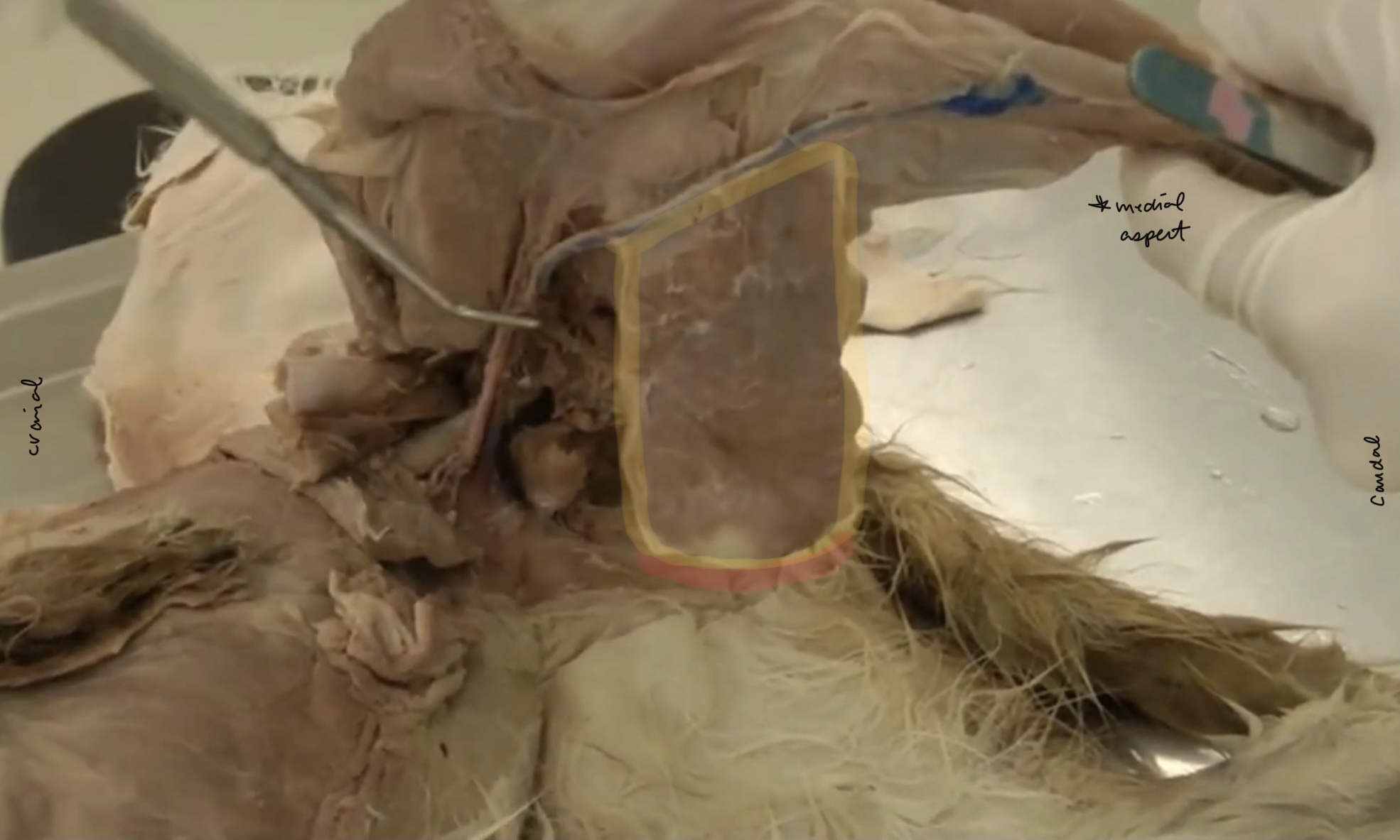
What is the yellow highlighted part?
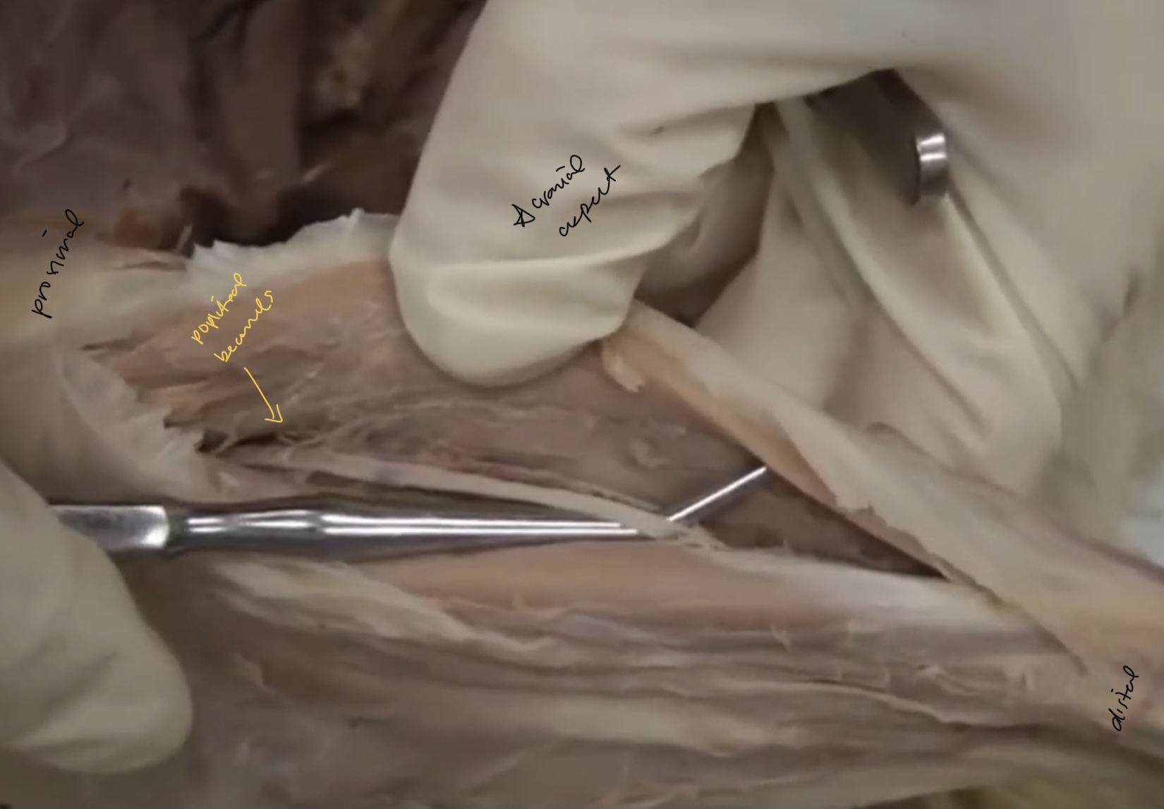
popliteal lymph node
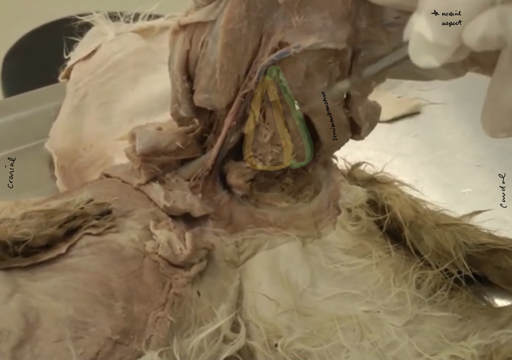
What is the yellow highlighted muscle?
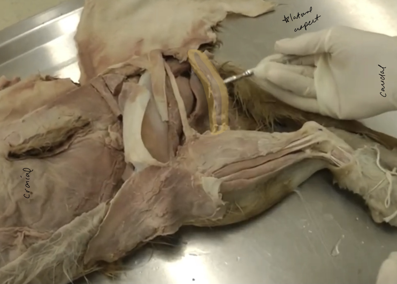
semimembranosus m.
What is the yellow highlighted muscle?
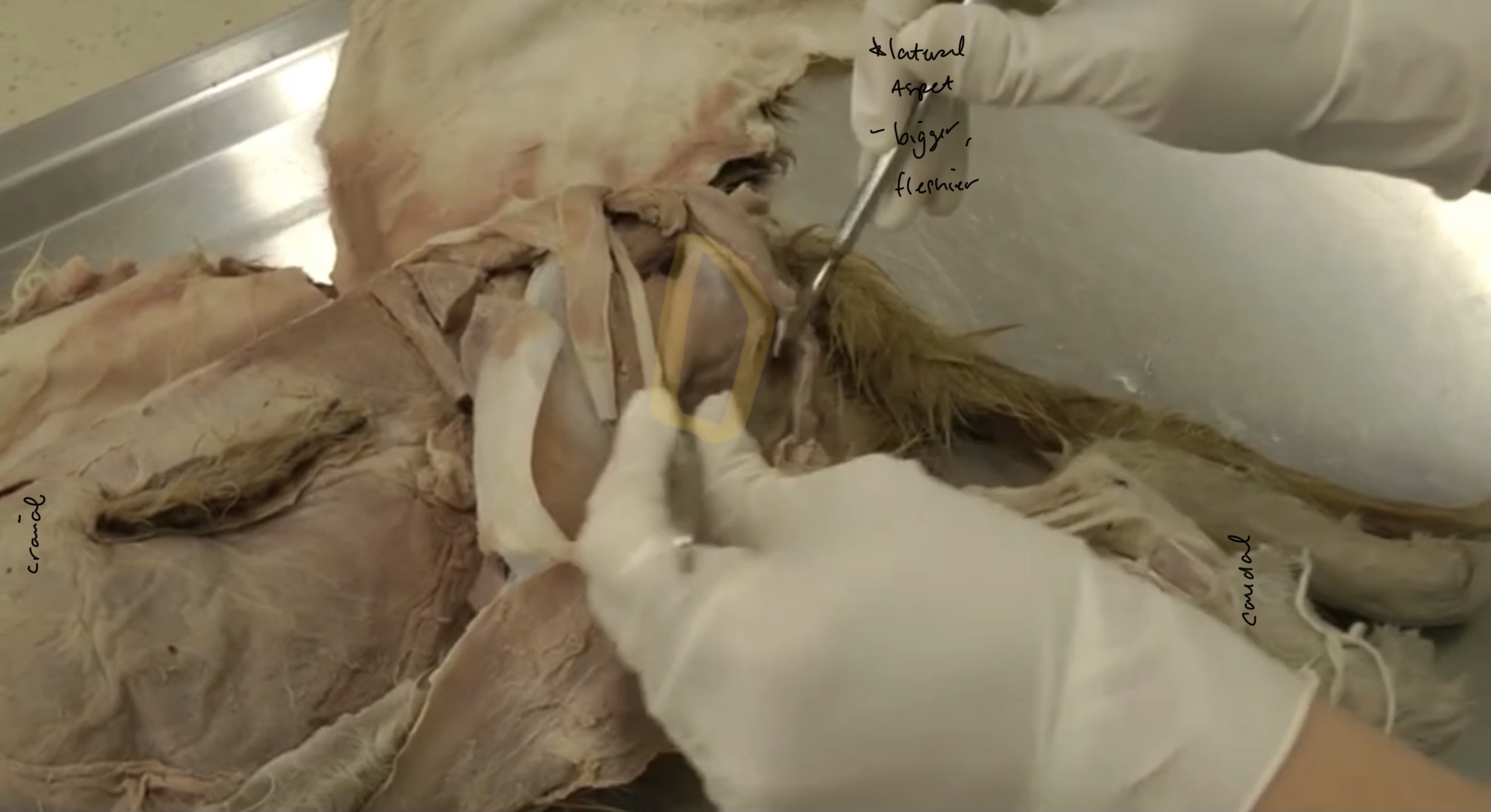
gluteofemoralis/caudofemoralis m.
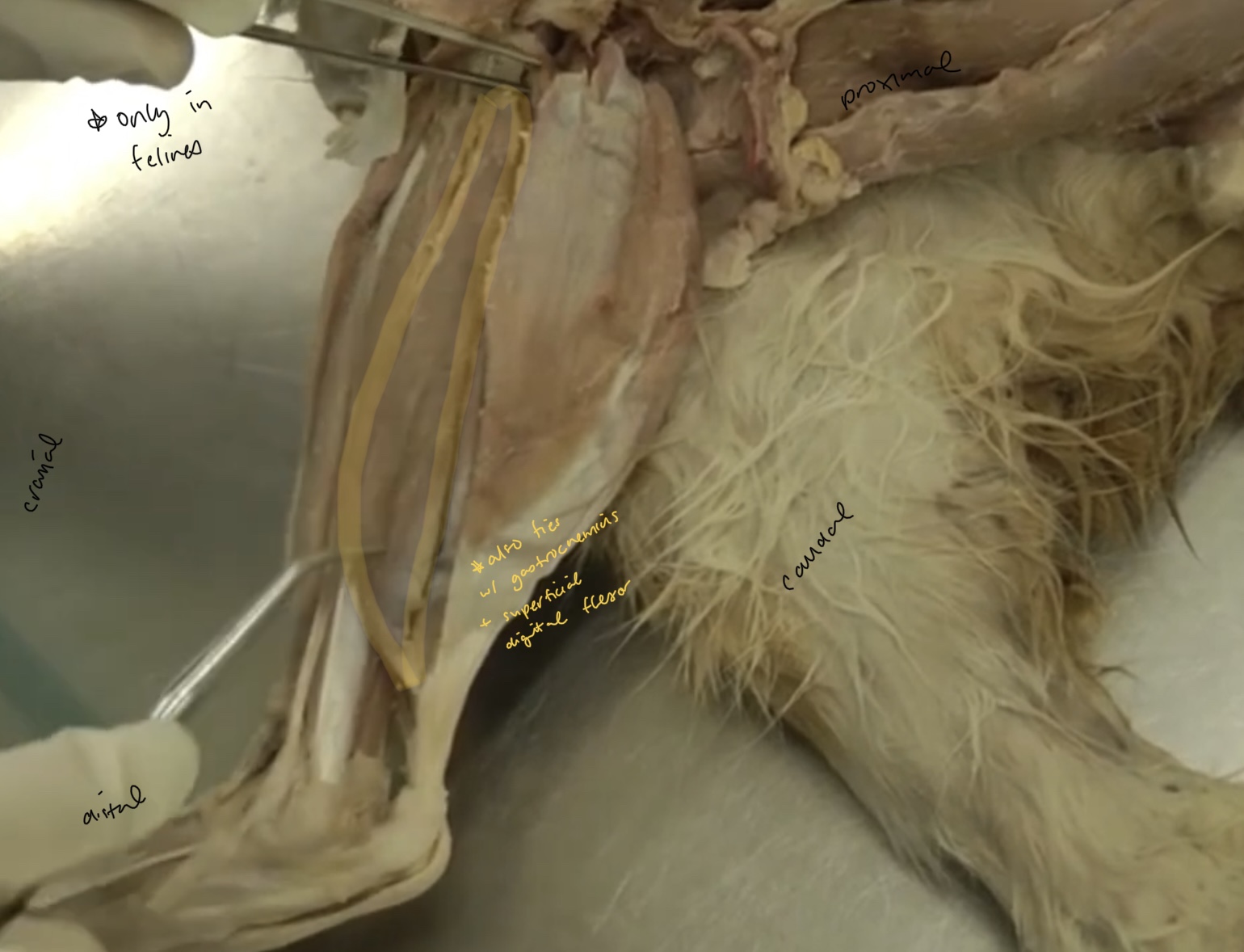
Which muscles can only be found in the cat?
gluteofemoralis/caudofemoralis m.
soleus m.
Which muscle is highlighted?
sartorius m.
Which muscle is highlighted?
pectineus m.
does the cat have 1 or 2 bellies of the sartorius delineated?
1
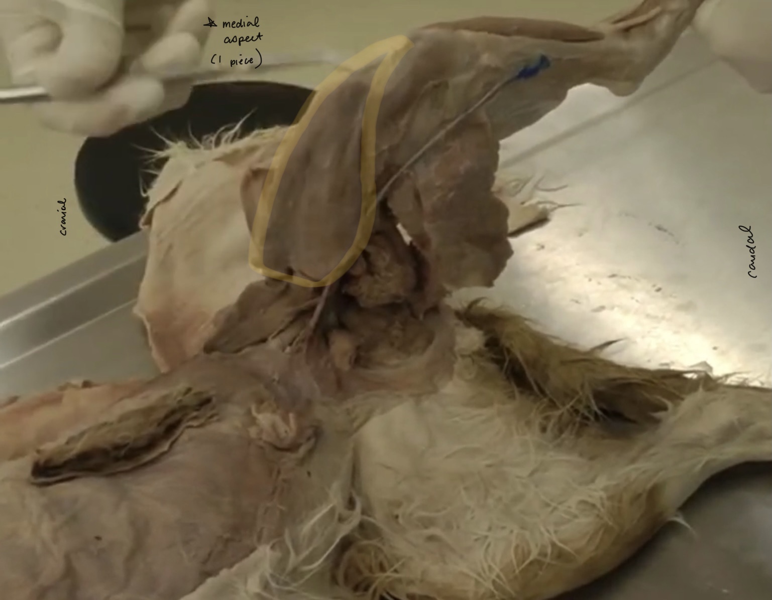
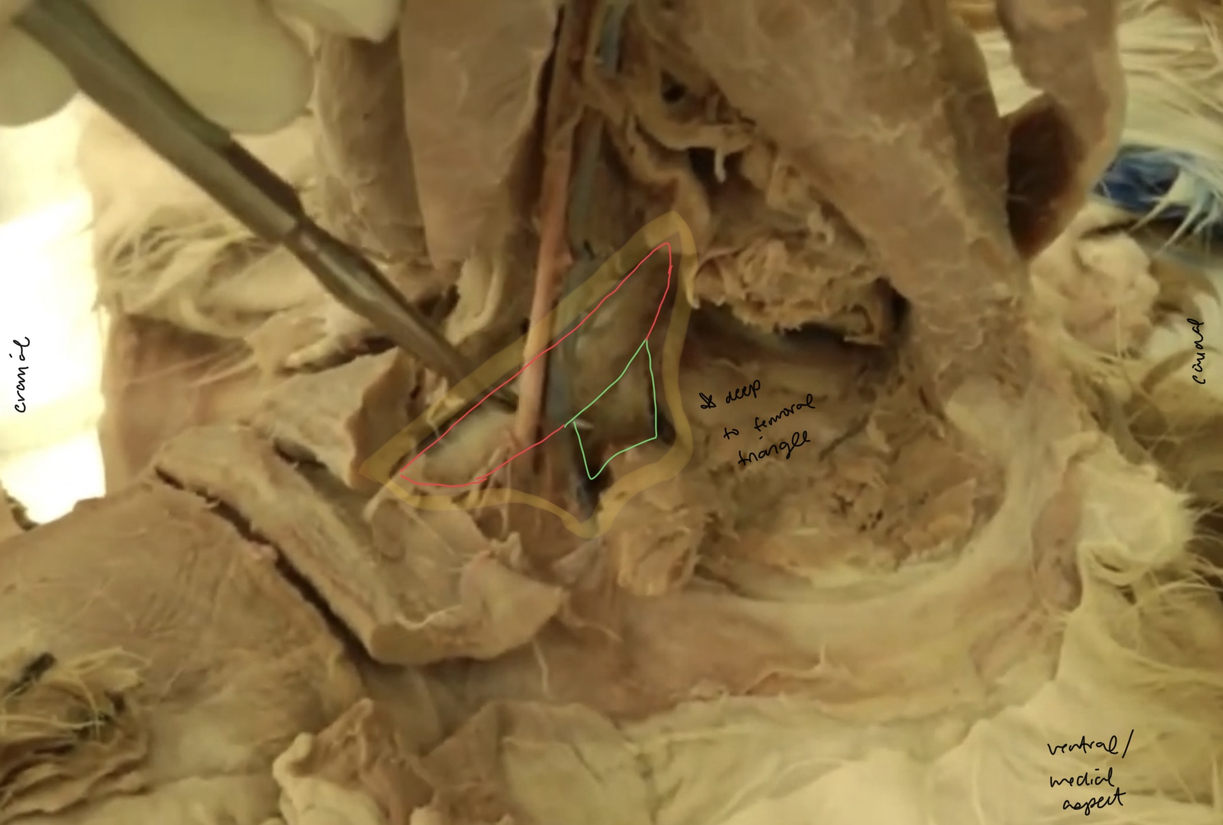
What is the highlighted area called?
femoral triangle
What is the yellow highlighted portion?
gracilis m.

What is the red highlighted portion? (not on terms list but good for landmarking)
symphyseal tendon
What do the green and yellow parts make up?
adductor m.

what is the yellow portion? (don’t necessarily need to know but good for landmarking)
adductor longus

what is the green portion? (don’t necessarily need to know but good for landmarking)
adductor magnus et brevis
What is the yellow highlighted portion
external obturator m.

what is the red highlighted part?
gluteofemoralis/caudofemoralis m.
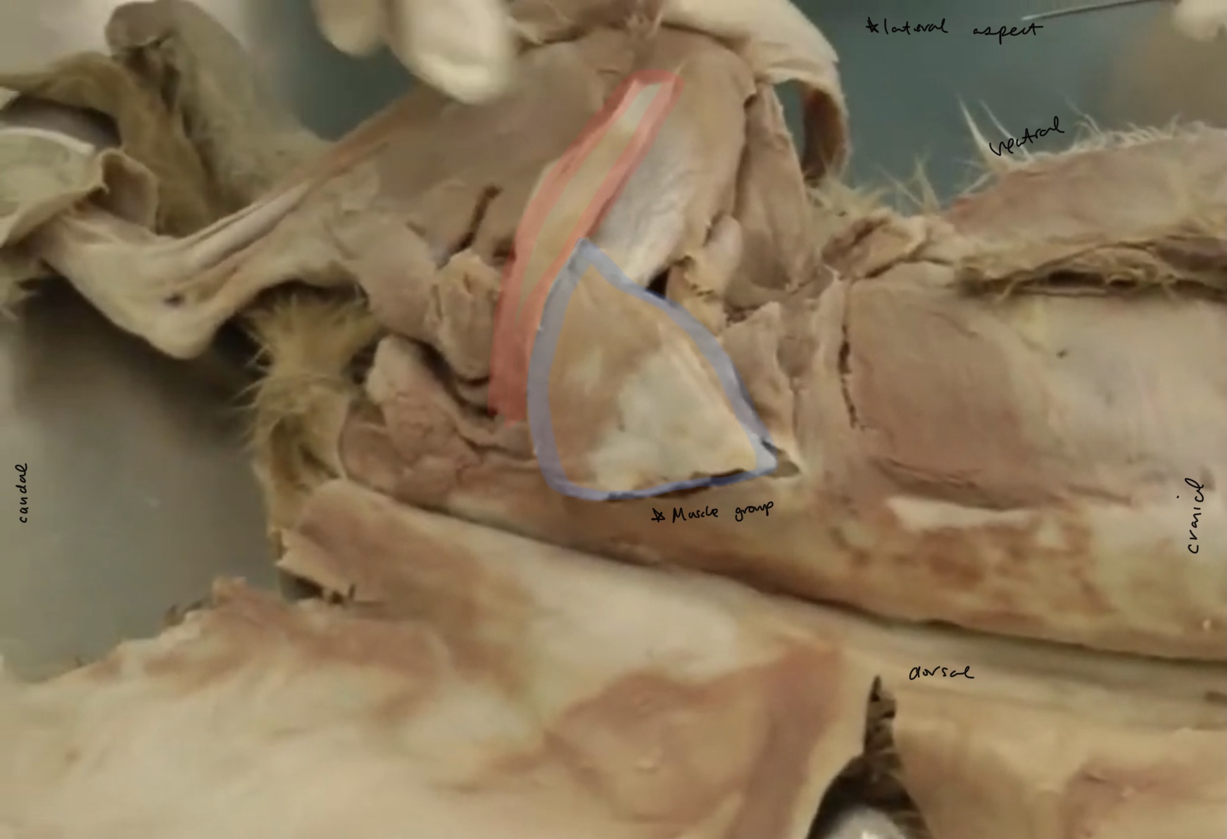
what MUSCLE GROUP is blue?
gluteal mm.

what is the yellow highlighted portion?
superficial gluteal m.
what is the green?
aponeurosis
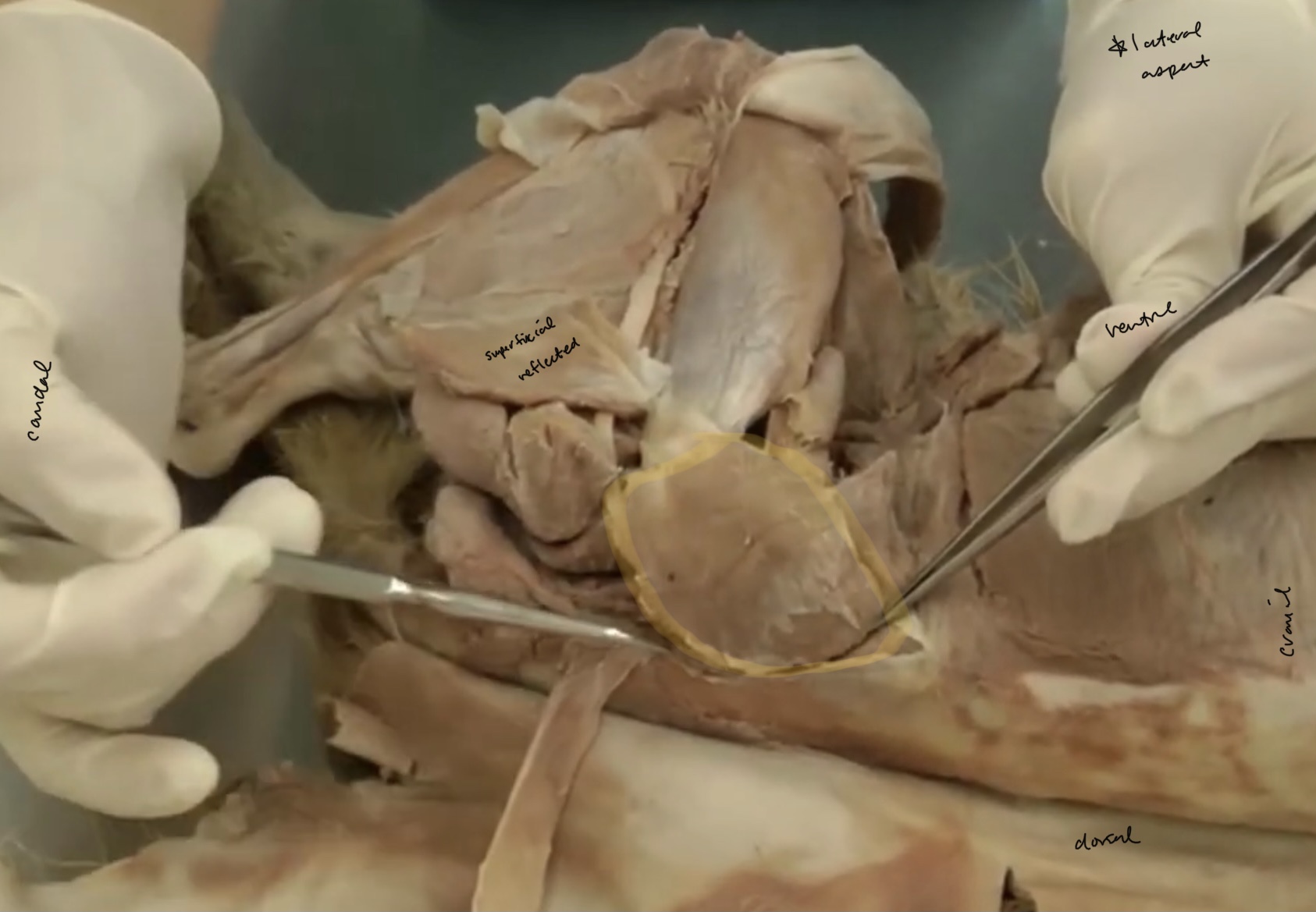
what is the highlighted portion?
middle gluteal m.
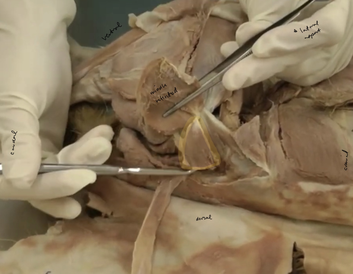
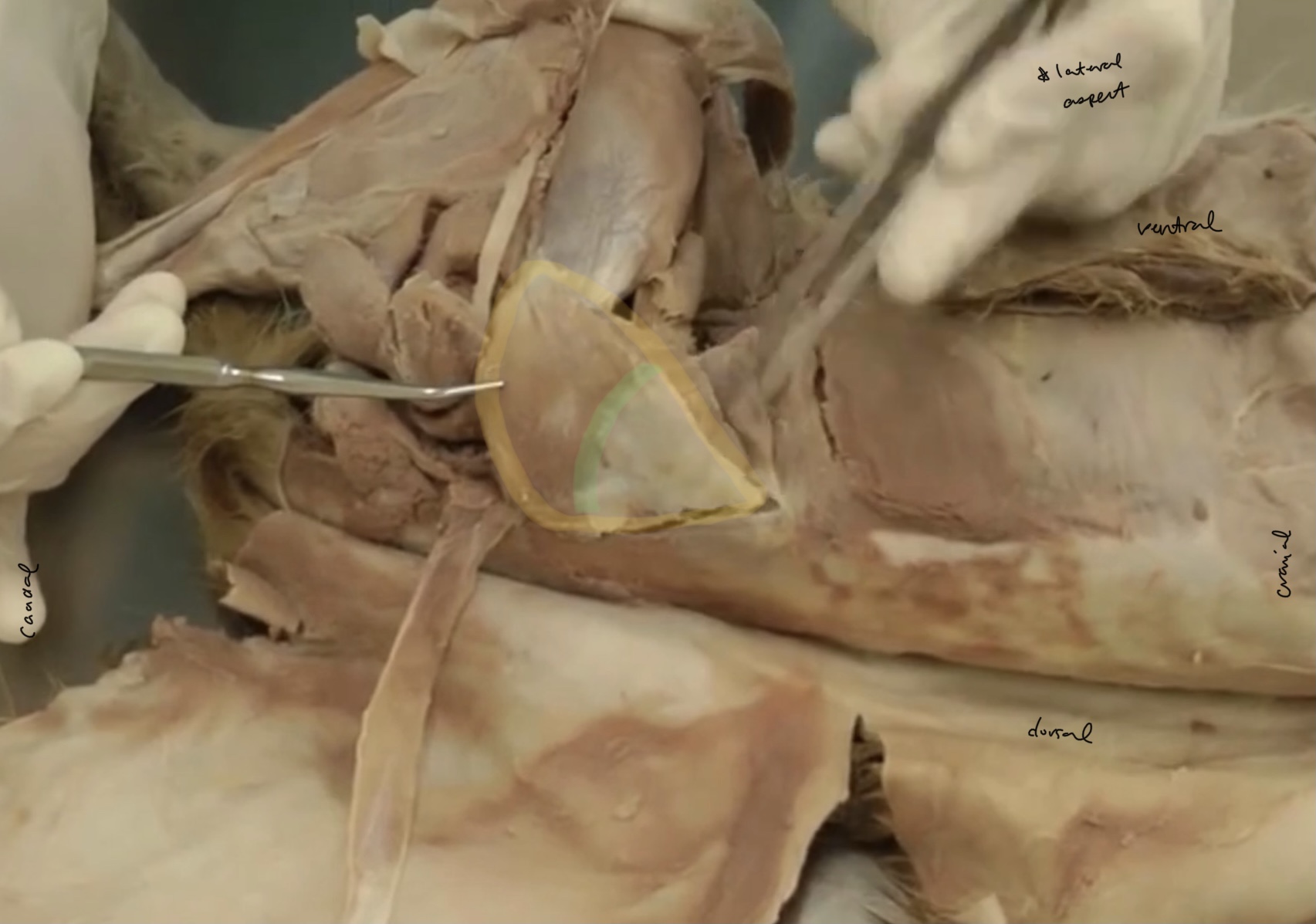
what is the highlighted portion?
piriformes m.
The piriformes m. is attached to?
middle gluteal m.
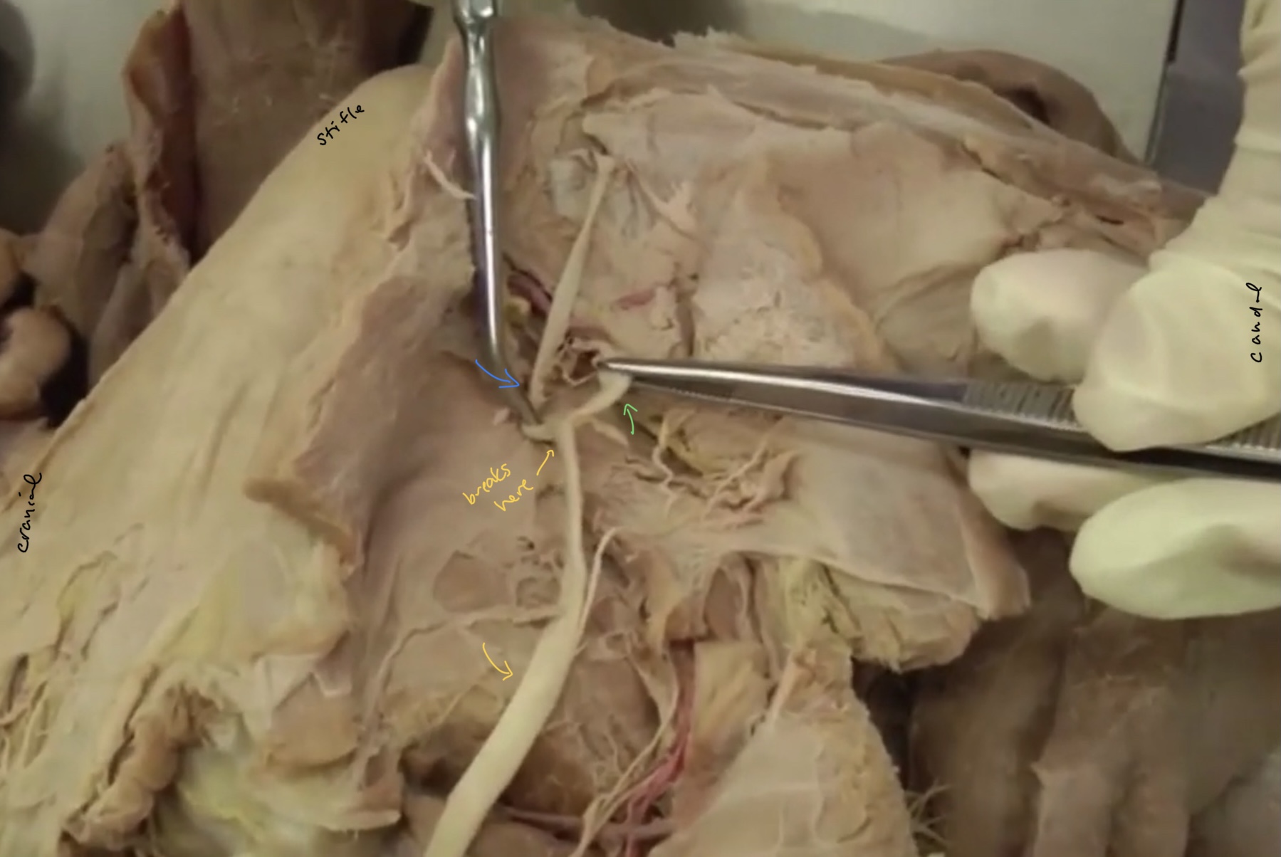
which ligament do dogs have that is not found in the cat?
sacrotuberous ligament
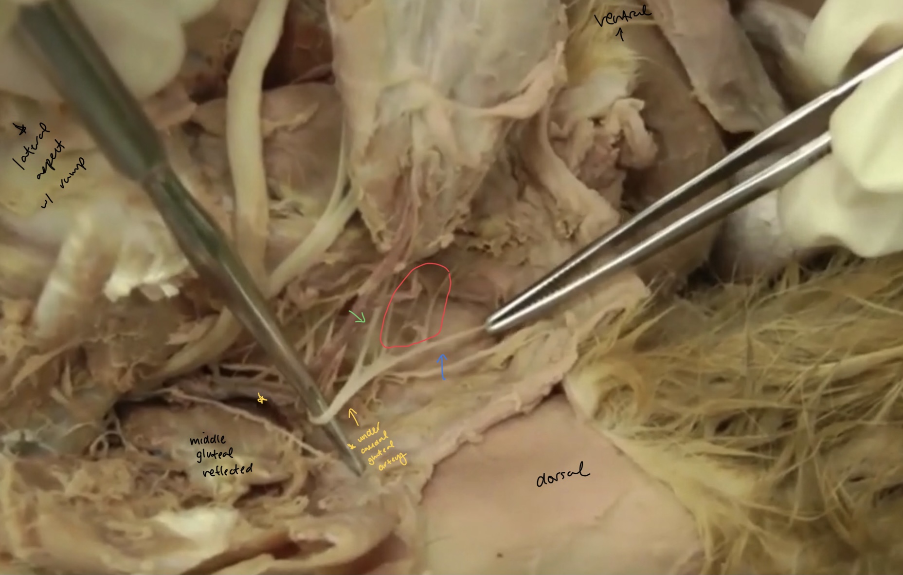
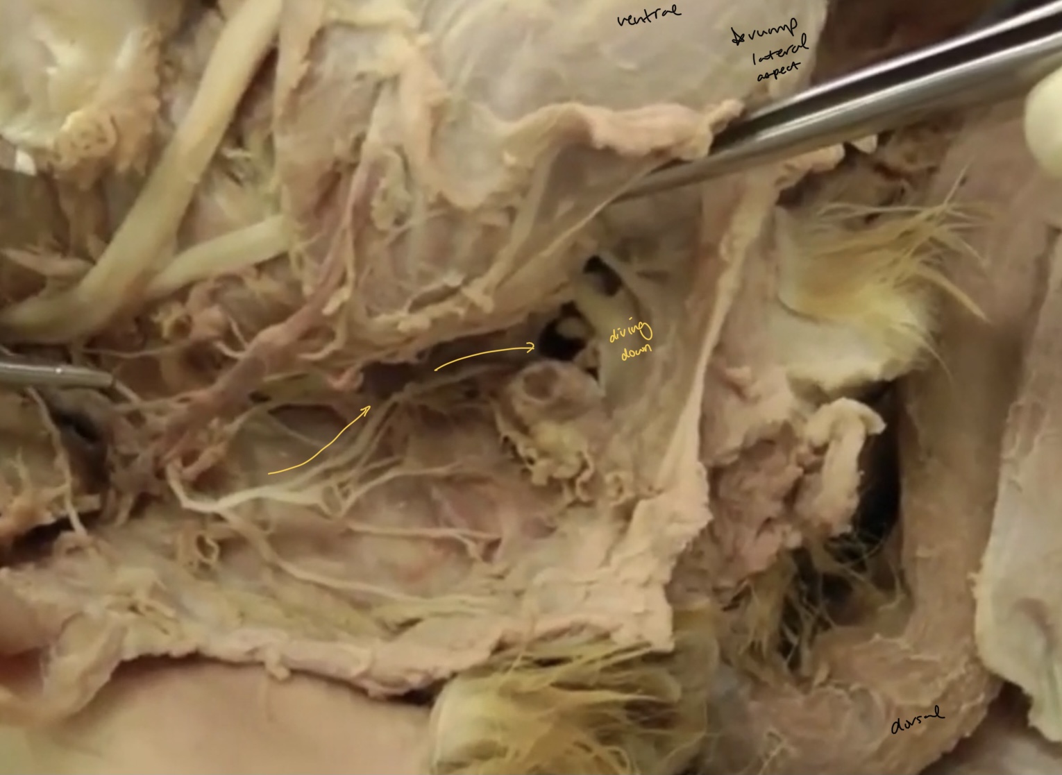
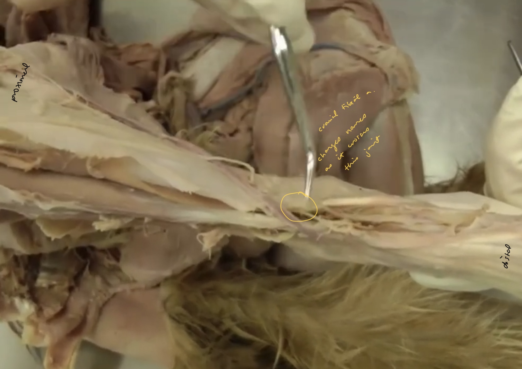
What is the name for the area of the lateral rump where the nerves are found?
ischiorectal fossa
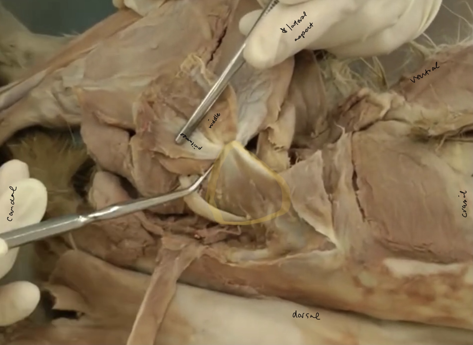
What is the highlighted portion called?
deep gluteal m.

what is the yellow portion called?
iliopsoas m.

What is the red portion called? (don’t think we need to know this but good for landmarking)
iliopsoas major
what is the green portion called? (don’t think we need to know but good for LM)
iliacus

What is this muscle in blue?
quadriceps femoris m.
What is the yellow portion?
rectus femoris m.

What is the red portion?
vastus intermedius m.
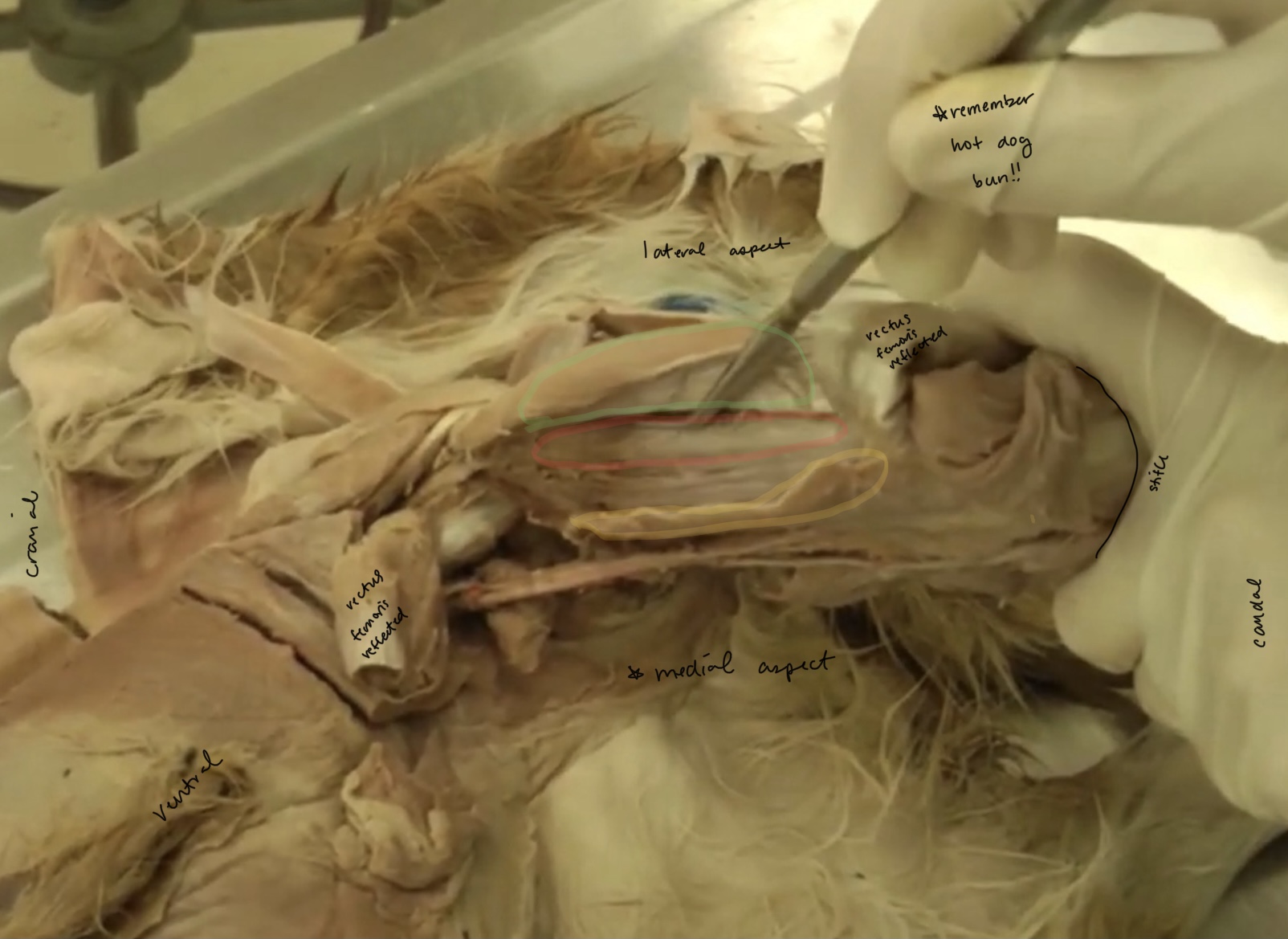
what is the yellow portion?
vastus medialis m.

what is the green portion?
vastus lateralis m.

what makes up the cranial border of the femoral triangle?
sartorius m.

what makes up the caudal border of the femoral triangle?
pectineus m.

what makes up the proximal/dorsal border of the femoral triangle?
abdominal wall, iliopsoas m.
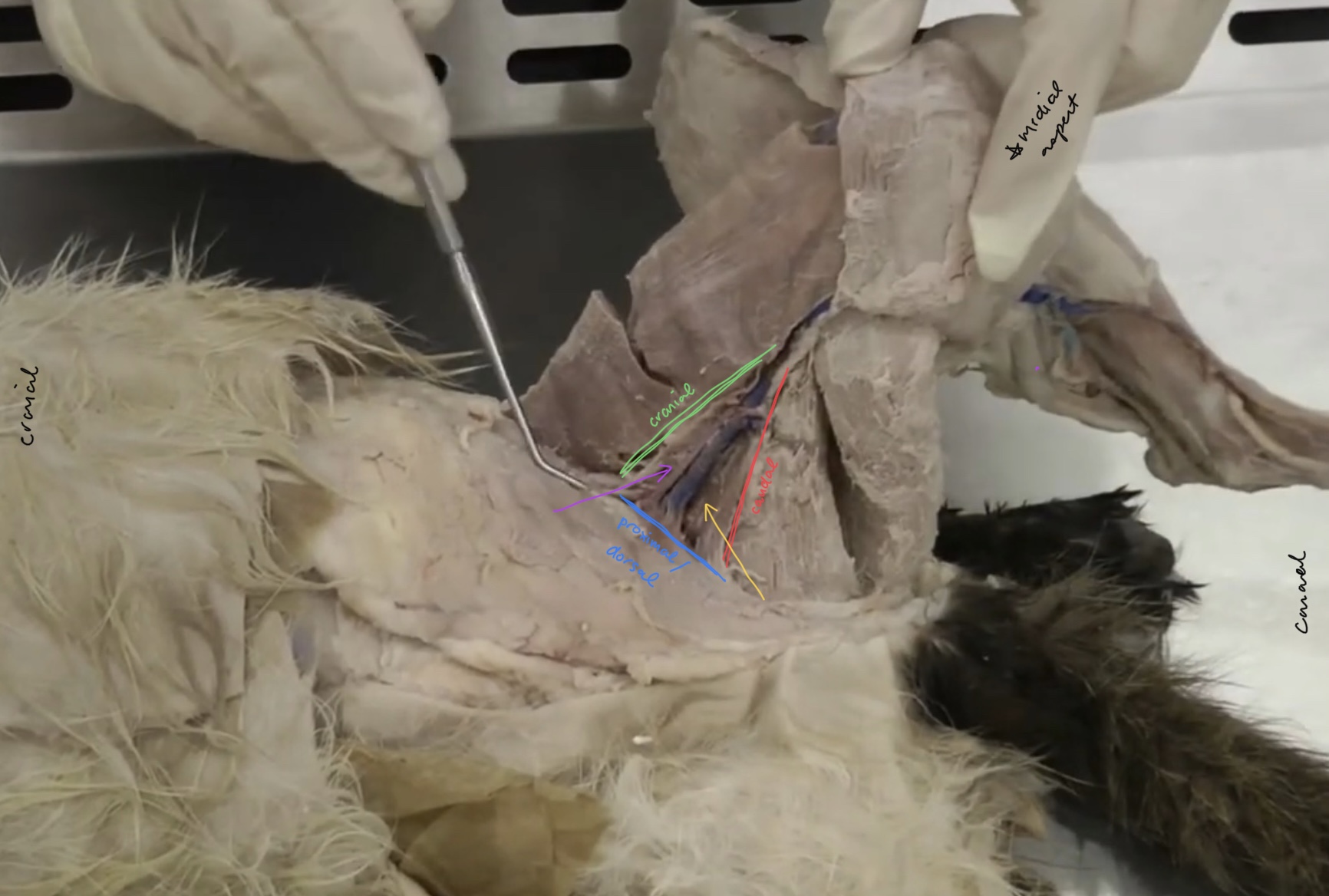
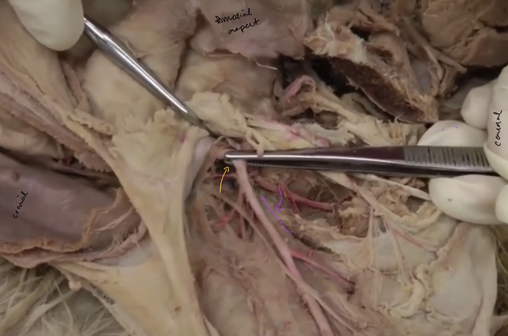
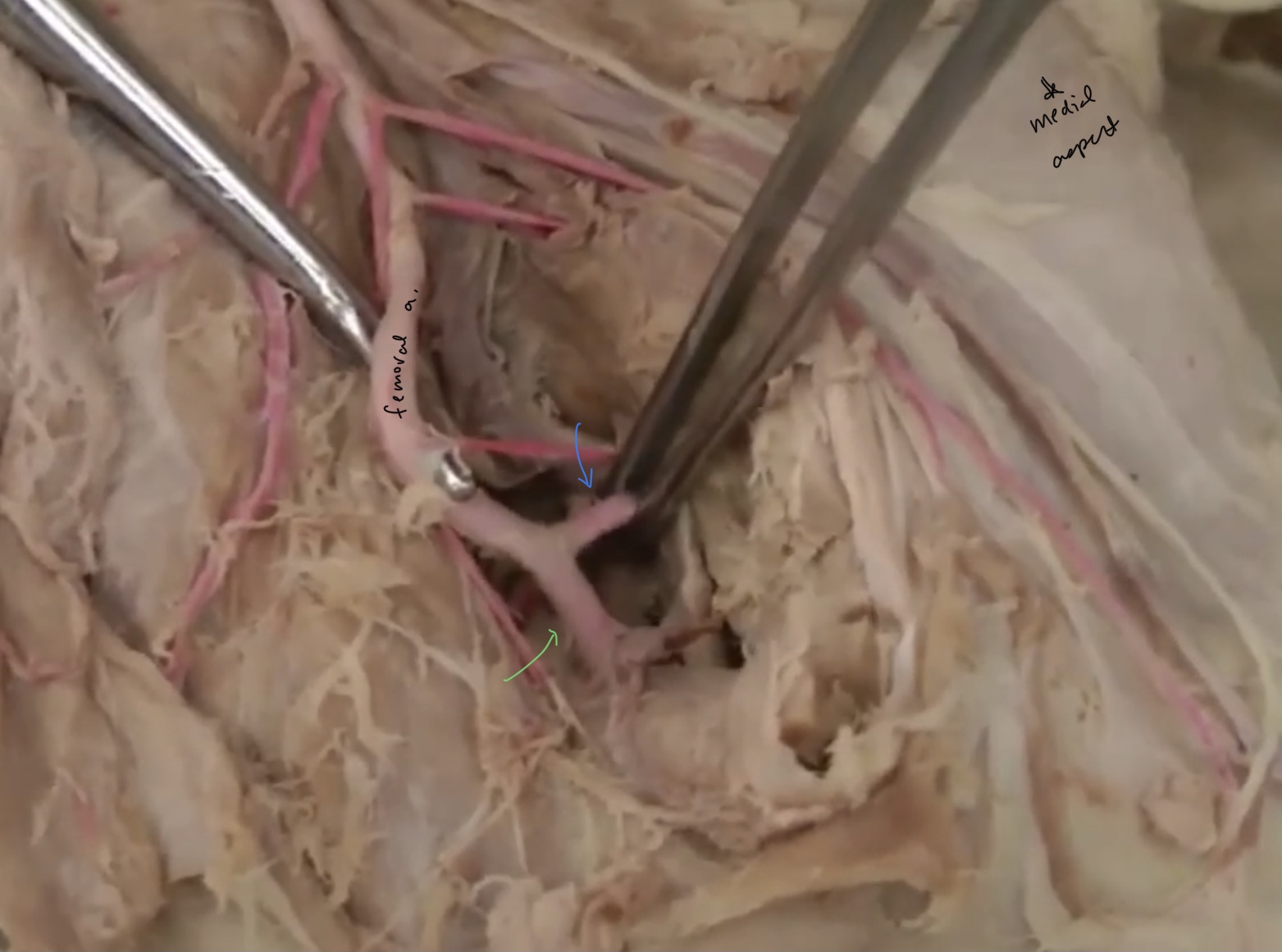
which vessel is the yellow pointing at?
femoral v.

which vessel is the purple pointing at?
femoral a.

which vessel is yellow pointing at?
femoral a.
which vessel is purple pointing at? (color washed out)
femoral v.

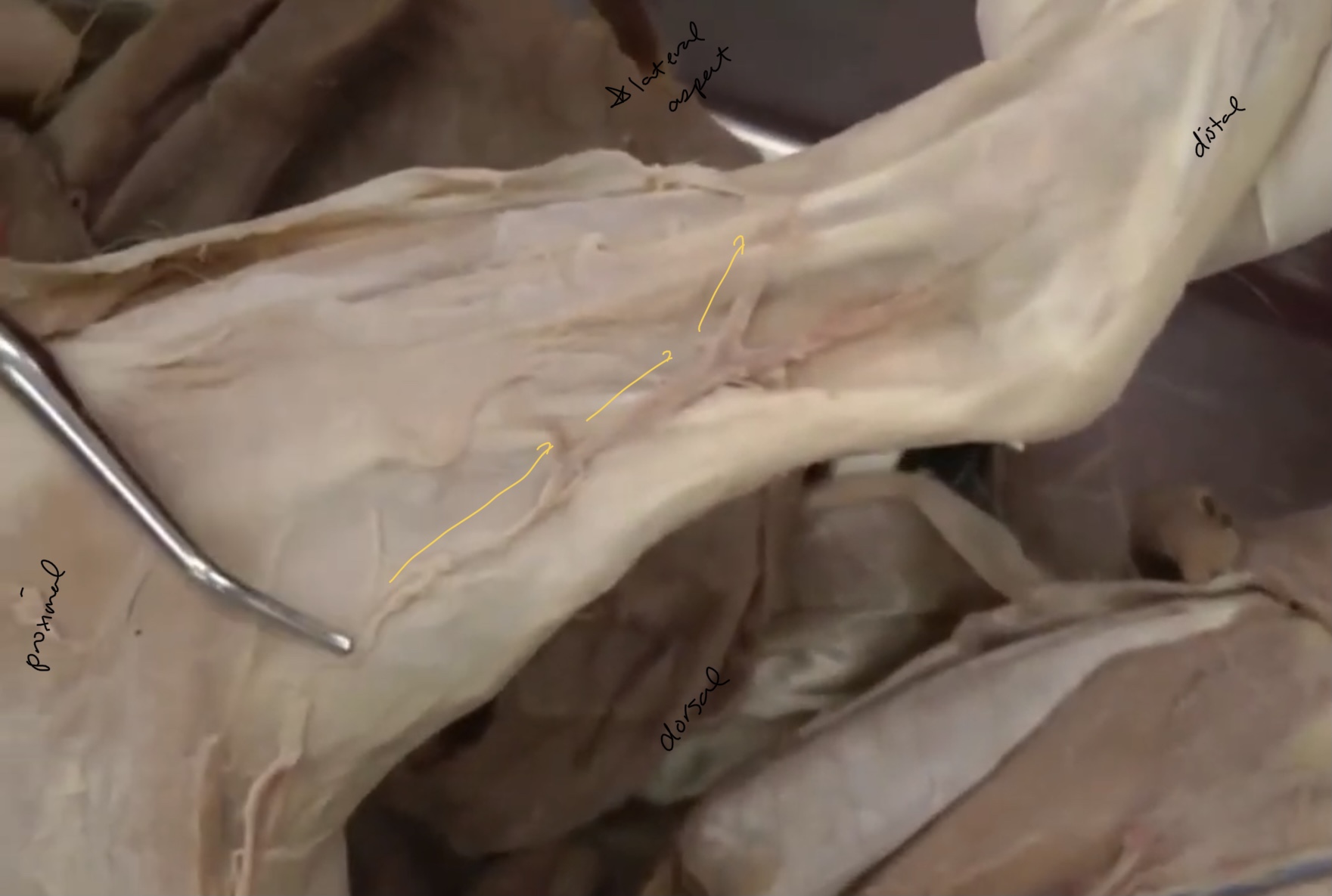
What is the blue pointing at?
saphenous n.
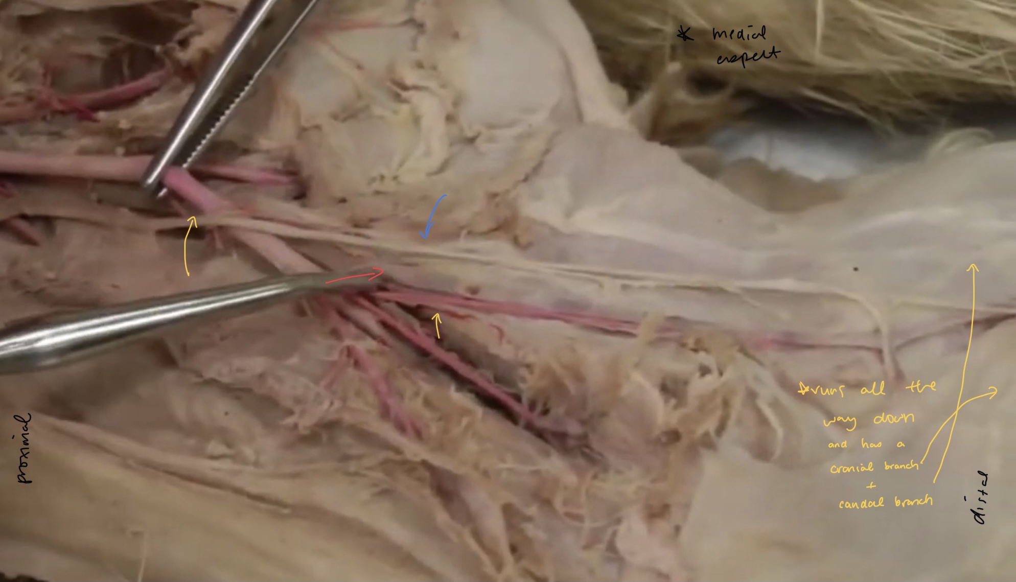
what is the yellow pointing at?
saphenous a.

what is red pointing at?
medial saphenous v.

what is the yellow portion?
femoral n.
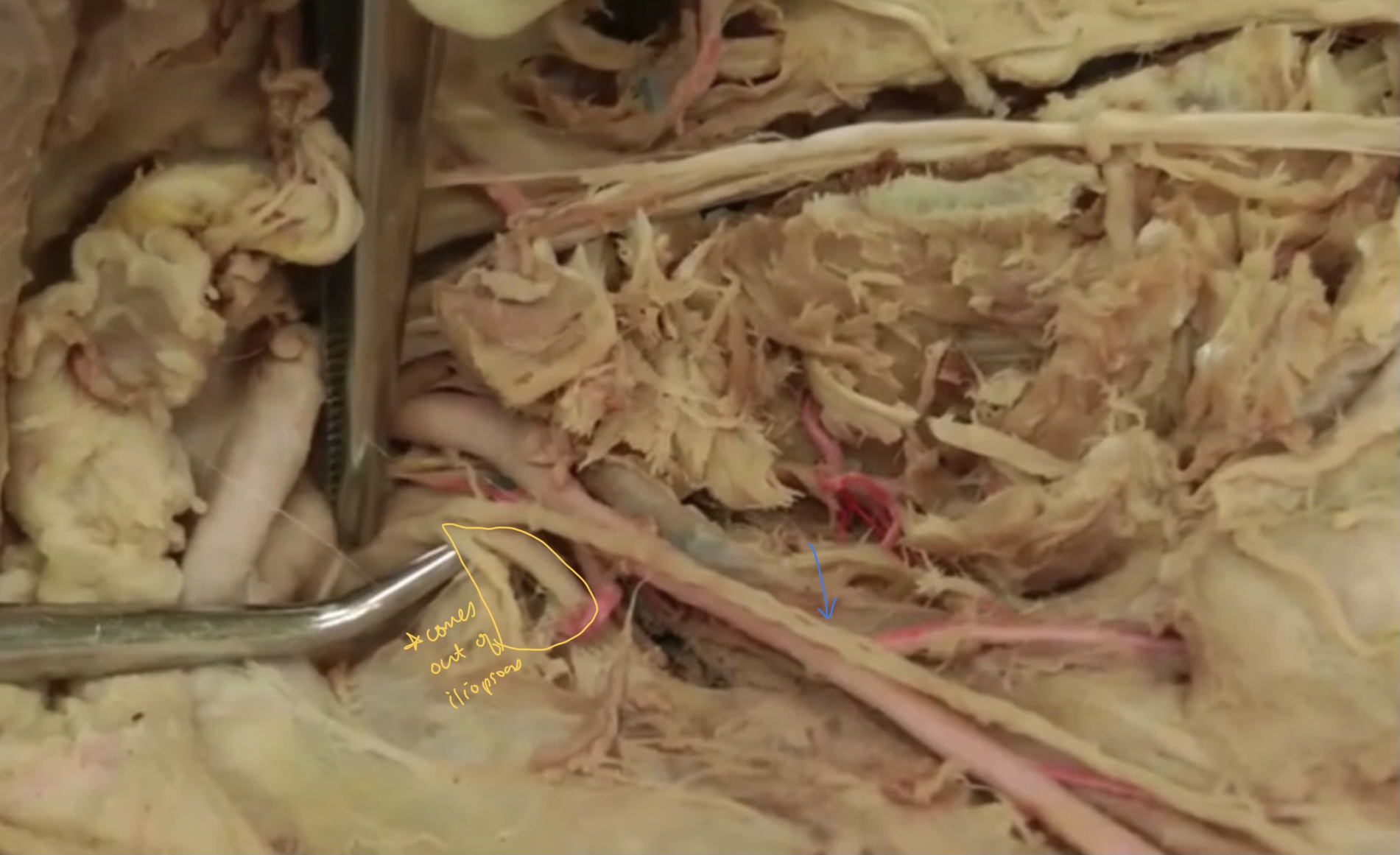
what is the blue pointing at?
saphenous n.

what is the yellow nerve? (I don’t think we need to know this?)
pudendal n.
what is the blue nerve? (I don’t think we need to know this?)
caudal rectal n.

what is the green nerve? (I don’t think we need to know this, but connects to penis)
dorsal n.

what is the red nerve? (I don’t think we need to know this?)
perineal nn.
What is this nerve?
dorsal n.

What is the yellow n.
sciatic n.

what is the blue n.
common fibular (peroneal) n.
what is the green n.
tibial n.
what are the yellow highlighted portions?
extensor retinacula

which retinaculum is the blue
crural retinaculum

which retinaculum is the red?
tarsal extensor retinaculum
Which muscle is highlighted?
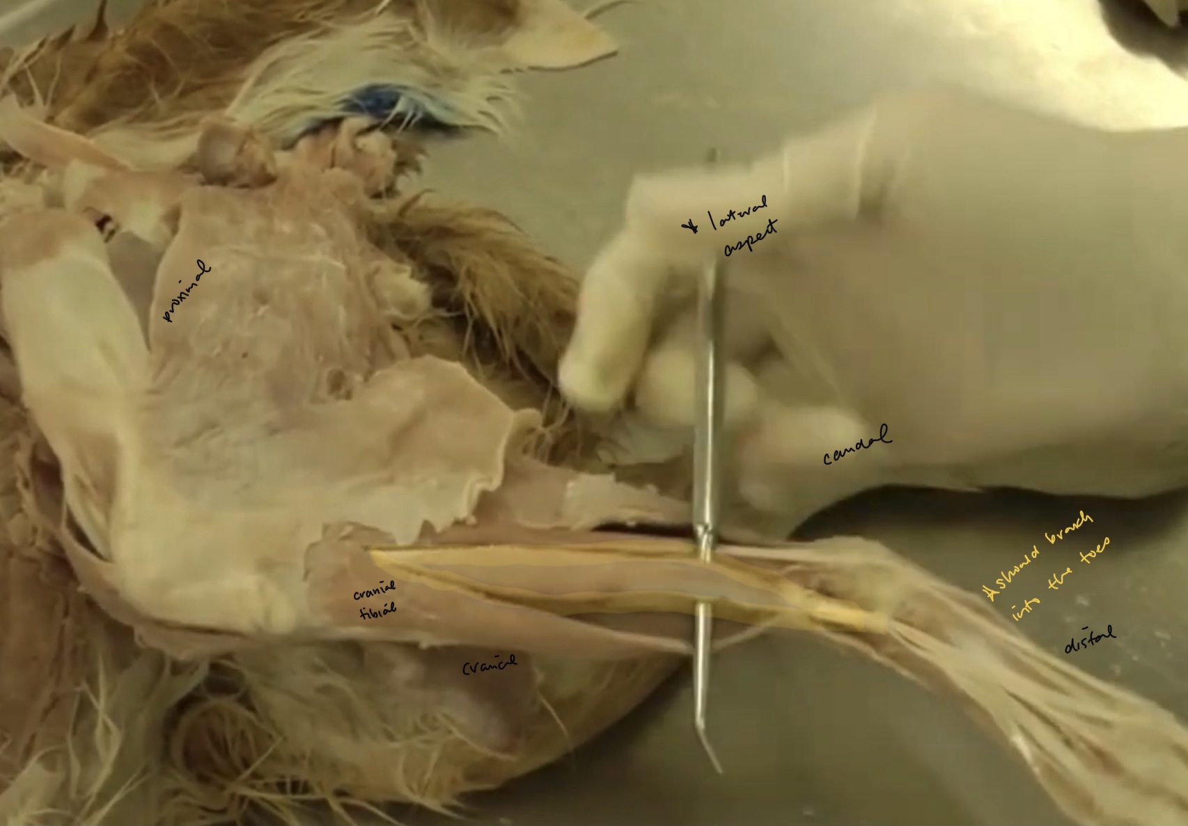
cranial tibial m.
The cranial tibial m. is a flexor or extensor of the tarsus?
flexor
What is the only extrinsic muscle of the pelvic limb?
iliopsoas m.
what is highlighted?
long digital extensor m.
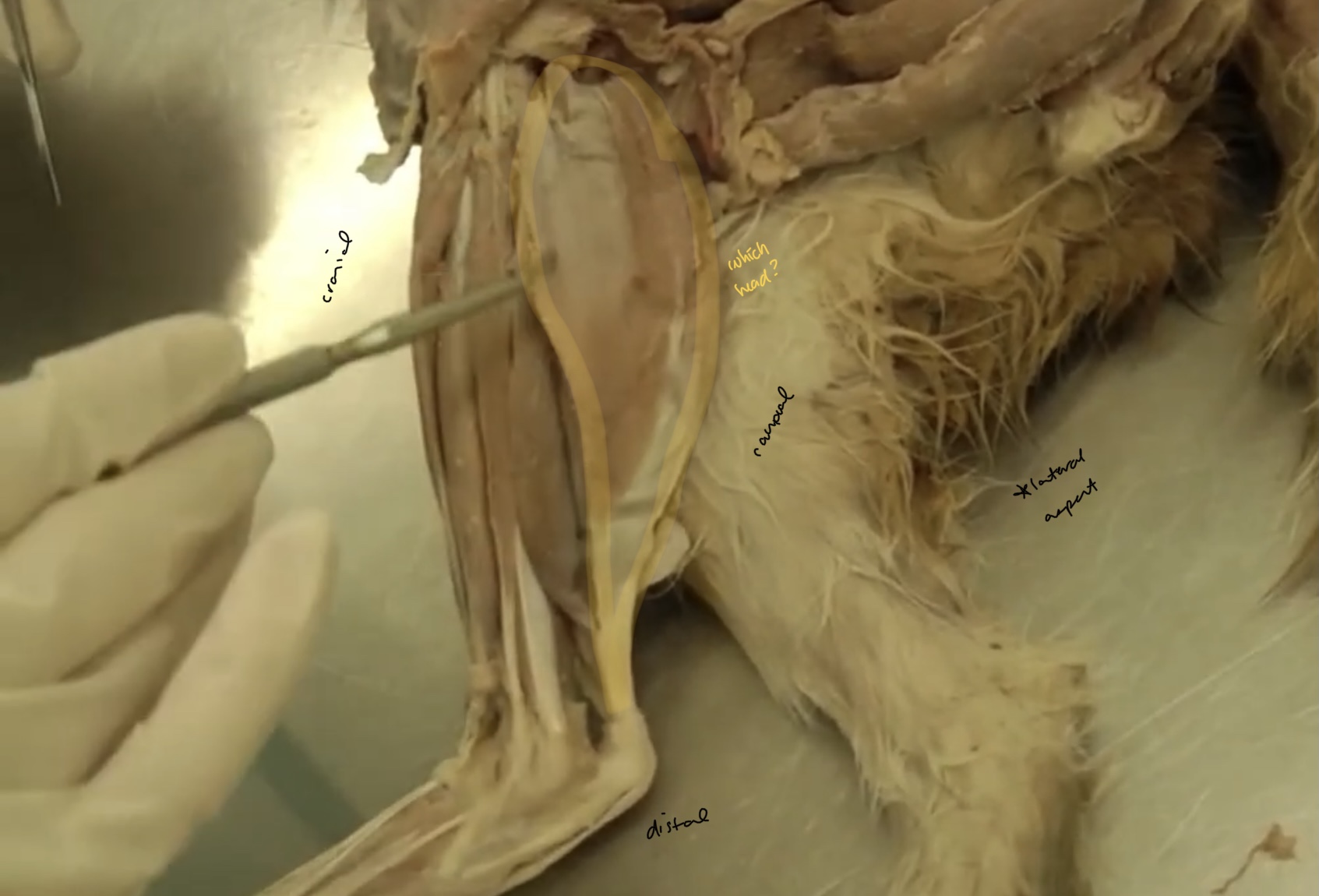
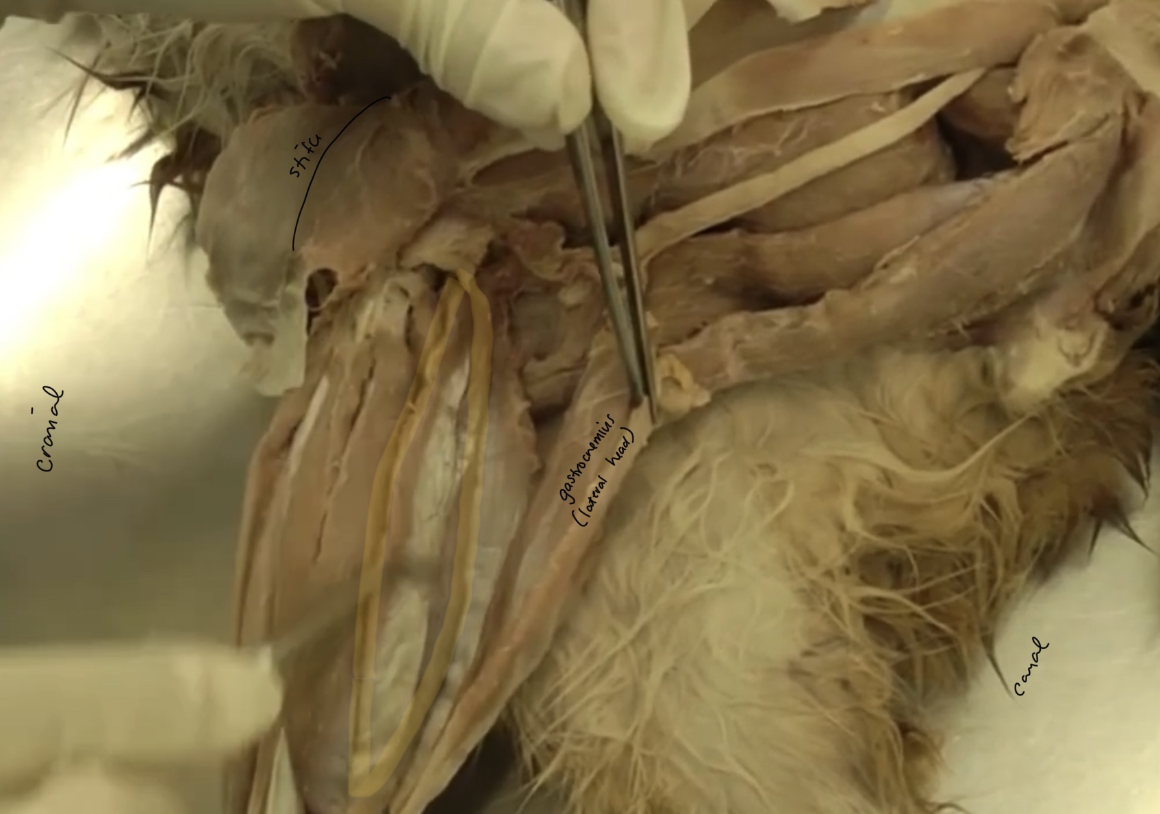
what is highlighted here? and which head?
gastrocnemius m. (lateral head)
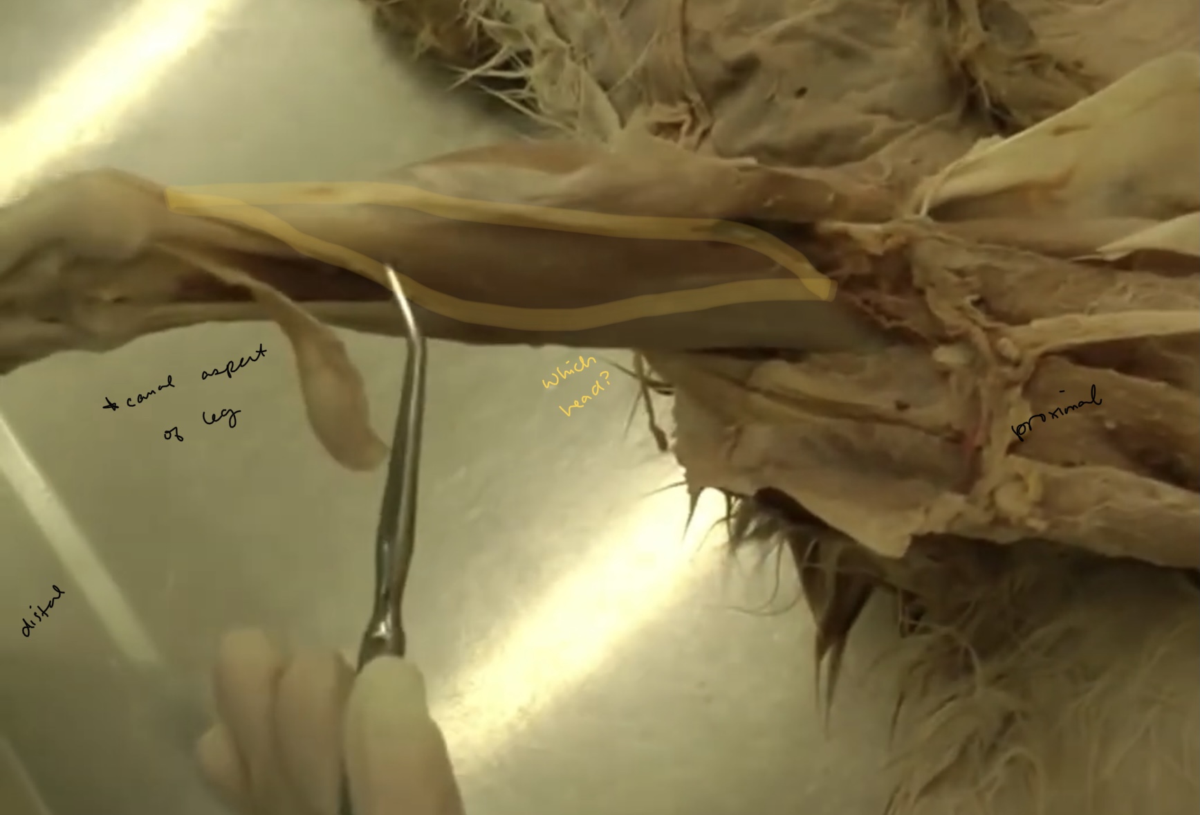
what is highlighted? which head?
gastrocnemius m. (medial head)

what is the yellow?
superficial digital flexor

what is blue?
gastrocnemius tendon
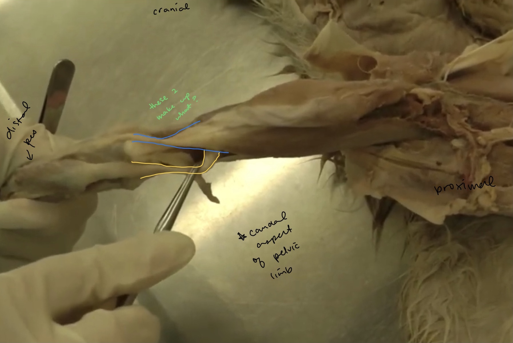
what do these make up?
common calcanean tendon
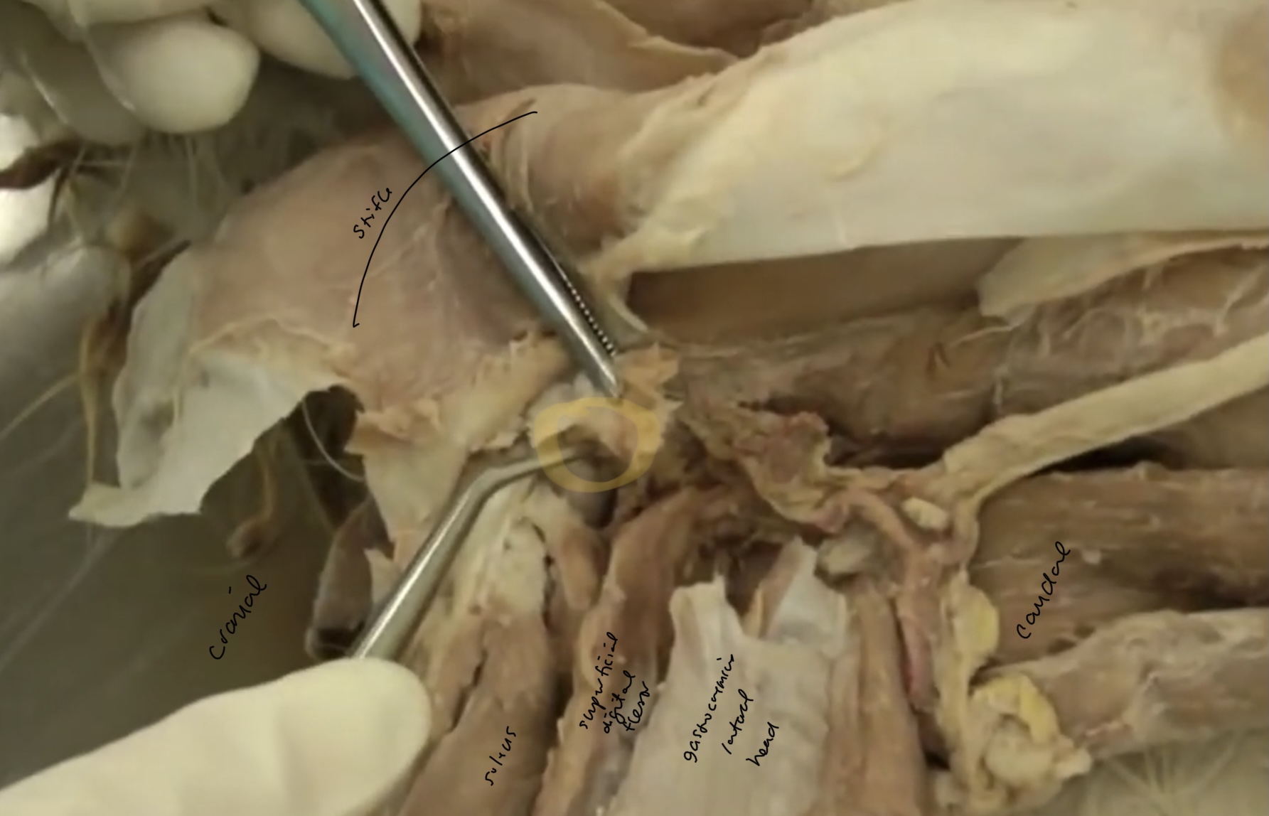
what are the extensors of the tarsus in cats?
gastrocnemius m.
soleus m.
what is being highlighted?
superficial digital flexor m.
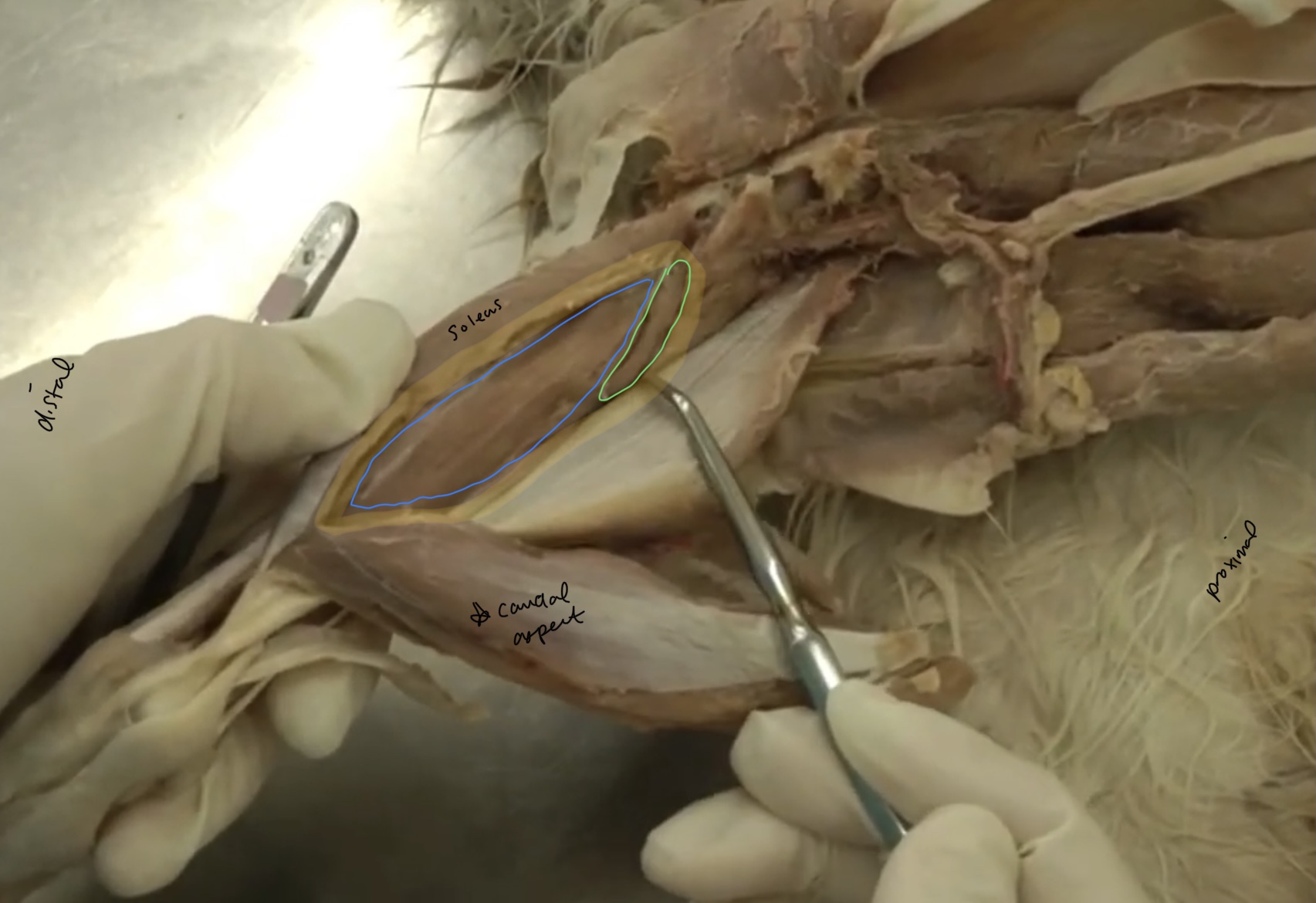
what is highlighted?
soleus m.
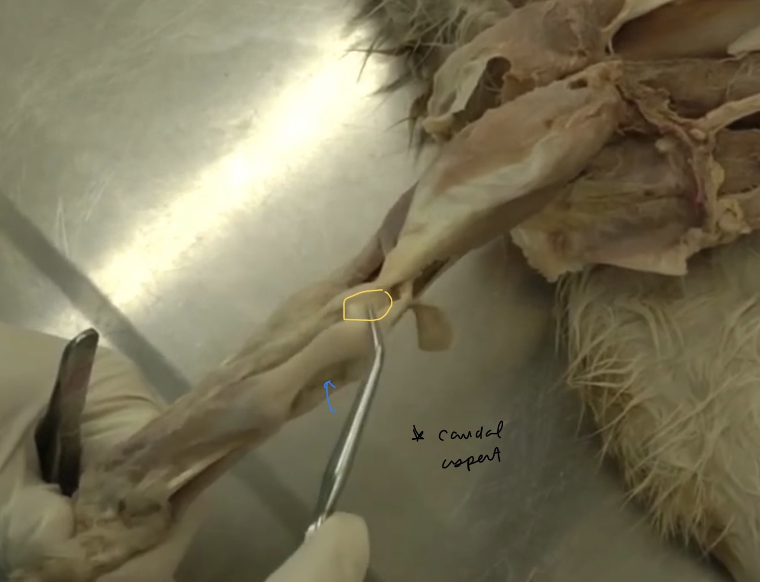
what is circled?
sesamoid (fabellae)
what is this structure in yellow?
calcaneal bursa

what is the yellow?
deep digital flexor m.
what is the head in blue?
lateral head of deep digital flexor m.

what head is green?
medial head of deep digital flexor m.
what is being pointed to in yellow? (not on list)
medial digital flexor
what is highlighted? (not in list)
lateral digital flexor
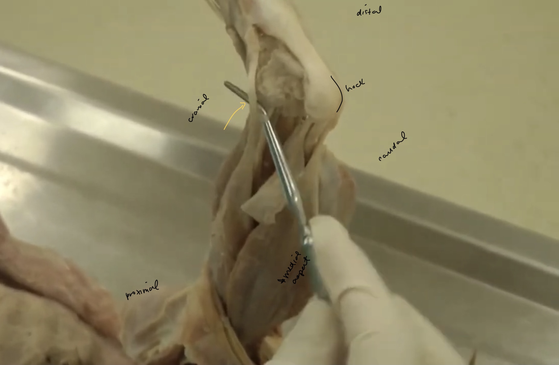
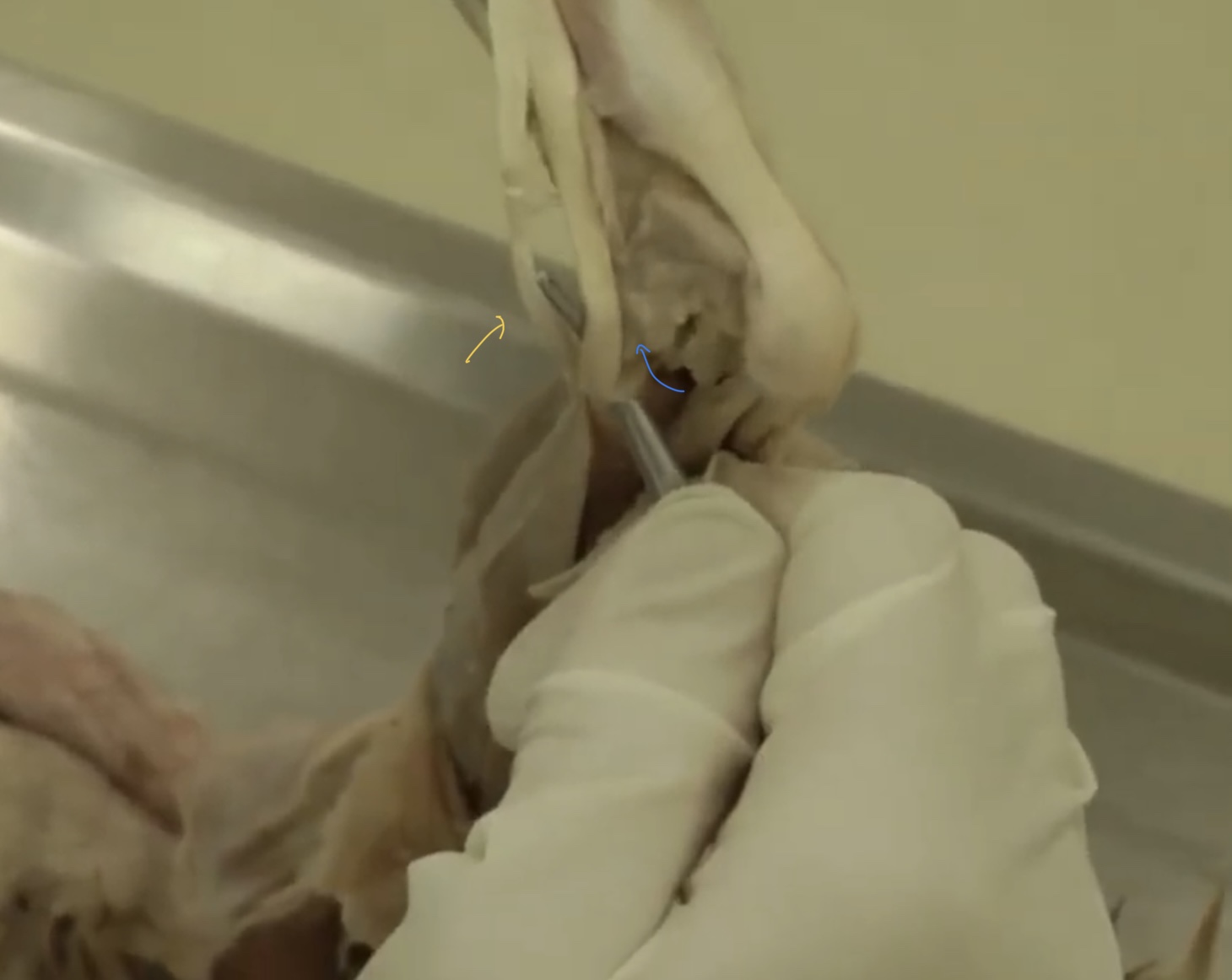
what is being pointed to?
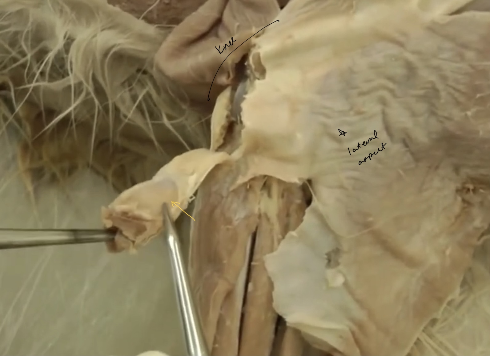
lateral collateral ligament
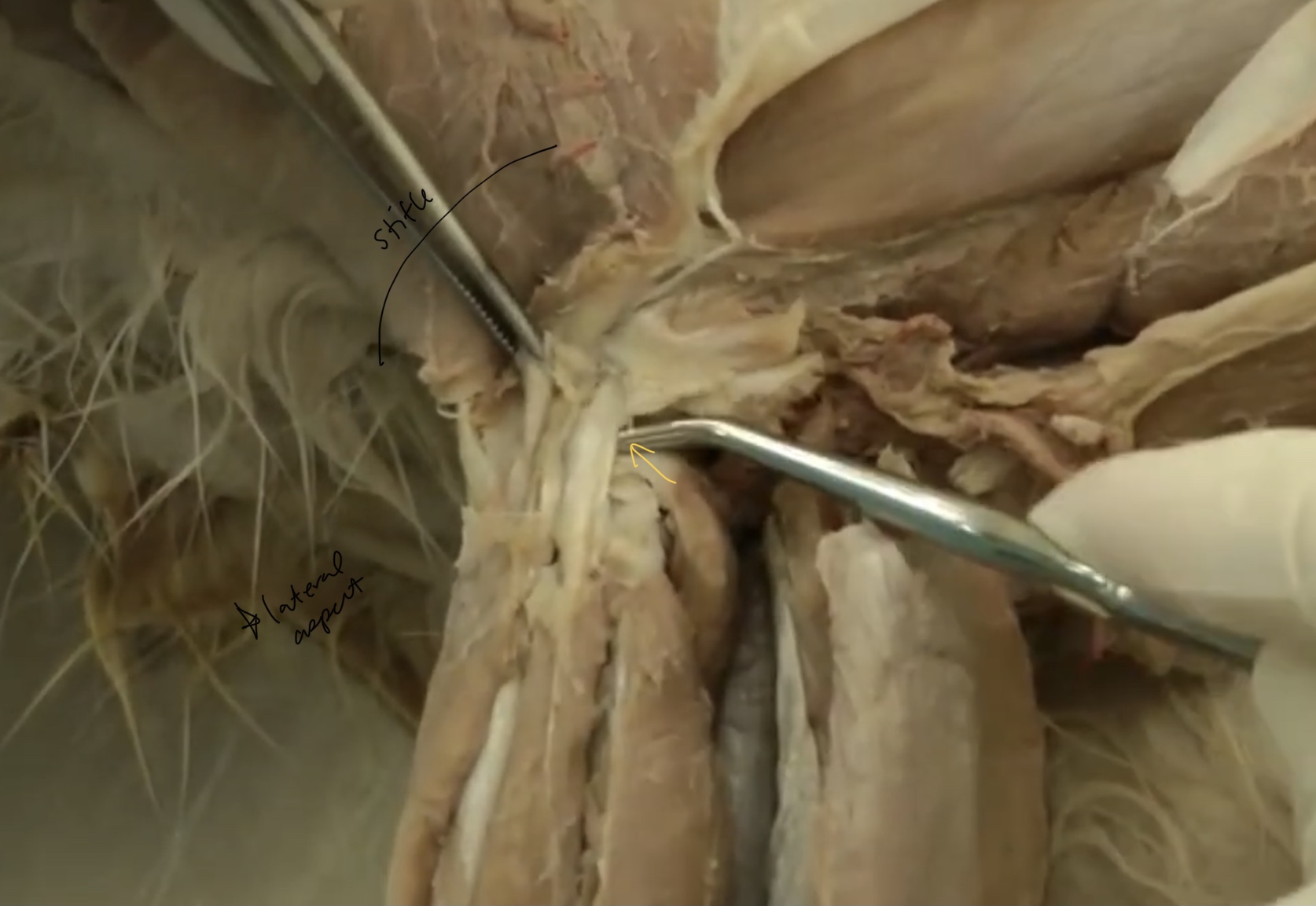
what is pointed at?
patella
what is the yellow?
menisci

what is the green?
lateral meniscus

what is blue?
medial meniscus

what is red?
cranial cruciate ligament

what is purple?
caudal cruciate ligament
what is this?
medial collateral ligament

what is blue? (not on list)
distal caudal femoral a.
what is green?
popliteal a.
what is this?
cranial tibial a.
what is this?
dorsal pedal a.

what is yellow? (not in list)
arcuate a.

what is blue? (not on list)
perforating branch
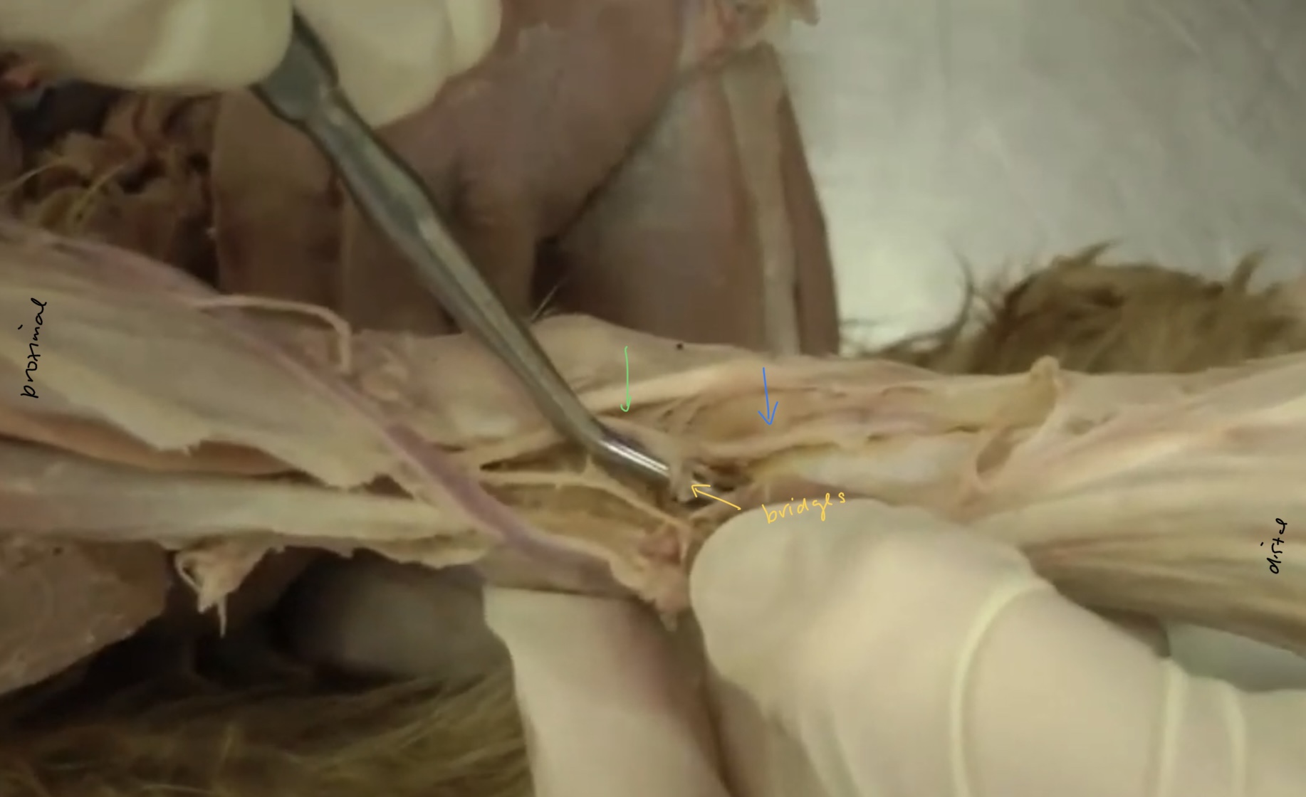
what is green?
dorsal pedal a.
what is this?
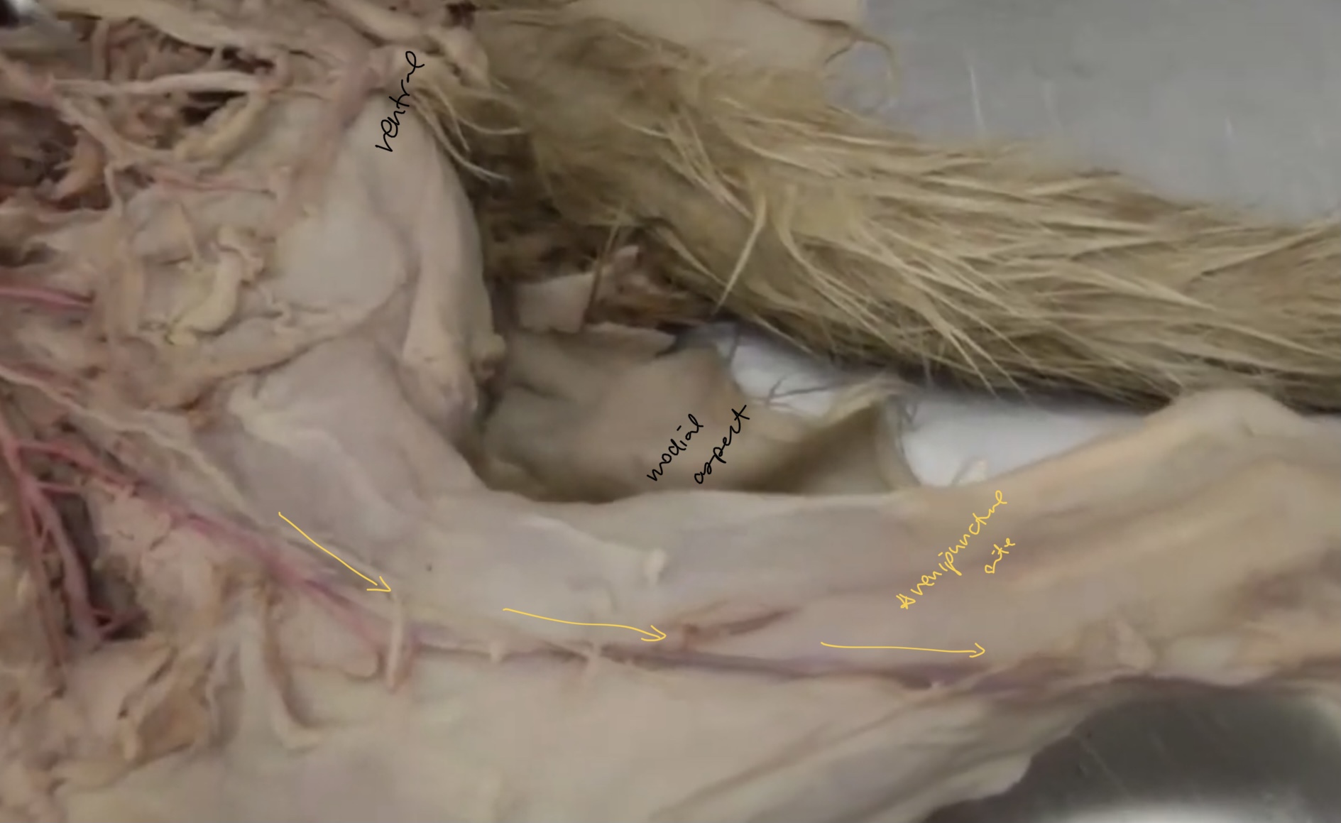
medial saphenous a.
what is this?
lateral saphenous artery
which vessel is used for venipuncture in the cats?
medial saphenous a.