MLS serology exam 2
 Call Kai
 Learn
Learn Practice Test
 Spaced Repetition
 Match
Match Flashcards
 Knowt Play
1/129

There's no tags or description
Looks like no tags are added yet.
Name | Mastery | Learn | Test | Matching | Spaced | Call with Kai | Chat |
|---|
No analytics yet
Send a link to your students to track their progress
130 Terms
First marker for Hepatitis B
Hepatitis B surface antigen (HBsAG), indicator of active infection, peaks during acute stages of infection, declines with development of host antibody response, remains elevated during chronic infection
Second marker for Hepatitis B
Hepatitis Be antigen (HBeAG), core of HBV, marker of active viral replication, appears shortly after HBsAG and levels decline before HBsAg, may remain elevated during chronic infection, indicates high degree of infectivity
Third marker of Hepatitis B
Anti-HBc, directed against hepatitis B core antigen, IgM anti-HBc appears first and indicated current or recent acute infection, consists mostly of IgG and can indicate current or past infection, can persist for a lifetime
Fourth marker of Hepatitis B
Anti-HBe, directed against HBeAg, indicates recovery from Hepatitis B
Last marker of Hepatitis B
Anti-HBs, directed against HBsAg, indicates immunity to hepatitis B
Which Hep B markers can persist for years
Anti-HBe and anti-HBs persist for years and help protect against subsequent infection
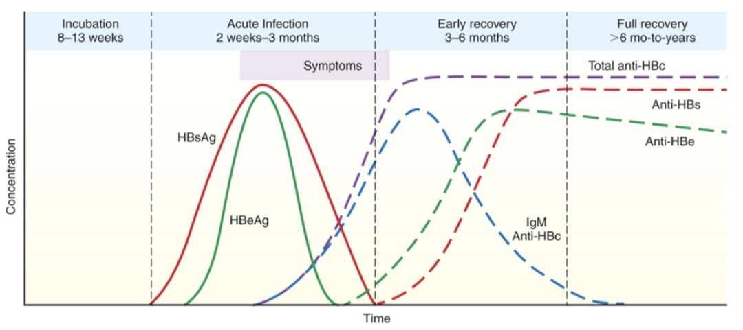
Acute HBV
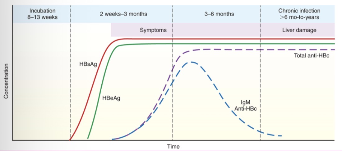
Chronic HBV
What markers are positive for past Hepatitis B infection
Anti-HBeAg and anti-HBs
Which hepatitis b marker means immune due to vaccination
Positive for Anti-HBs
What are the early antigen markers for mono
EA-D- anti-EA-D present in acute IM and EA-R
What are the late markers for mono
Viral capsid antigens (VCA), IgM anti-VCA (best marker for IM) appears at the onset of clinical symptoms and persists for 2 months, IgG anti-VCA (acute or past IM)
What are the latent-phase markers for mono
EBV nuclear antigens (EBNA), anti-EBNA (convalescent IM), latent membrane proteins (LMP-1, LMP-2A, LMP-2B)
Serological test result for acute EBV
Anti-VCA IgM, anti-VCA IgG, anti-EA-D, could be anti-VCA IgA and heterophile antibody IgM
Serological test results for past EBV
Anti-VCA, anti-EBNA
Serological test results for reactivated EBV
Anti-VCA IgG, anti-EBNA, could be anti-EA-R or heterophile antibody IgM
Serological test results for never infected EBV
All negative
Serological test results for chronic EBV
Very positive for Anti-VCA IgG, anti-EA-D, anti-EA-R, could be anti-EBNA
Transmission for CMV
Close, prolonged contact with infectious body secretions, sexual contact, blood transfusions and solid organ transplant, mother to infant
Symptoms for CMV
Healthy people: asymptomatic to mild IM-like illness, immunocompromised/suppressed: severe disease involving GI tract, CNS, blood, virus remains dormant but can be reactivated later in life
Complications in CMV
Infection is dangerous for immunocompromised patients, transplant patients have an increased risk of reactivation and previously CMv negative patients have high risk of GVHD, major opportunistic pathogen in AIDs patients, in neonates congenital infection, platelet dysfunction, CNS involvement, hearing loss
Testing for CMV
Organs screened for CMV, neonates receive CMV negative blood, molecular methods to detect CMV DNA, serological assays for CMV IgG to detect path infections, assays for IgM have limited value
What is measles?
Morbillivirus paramyoxoviridae, rubeola, RNA virus
Symptoms of measles
Subacute sclerosing panecephalitis (SSPE)
What is transmission in measles
Respiratory droplets
What is the testing for measles
RT-PCR to detect virus, capture ELISA to detect antibodies IgM or IgG, titers for healthcare works to confirm immunity
What is Mumps?
Rubulavirus paramyxoviridae, single stranded RNA virus
Mumps transmission
Respiratory droplets
What does mumps cause
Parotitis
What is the testing for mumps
RT-PCR or viral culture to detect virus, ELISA to determine current infection, recent infection, or immunity, PCR is used to monitor viral load for HTLV I/II
What kind of virus is Varicella Zoster?
DNA, Herpesviridae
Transmission of Varicella zoster
Primarily through inhalation of infected respiratory secretions, contact with or inhalation of aerosols from skin lesions, mother to fetus
What diseases does varicella zoster cause
Varicella (chickenpox), herpes zoster (shingles)
What is varicella zosters life long laten state?
Dorsal root, autonomic, and cranial ganglia, T cell-mediated immune response keeps virus in latency
Lab diagnosis for varicella zoster
Based on characteristic clinical findings, real-time PCR is main method to detect VZV DNA, titers (health-care workers, pregnant women, organ transplant candidates), ELISA detects immunity- total VZV IgG and automation helps provide objective results
What is Rubella
Rubrivirus togaviridae, RNA virus
What does Rubella cause
German measles
Transmission of Rubella
Respiratory droplets, mom to fetus (across placenta), CRS= congenital rubella syndrome
Testing for rubella
RT-PCR to detect viral antigens, serological assays to determine immunity or current infection, ELISA- best sensitivity and specificity, IgM- primary infection, IgG- past infection or immunity
What kind of virus is Human T-Cell lymphotropic virus (HTLV)
HTLA I/II are closely related retroviruses, RNA, posses reverse transcriptase
Transmission of HTLV
Bloodborne, sexual contact, mother to child (primarily breast feeding)
What disease is HTLV-I associated with
Adult T-cell leukemia/lymphoma
Testing for HTLV
ELISA used to screen, western blot used for confirmatory, all blood donors screen for HTLV I/II
Which cell is the main target for HIV infection?
CD4+ T cells- macrophages, monocytes, dendritic cells, Langerhans cells, and microglial brain cells can also be affected due to CD4 expression
How does the HIV virus attach to the target cell?
Coreceptor required (chemokines), CXCR4- T lymphocytes, CCR5- macrophages
Primary infection for HIV infection
Acute, early infection, may be asymptomatic or have flu-like syndrome that resolves, high level of viremia and decrease in CD4 T cell-number
Clinical latency of HIV infection
Absence of clinical symptoms, decrease in viremia and increase in CD4 T cell number, long-term nonprogressors (LTMP)- patients that remain asymptomatic for more than 10 years without antiviral therapy, normal or mildly decreased CD4+ counts, and low viral load
Clinical course of AIDs
Resurgence of viremia and decrease in CD4 T cell number, profound immunosuppression, with appearance of life-threatening opportunistic infections and malignancies
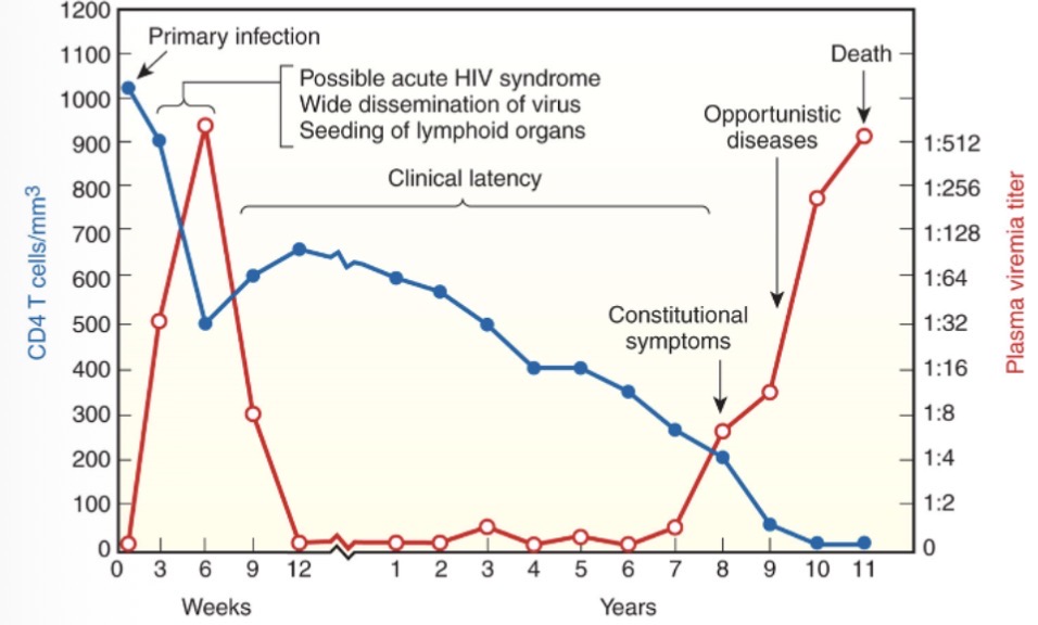
Clinical course of HIV
What is stage 0 for HIV
Patient with early HIV infection, have one positive HIV test, followed by negative or indeterminate test within 6 months, reclassified into one of the other categories 180 days or more after initial diagnosis
What is stage 1 of HIV staging
CD4 count: >/= 500 cells/microliter, CD4 count/= 26%
What is stage 2 of HIV
CD4 count: 200-499 cells/microliter, CD count: 14-25%
What is stage 3 of HIV
CD4 count: </= 200 cells/microliter, CD4 count: </= 14%, indicative of any opportunistic illnesses present in patient
What is unknown stage of HIV
When CD4 data is missing
What test methodology is used to stage HIV infection?
CD4 T cell enumeration through flow cytometry, HIV viral load through PCR and branched chain DNA, drug resistance testing through genotype and phenotype resistance assay
What is the fourth generation ELISA test for HIV being tested for?
Measures the optical absorbance of a patient HIV1/HIV2 antibodies and p24
What is the conjugate used in fourth generation ELISA test for HIV?
Enzyme labeled anti-p24 and HIV-1/HIV-2 antigens
Which type of ELISA is the fourth generation test for HIV?
Combination, sandwich ELISA
List the bands that must be present for a positive Western blot HIV test?
P24, gp41, gp120/gp160
List the test methods used to test for resistance in drugs used to treat HIV?
Genotype resistance assay: detects mutation in reverse transcriptase and protease from RNA in patient plasma, amplified RT-PCR, nucleotide sequenced products entered into database and analyzed with software for mutations, reported as resistance, possible resistance, no level resistance
Phenotype resistance assays: determine ability of HIV from clinical samples to grow in presence of antiretroviral drugs, direct measure of drug susceptibility, create recombinant virus from patient plasma and reference lab strain using mammalian cells, advanced procedure
Discuss HIV testing in infants
Less than 18 months of age, maternal antibodies in infant serum can complicate serologic test results, use molecular methods of diagnosis, qualitative HIV-1 DNA PCR (preferred), serologic testing at 12-18 months of age used to confirm
Describe the test methodology for the fluorescent ANA test
HEp-2 cells are fixed on the slide patient serum added to the slide and incubated, wash, add secondary fluorescent marker, wash, examine under microscope. If patient has the ANA it will bind w/the HEp-2 cells and the fluorescent marker will bind to this structure and glow. If the patient does not have ANA, it will not bind and everything will be washed away with no glowing
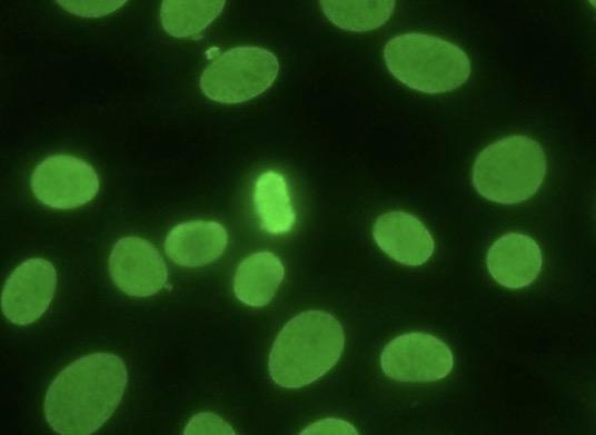
Homogenous pattern; SLE, drug-induced lupus, autoimmune hepatitis, juvenile idiopathic arthritic, antibodies to dsDNA, histones, nucleosome
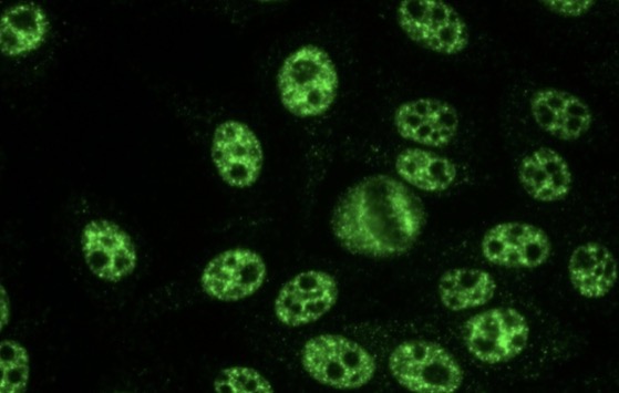
Speckled pattern; SLE, sjogren’s syndrome, SSc, other SARDs, antibodies to ENAs, DFS70/LEDFG
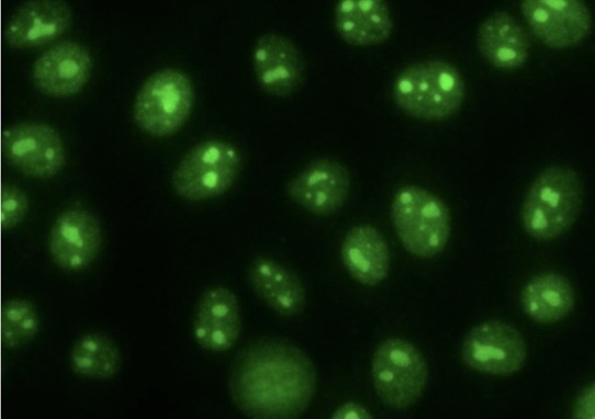
Nucleolar pattern; antibodies to RNA and RNP, SSc and other SARDs
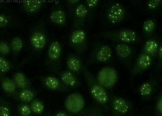
Nuclear dot pattern; broad spectrum autoimmune disease
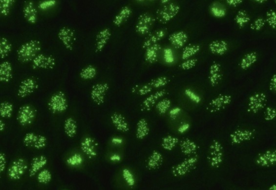
Centromere pattern; antibodies to centromeres of chromosome, CREST syndrome (limit cutaneous SSc)
What is systemic lupus erythematosus (SLE)
Inflammatory disease that affects multiple organs including skin, joints, renal, neurological, and cardiac, anti-ds DNA (lupus-specific), anti-ss DNA, anti-histones and nucleosomes detected by immunofluorescent assay immunoblotting (western blot), EIA
What does systemic lupus erythematosus have antibodies to?
Centromere or nucleolar components, anti-ENA (anti-SM specific for lupus detects by IIF, immunodiffusion, immunoblotting, immunoprecipitation, EIA), anti-RNP, anti-SS-A, anti-SS-B
What plays a key role in rheumatoid arthritis
TNF-alpha plays a key role in process
What organs are affected in rheumatoid arthritis
Joints and bones
What is tested for in rheumatoid arthritis
Rheumatoid factor- IgM autoantibodies reacts with Fc portion of IgG, anti-CCp- autoantibody against cyclic citrullinated peptide and highly specific, ANAs, ESR, CRP, and complement assays
What does graves’ disease cause?
Hyperthyroidism
In graves’ disease what does TRAbs produce?
Autoantibodies to thyroid stimulating hormone (anti-TSHR)
Lab results of graves disease
Low TSH, high FT4, antibodies to TPO and Tg, antibodies to TSHR
What happens in Hashimotos thyroiditis?
Immune destruction of the thyroid gland produces hypothyroidism
Lab results of hashimotos thyroiditis
Normal or high TSH, low free T4, antibodies-TPO, antibodies-Tg
What are autoantibodies directed against in hashimoto’s thyroiditis
Thyroglobulin (anti-thyroglobulin)
What part of the body does myasthenia gravis affect
Neuromuscular junction = weak skeletal muscle
What are antibodies against in myasthenia gravis
Antibodies to acetylcholine receptors and block binding of ACH to its receptor and transmission of nerve impulses that activate muscle, anti-ACHR autoantibodies can be detected
What is affected in multiple sclerosis
Inflammation and destruction of the central nervous system by destroying the myelin sheath
What are antibodies against multiple sclerosis
Against myelin basic protein
Lab findings in multiple sclerosis
Lesions seen on magnetic resonance imaging, increased immunoglobulins in spinal fluid and increased IgG index, oligoclonal bands on protein electrophoresis of CSF
What part of the body is affected in goodpasture’s syndrome
Basement membranes lining the renal glomeruli and lung alveoli
What are antibodies directed to in goodpasture’s syndrome?
Basement membranes
What is Type I diabetes mellitus?
Endocrine disorder characterized by hyperglycemia
What are antibodies directed against in type I diabetes mellitus
Autoantibodies destroy beta cells in pancreas causing insulin deficiency
Laboratory results in type I diabetes mellitus
Increased blood glucose level, elevated HGBA1c, autoantibodies to glutamic acid decarboxylase (GAD), insulinoma antigen 2 (IA-2), islet cell antibodies(ICA)
What part of the body is affected in celiac disease
Affects small intestine and other organs
What autoantibodies form in celiac disease
HLA-DQ2 or HLA-DQ8- gliadin (component of gluten) and DGPs, tissue transglutaminase (tTg IgA), endomysium (EMA)
Mechanism of type I hypersensitivity
Sensitization phase: APCs process allergens and present them to help T cells, helper T cells induce production of allergen-specific IgE, IgE binds to high-affinity receptors on mast cells and basophils
Activation phase:allergen cross-links adjacent cell-bound IgE, mast cells and basophils degranulate, chemical mediators are released and bind to target organs, allergy symptoms are produced
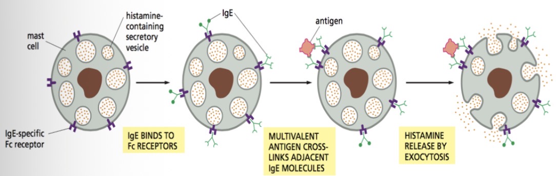
Type I hypersensitivity mechanism
Example of type I hypersensitivity
Allergies
Examples of Type II hypersensitivity
Transfusion reactions, HDFN, autoimmune hemolytic anemia, goodpasture’s syndrome, hashimoto’s disease
Mechanism of Type II hypersensitivity
Triggered by antigen found on cell surface (IgG or IgM, altered self antigen or heteroantigens), antibody binds to macrophages, neutrophils, eosinophils, NK cells, results in cell destruction and lysis
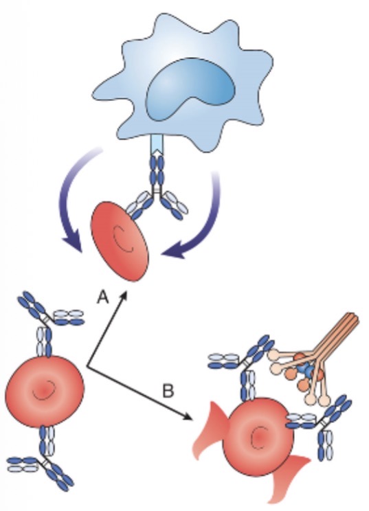
Type II hypersensitivity
Type III hypersensitivity reaction examples
Arthus reaction, serum sickness, SLE, RA, reaction to bee stings, drug reactions, sequelae to infections
Mechanism of type III hypersensitivity
Antigen-antibody complexes precipitate out and deposit in tissues, immune system in overwhelmed, complexes deposit in tissues where they bind complement, immune complex deposition typically occurs in glomerular basement membranes, vascular endothelium, joint lining, pulmonary alveolar membranes, complement binds to complexes in tissues
Examples of type IV hypersensitivity
Infections with intracellular pathogens, skin test for TB, contact dermatitis, hypersensitivity pneumonitis, mycobacterium tuberculosis, mycobacterium leprae, pneumocystsis carinii, leishmania species, herpes simplex virus
Mechanism in type IV hypersensitivity
Delayed cell mediated hypersensitivity, involved sensitized Th1 cells and macrophages, antibody and complement are not involved, Th1 cells release cytokines that attract and activate macrophages, macrophage induce inflammation, cytotoxic T lymph are recruited and destroy target cells, hypersensitivity peaks 48 to 72 hours after antigen exposure