Hand Pathology
 Call Kai
 Learn
Learn Practice Test
 Spaced Repetition
 Match
Match Flashcards
 Knowt Play
1/8

There's no tags or description
Looks like no tags are added yet.
Name | Mastery | Learn | Test | Matching | Spaced | Call with Kai | Chat |
|---|
No analytics yet
Send a link to your students to track their progress
9 Terms
Bone Pathology
Fractures
Metacarpal/Phalangeal Fractures: Common in trauma (e.g., boxer’s fracture of 5th metacarpal).
MRI Findings: Low-signal fracture line on T1; bone marrow edema on T2 FS/STIR.
Avulsion Fractures: Often at tendon/ligament insertions.
Osteomyelitis
MRI Findings:
Bone marrow edema
Cortical destruction
Periosteal reaction
Soft tissue abscess (T2 hyperintense, enhances with contrast on T1 - Fat/Sat helps to visualize this).
Bone Tumors
Enchondroma: Common benign tumor in phalanges.
MRI Findings: Lobulated, T2-hyperintense lesion with chondroid matrix.
Joint and Ligament Pathology
Osteoarthritis
1st CMC Joint: Common site due to thumb mobility.
MRI Findings: Joint space narrowing, osteophytes, cartilage loss, subchondral cysts/edema.
Rheumatoid Arthritis
MRI Findings:
Synovitis (T2 hyperintense, enhances with contrast)
Erosions
Joint effusion
Collateral Ligament Tears
MRI Findings:
Discontinuity or abnormal signal in collateral ligaments, often at MCP joints (e.g., gamekeeper’s thumb at 1st MCP ulnar collateral ligament).
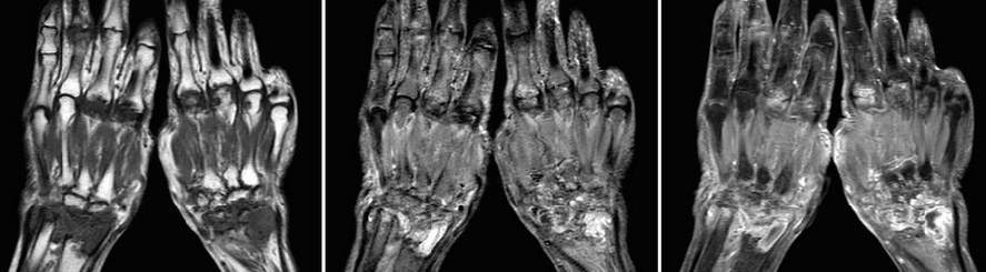
rheumatoid arthritis
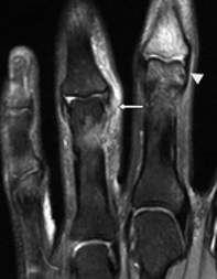
Collateral Ligament Tears
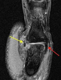
Tendon Injuries
Tenosynovitis
An inflammation of the synovium, the sheath that surrounds and protects tendons.
It can occur in any tendon in the body, but it is most common in the wrists, hands, and feet.
MRI Findings: Fluid in tendon sheath (T2 hyperintense), tendon thickening, synovial enhancement.
Common in flexor tendons (e.g., trigger finger due to A1 pulley thickening).
Tendon Tears
MRI Findings: Partial or complete discontinuity, increased T2 signal, retraction in complete tears.
Common in flexor digitorum or extensor pollicis longus.
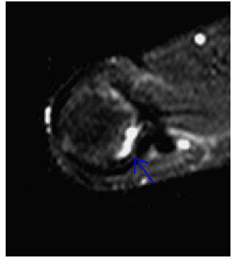
Gamekeeper Thumb
Gamekeeper thumb is an injury to the ulnar collateral ligament of the thumb at the level of the first metacarpophalangeal joint. This injury can occur from skiing or falling on to an outstretched hand.
Appearance on MRI:
•
May show avulsion fracture at the attachment site of the ulnar collateral ligament (one-third of cases).
•
Shows tear of the ulnar collateral ligament as a thin, low-signal ligament or discontinuity of the ligament with hemorrhage and edema surrounding the torn ends of the ligament.
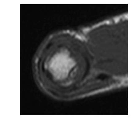
Giant Cell Tumor of Tendon Sheath
Giant Cell Tumor of Tendon Sheath
Common benign tumor.
MRI Findings: T1/T2 hypointense due to hemosiderin, enhances with contrast.
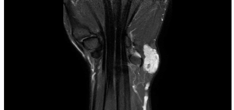
Lipoma or Hemangioma
image is stir demonstrating hemangioma
Lipoma or Hemangioma
Fat mass or growth of extra blood vessels.
MRI Findings: Lipoma (T1 hyperintense, suppresses on fat-sat); hemangioma (T2 hyperintense, vascular flow voids).
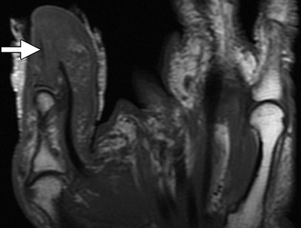
Soft Tissue Infections
Soft Tissue Infections
MRI Findings:
Soft tissue edema
Abscess formation (T1 hypointense, T2 hyperintense, rim-enhancing)
Possible bone involvement.