Blood cells - applied microscopic anatomy
1/31
Earn XP
Description and Tags
Name | Mastery | Learn | Test | Matching | Spaced |
|---|
No study sessions yet.
32 Terms
used to focus the specimen
What is this knob used to do as part of Kohler illumination (and in general)?
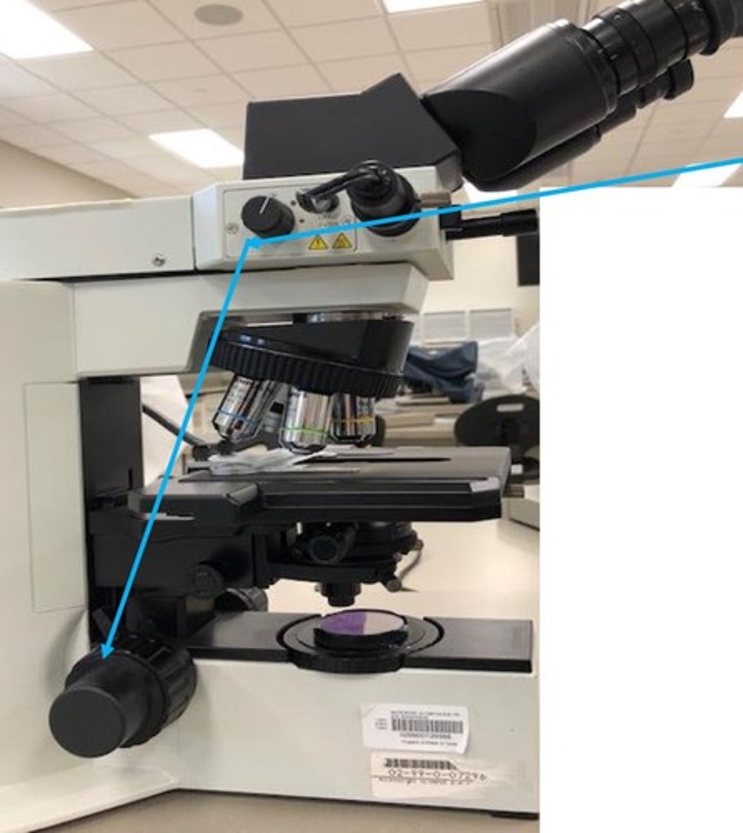
this is the field diaphragm. it is closed at the beginning (what gives you the one bright spot in your view) and opened at the end
What is this doohicky and what parts of Kohler illumination is it involved in?
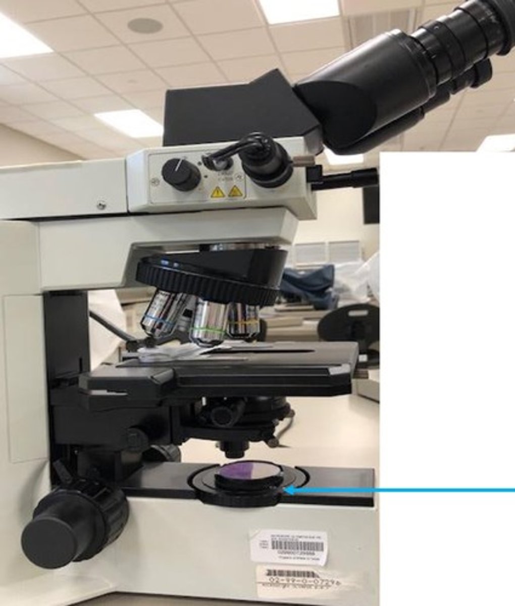
closed!
Is this field diaphragm opened or closed?
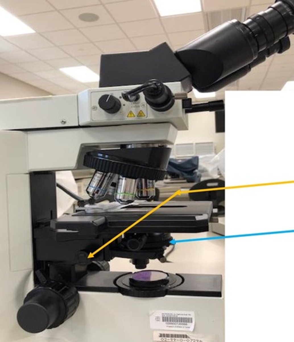
yellow- used to focus the condenser, blue - used to center the condensor
What are these two knobs used to do?
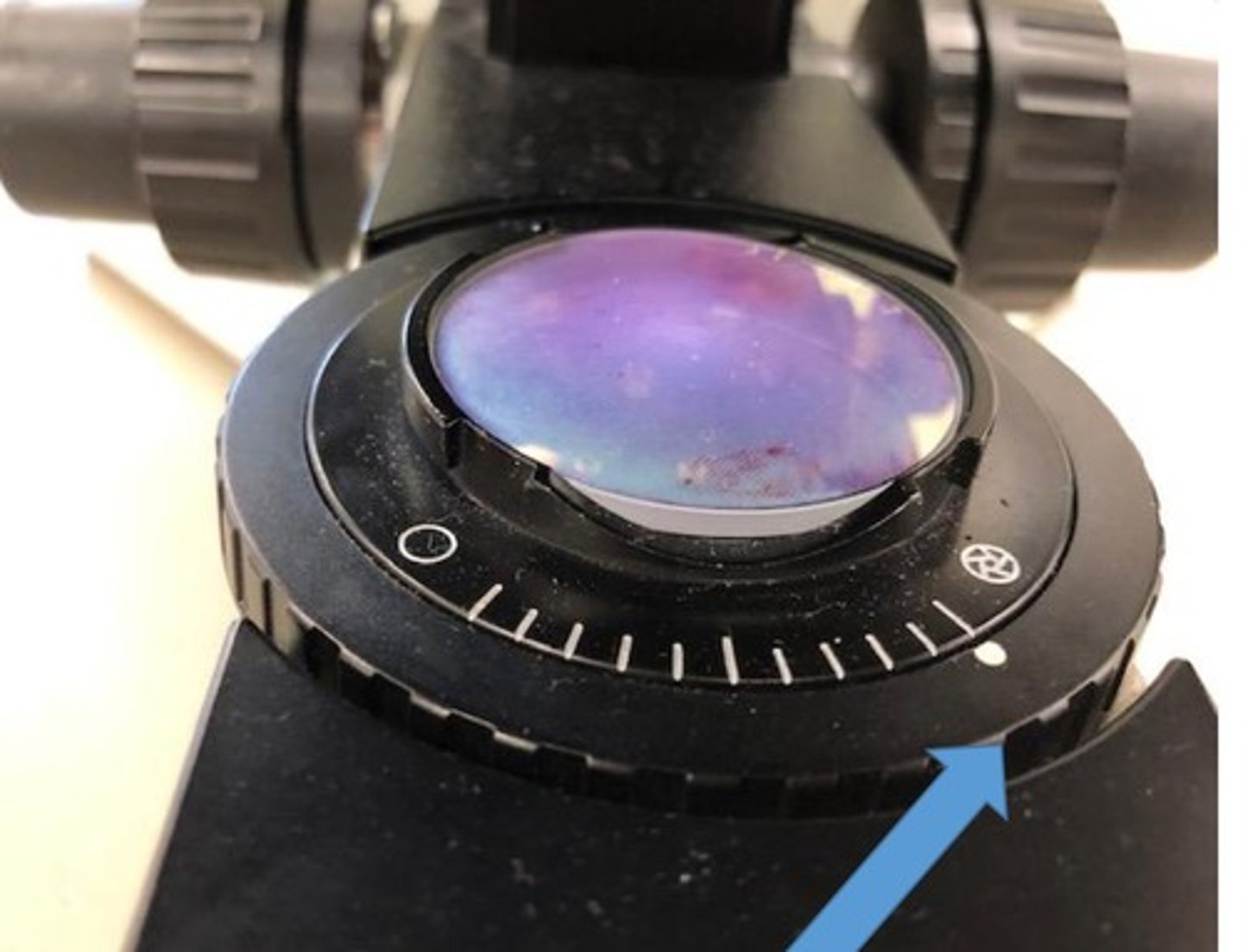
opened (just outside of view), focused, centered
This image through the microscope is indicative that the field diaphragm is _________, the condenser is _______ & ___________.
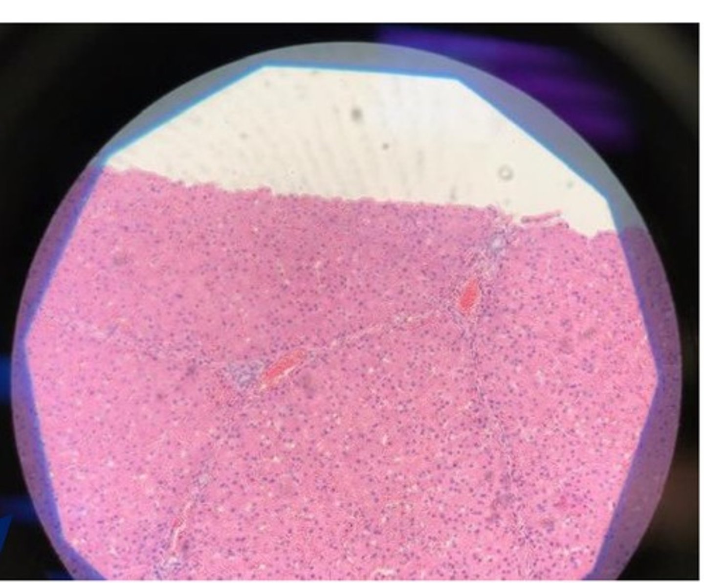
40x objectives - because they do not require the use of immersion oil
What objective on the microscopes are considered "high-dry"? Why are they referred to as such?
Yes! they'll just be a little out of focus - it would be best to view these slides with immersion oil on the 100x objective
Can slides without a coverslip be viewed with 40x objective?
1) the slide will be blurry and 2) you gonna have to clean those objectives with lens cleaner
What is the consequence of using immersion oil and viewing with the 10x, 20x, and 40x objectives?
YES - and the slide you're viewing does not have to have a coverslip to use oil
Do you have to use immersion oil if viewing a slide on 100x objective?
to gather light from the microscope light source and concentrate it into a cone of light that illuminates the specimen with uniform intensity over the entire view field.
What is the purpose of the condenser on a microscope?
properly adjusting the condenser light cone so that the intensity and angle of light entering the objective front lens is optimum for viewing your specimen.
What even is Kohler illumination?
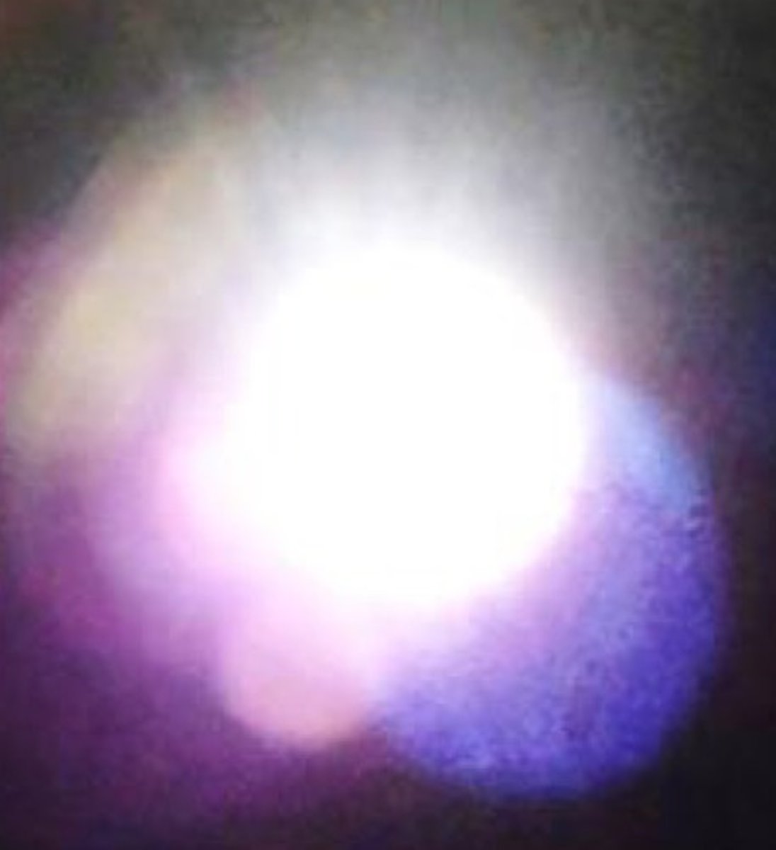
Next, I'll need to focus the condenser, slightly open the field diaphragm, and center the condenser so the "octagon shape" is crisp (should see blue rim) and in the center of the light field.
Knowing that you've already focused the specimen and closed field diaphragm, you see this through the microscope - what are your next steps in completing this Kohler illumination?
the monolayer!
What part of a blood smear do you look at to evaluate blood cells?
here's a review of that: https://quizlet.com/_70yc65?x=1jqt&i=ianvd Histology >blood cells in tissue
Identify leukocytes found in peripheral blood: neutrophil, monocyte, lymphocyte.
1) count 100 leukocytes 2) of these 100, keep track of how many are neutrophils, lymphocytes, monocytes, eosinophils, basophils 3) multiply the percentage of each leukocyte by the total WBC
Describe how you would perform a white blood cell count differential from a blood smear
Absolute = (.92)*46,700 = 42,694. neutrophilia
While performing a white blood cell differential, you observe 92% of your 100 leukocytes to be neutrophils. If the total WBC is 46,700/microliter, what is your absolute count of neutrophils? How would you interpret this knowing a normal neutrophil range is 3,000-11,500/microliter?
increased numbers of white blood cells
define leukocytosis
decreased numbers of white blood cells
define leukopenia
increased numbers of neutrophils in peripheral blood
define neutrophilia
decreased numbers of neutrophils in peripheral blood
define neutropenia
increased number of eosinophils in peripheral blood
define eosinophilia
decreased numbers of eosinophils in peripheral blood
define eosinopenia
increased number of basophils in peripheral blood
define basophilia
increased numbers of monocytes in peripheral blood
define monocytosis
decreased numbers of monocytes in peripheral blood
define monocytopenia
increased numbers of lymphocytes in peripheral blood
define lymphocytosis
decreased numbers of lymphocytes in peripheral blood
define lymphopenia
the 100x objective with immersion oil!
Which objective is used to evaluate erythrocyte morphology and platelet numbers?
10, 15
In the monolayer of the blood smear, there should be approximately ____ to ____ platelets per field.
looking at 5 to 10 different fields, count the number of platelets you see in each field and calculate an average. From this number, multiply it by 15,000 and 20,000 to obtain an estimated range of platelet numbers in this animal.
Assuming you've already performed kohler illumination, you are in the monolayer of a blood smear slide, and using the 100x objective how would you perform a platelet count?
range = 8 x 15,000 - 8 x 20,000 = 120,000 - 160,000. these numbers are low!
You've looked at 5 different fields of the monolayer of a blood smear and calculated there to be an average of 8 platelets per each of these fields. What is your estimated range of platelet numbers for the animal this blood came from? Are these platelet numbers low, adequate or high?
200,000-500,000 per microliter
What is the reference interval for platelet numbers?