Anatomical Landmarks on Panoramic Images​

studied byStudied by 0 people
0.0(0)
learn
LearnA personalized and smart learning plan
exam
Practice TestTake a test on your terms and definitions
spaced repetition
Spaced RepetitionScientifically backed study method
heart puzzle
Matching GameHow quick can you match all your cards?
flashcards
FlashcardsStudy terms and definitions
Get a hint
Hint

Articular Eminence​

1 / 37

encourage image

There's no tags or description

Looks like no one added any tags here yet for you.

38 Terms

1

Articular Eminence​

  • Rounded projection of the temporal bone located anterior to the glenoid fossa.
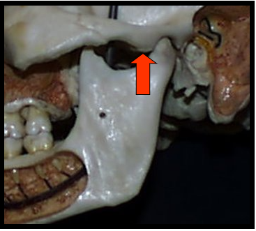
<ul><li><p><span>Rounded projection of the temporal bone located anterior to the glenoid fossa.</span></p></li></ul><p></p>
New cards
2

Glenoid Fossa​

  • Concave depressed are of the temporal bone.

  • The mandibular condyle rests in this when the teeth are in maximum intercuspation or clenched together.
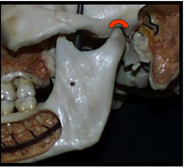
<ul><li><p><span>Concave depressed are of the temporal bone. </span></p></li><li><p><span>The mandibular condyle rests in this when the teeth are in maximum intercuspation or clenched together.</span></p></li></ul><p></p>
New cards
3

Articular Eminence and Glenoid Fossa​

  • One appears as a concave radiopacity superior to the mandibular condyle.

  • One appear as a rounded radiopaque projection of the bone located anterior to the glenoid fossa.
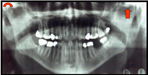
<ul><li><p><span>One appears as a concave radiopacity superior to the mandibular condyle.</span></p></li><li><p><span>One appear as a rounded radiopaque projection of the bone located anterior to the glenoid fossa.</span></p></li></ul><p></p>
New cards
4

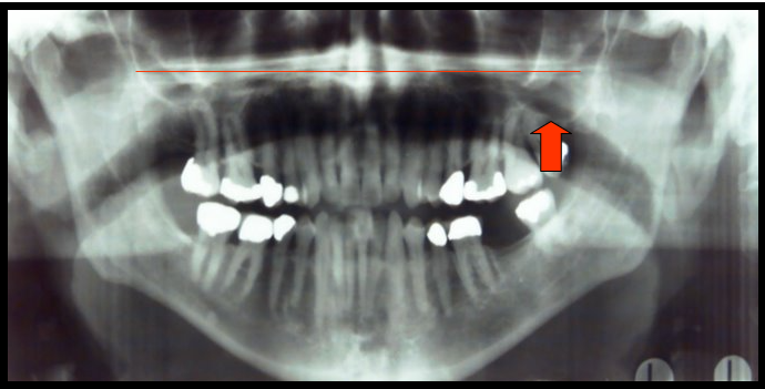
Maxillary Sinus​

Paired cavities or compartments of bone that are located within the maxilla, superior to the maxillary posterior teeth.
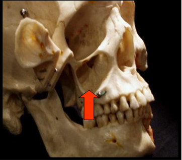
<p><span>Paired cavities or compartments of bone that are located within the maxilla, superior to the maxillary posterior teeth.</span></p>
New cards
5

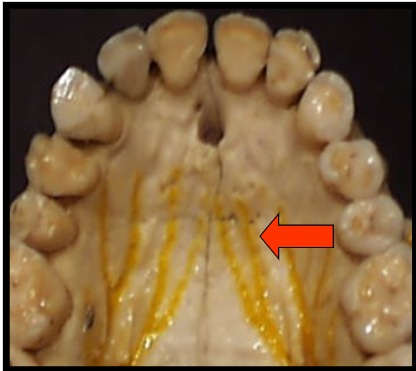
Hard Palate​

The bony wall that separates the nasal cavity from the oral cavity.
<p><span>The bony wall that separates the nasal cavity from the oral cavity.</span></p>
New cards
6

Maxillary Sinus and Hard Palate​

  • One appears as a horizontal radiopaque band superior to the apices of maxillary teeth.

  • One appears as paired radiolucent areas located superior to the apices of maxillary premolars and molars.
<ul><li><p><span>One appears as a horizontal radiopaque band superior to the apices of maxillary teeth.</span></p></li><li><p><span>One appears as paired radiolucent areas located superior to the apices of maxillary premolars and molars.</span></p></li></ul><p></p>
New cards
7

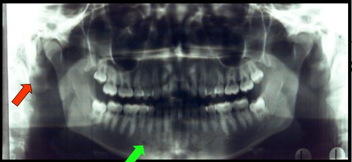
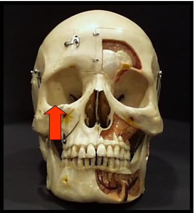
Orbit​

The bony cavity that contains the eyeball.
<p><span>The bony cavity that contains the eyeball.</span></p>
New cards
8

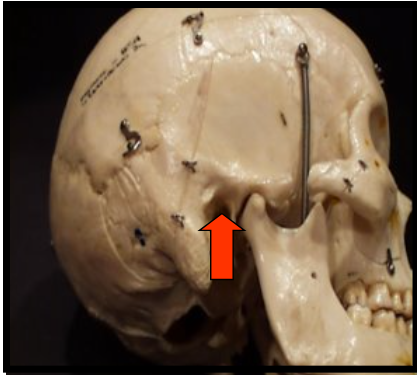
External Auditory Meatus​

Is a hole, or opening, in the temporal bone located superior and anterior to the mastoid process.
<p><span>Is a hole, or opening, in the temporal bone located superior and anterior to the mastoid process.</span></p>
New cards
9

Orbit and External Auditory Meatus​

  • One appears as a round radiolucent compartment with radiopaque borders located superior to the maxillary sinuses.

    • On most panoramic images only the inferior border of this is visible

  • One appears as a round or ovoid radiolucency anterior and superior to the mastoid process.
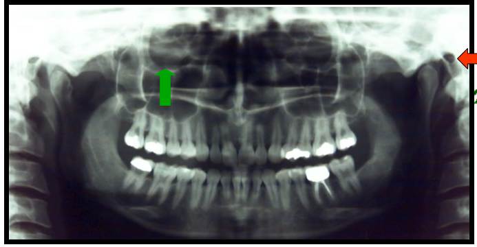
<ul><li><p><span>One appears as a round radiolucent compartment with radiopaque borders located superior to the maxillary sinuses.</span></p><ul><li><p><span>On most panoramic images only the inferior border of this is visible</span></p></li></ul></li><li><p><span>One appears as a round or ovoid radiolucency anterior and superior to the mastoid process.</span></p></li></ul><p></p>
New cards
10

Nasal Fossa and Inferior Nasal Conchae​

  • A pear-shaped compartment of bone located superior to the maxilla

  • Are wafer-thin, curved plates of bone that extend from the lateral walls of the nasal cavity.
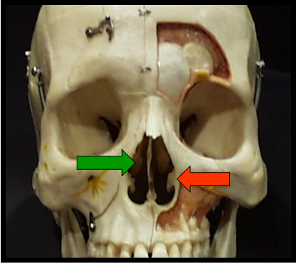
<ul><li><p><span>A pear-shaped compartment of bone located superior to the maxilla</span></p></li><li><p><span>Are wafer-thin, curved plates of bone that extend from the lateral walls of the nasal cavity.</span></p></li></ul><p></p>
New cards
11

Nasal Septum​

Vertical bony wall or partition that divides the nasal cavity into the right and left nasal fossa
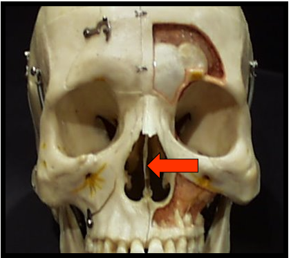
<p><span>Vertical bony wall or partition that divides the nasal cavity into the right and left nasal fossa</span></p>
New cards
12

Nasal Fossa, Inferior Nasal Conchae and Nasal Septum​

  • Appears as a large radiolucent area superior to the maxillary incisors divided into two compartments or fossa

  • Appears as a vertical radiopaque partition that divides the nasal cavity

  • Appears as diffuse radiopaque masses or projections within the nasal cavity.
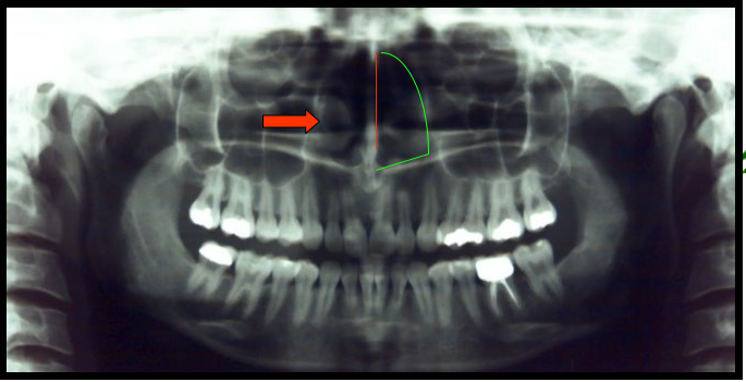
<ul><li><p><span>Appears as a large radiolucent area superior to the maxillary incisors divided into two compartments or fossa</span></p></li><li><p><span>Appears as a vertical radiopaque partition that divides the nasal cavity</span></p></li><li><p><span>Appears as diffuse radiopaque masses or projections within the nasal cavity.</span></p></li></ul><p></p>
New cards
13

Anterior Nasal Spine​

A sharp bony projection of the maxilla located at the anterior and inferior portion of the nasal cavity.
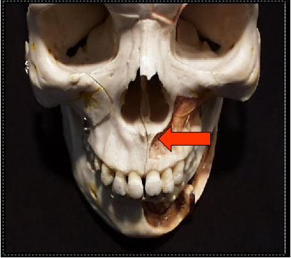
<p><span>A sharp bony projection of the maxilla located at the anterior and inferior portion of the nasal cavity.</span></p>
New cards
14

Pterygomaxillary Fissure​

A narrow space that separates the lateral pterygoid plate and the maxilla
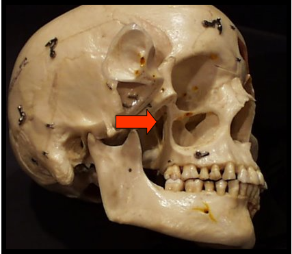
<p><span>A narrow space that separates the lateral pterygoid plate and the maxilla</span></p>
New cards
15

Anterior Nasal Spine and Pterygomaxillary Fissure​

  • Appears as a V-shaped radiopaque area located at the intersection of the floor of the nasal fossa and the nasal septum.

  • Appears as a radiolucent area between the lateral pterygoid plate and the maxilla.
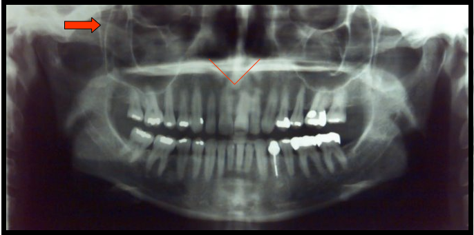
<ul><li><p><span>Appears as a V-shaped radiopaque area located at the intersection of the floor of the nasal fossa and the nasal septum.</span></p></li><li><p><span>Appears as a radiolucent area between the lateral pterygoid plate and the maxilla.</span></p></li></ul><p></p>
New cards
16

Mandibular Canal​

  • A tubelike passageway through bone that travels within the body or length of the mandible.

  • Extends from the mandibular foramen to the mental foramen and houses the inferior alveolar nerve and blood vessels
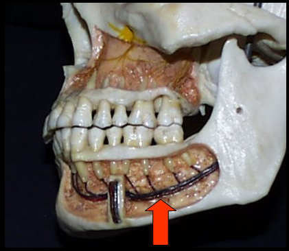
<ul><li><p><span>A tubelike passageway through bone that travels within the body or length of the mandible.</span></p></li><li><p><span>Extends from the mandibular foramen to the mental foramen and houses the inferior alveolar nerve and blood vessels</span></p></li></ul><p></p>
New cards
17

External Oblique Ridge​

A linear prominence of bone located on the external surface of the mandible that extends downward and forward from the ramus to the molar region.
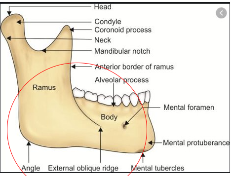
<p><span>A linear prominence of bone located on the external surface of the mandible that extends downward and forward from the ramus to the molar region.</span></p>
New cards
18

Mandibular Canal and External Oblique Ridge​

  • Appears as a radiolucent band outlined by two thin radiopaque lines representing the cortical walls of the canal.

  • Appears as a dense radiopaque band that extends downward and forward from the ramus to the molar region.
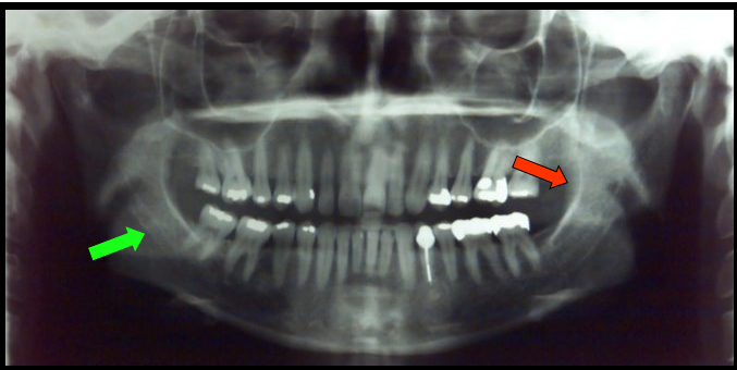
<ul><li><p><span>Appears as a radiolucent band outlined by two thin radiopaque lines representing the cortical walls of the canal.</span></p></li><li><p><span>Appears as a dense radiopaque band that extends downward and forward from the ramus to the molar region.</span></p></li></ul><p></p>
New cards
19

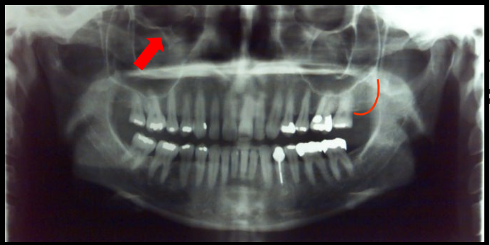
Zygomatic Process​

A bony projection of the maxilla that articulates with the zygoma or cheekbone.
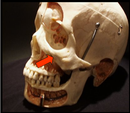
<p><span>A bony projection of the maxilla that articulates with the zygoma or cheekbone.</span></p>
New cards
20

Zygomatic Arch​

The cheekbone and it articulates with the zygomatic process of the maxilla.
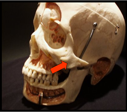
<p>The cheekbone and it articulates with the zygomatic process of the maxilla.</p>
New cards
21

Zygomatic Process and Arch​

  • Appears as a J or U-shaped radiopacity located superior to the maxillary first molar region.

  • Appear as a diffuse radiopaque band that extends posteriorly from the zygomatic process of the maxilla.
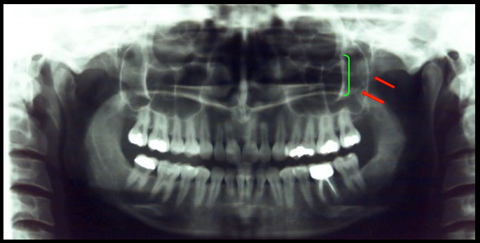
<ul><li><p><span>Appears as a J or U-shaped radiopacity located superior to the maxillary first molar region.</span></p></li><li><p><span>Appear as a diffuse radiopaque band that extends posteriorly from the zygomatic process of the maxilla.</span></p></li></ul><p></p>
New cards
22

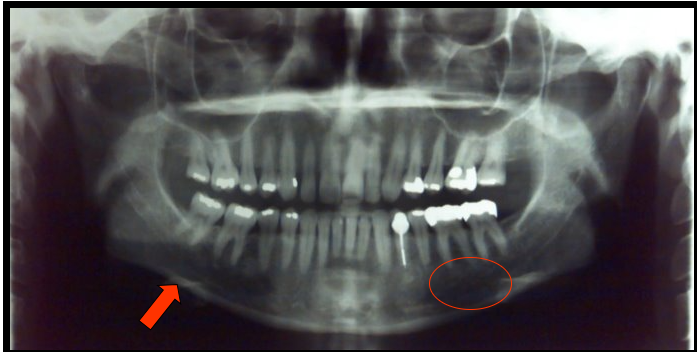
Inferior Border of Mandible​

A thick, linear prominence of cortical bone that defines the lower border of the mandible.
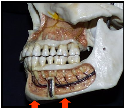
<p><span>A thick, linear prominence of cortical bone that defines the lower border of the mandible.</span></p>
New cards
23

Submandibular Fossa​

A scooped-out depressed are of bone located on the internal surface of the mandible, inferior to the mylohyoid ridge.
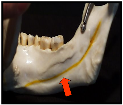
<p><span>A scooped-out depressed are of bone located on the internal surface of the mandible, inferior to the mylohyoid ridge.</span></p>
New cards
24

Inferior Border of the Mandible and Submandibular Fossa​

  • appears as a dense radiopaque band that outlines the lower border of the mandible.

  • appears as a diffuse radiolucent area in the molar region.

  • These landmarks can be seen on intraoral images.​

<ul><li><p><span>appears as a dense radiopaque band that outlines the lower border of the mandible.</span></p></li><li><p><span>appears as a diffuse radiolucent area in the molar region.</span></p></li><li><p><span>These landmarks can be seen on intraoral images.​</span></p></li></ul><p></p>
New cards
25

Mental Foramen​

An opening or hole in bone located on the external surface of the mandible in the region of the mandibular premolars.
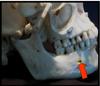
<p><span>An opening or hole in bone located on the external surface of the mandible in the region of the mandibular premolars.</span></p>
New cards
26

Soft Tissue of the Ear​

outer portion of the ear is composed of cartilage with a thin covering of connective tissue and skin
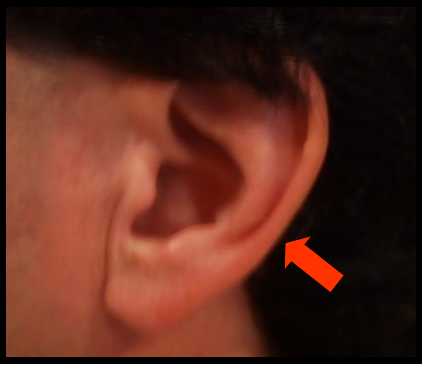
<p><span>outer portion of the ear is composed of cartilage with a thin covering of connective tissue and skin</span></p>
New cards
27

Mental Foramen and Soft Tissue of the Ear​

  • appears as a small, ovoid or round radiolucency located in the apical region of the mandibular premolars.

    • This landmark can be seen in intraoral images.

  • appears as a radiopaque shadow posterior to the condyle.

    • This landmark is not visible on intraoral images.​

<ul><li><p><span>appears as a small, ovoid or round radiolucency located in the apical region of the mandibular premolars.</span></p><ul><li><p><span>This landmark can be seen in intraoral images.</span></p></li></ul></li><li><p><span>appears as a radiopaque shadow posterior to the condyle.</span></p><ul><li><p><span>This landmark is not visible on intraoral images.​</span></p></li></ul></li></ul><p></p>
New cards
28

Incisive Foramen​

an opening, or hole, in bone that is located at the midline of the anterior portion of the hard palate directly posterior to the maxillary central incisors.
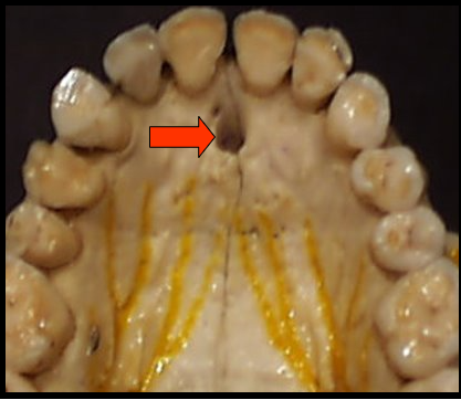
<p><span>an opening, or hole, in bone that is located at the midline of the anterior portion of the hard palate directly posterior to the maxillary central incisors.</span></p>
New cards
29

Hyoid Bone​

  • A horseshoe-shaped bone that lies below the mandible, between the chin and thyroid cartilage.

  • Ligaments and muscles located inferior to the mandible support this
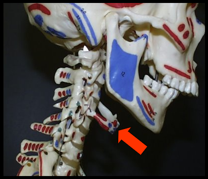
<ul><li><p><span>A horseshoe-shaped bone that lies below the mandible, between the chin and thyroid cartilage.</span></p></li><li><p><span>Ligaments and muscles located inferior to the mandible support this</span></p></li></ul><p></p>
New cards
30

Incisive Foramen and Hyoid Bone​

  • appears as a small, ovoid or round radiolucency located between the root of the maxillary central incisors.

  • appears as a “floating” curved radiopacity at or below the inferior border of the mandible.

    • is at the midline and as a result, appears as a double image and is seen on both the right and left sides of the panoramic image.
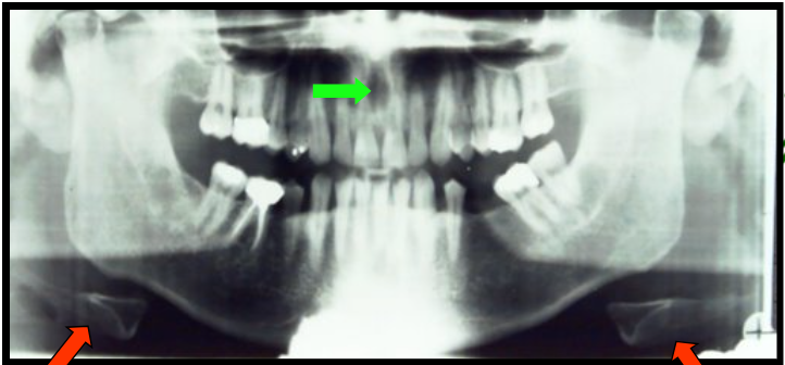
<ul><li><p><span>appears as a small, ovoid or round radiolucency located between the root of the maxillary central incisors.</span></p></li><li><p><span>appears as a “floating” curved radiopacity at or below the inferior border of the mandible.</span></p><ul><li><p><span>is at the midline and as a result, appears as a double image and is seen on both the right and left sides of the panoramic image.</span></p></li></ul></li></ul><p></p>
New cards
31

Maxillary Tuberosity​

A rounded prominence of bone that extends posterior to the third molar region.
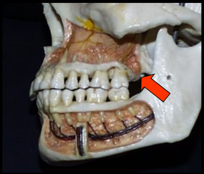
<p><span>A rounded prominence of bone that extends posterior to the third molar region.</span></p>
New cards
32

Infraorbital Foramen​

A hole, or opening in bone inferior to the border of the orbit.
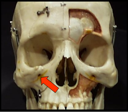
<p><span>A hole, or opening in bone inferior to the border of the orbit.</span></p>
New cards
33

Maxillary Tuberosity and Infraorbital Foramen​

  • appears as a radiopaque bulge distal to the third molar region.

    • This landmark is seen in intraoral images.

  • appears as a round or ovoid radiolucency inferior to the orbit.

    • It is not seen on intraoral images.

<ul><li><p><span>appears as a radiopaque bulge distal to the third molar region.</span></p><ul><li><p><span>This landmark is seen in intraoral images.</span></p></li></ul></li><li><p><span>appears as a round or ovoid radiolucency inferior to the orbit.</span></p><ul><li><p><span>It is not seen on intraoral images.</span></p></li></ul></li></ul><p></p>
New cards
34
<p><span>Vertibral Column​</span></p>

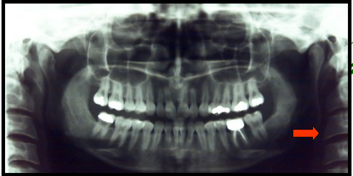
Vertibral Column​

  • The vertebrae and intravertebral discs form this

  • It is flexible column that supports the head, neck and body and allows for movement.

  • It also protects the spinal cord which passes through the openings in the vertebrae.

  • appears as a long radiopaque column on the left and right edges of a panoramic image.
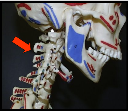
<ul><li><p><span>The vertebrae and intravertebral discs form this</span></p></li><li><p><span>It is flexible column that supports the head, neck and body and allows for movement.</span></p></li><li><p><span>It also protects the spinal cord which passes through the openings in the vertebrae.</span></p></li><li><p><span>appears as a long radiopaque column on the left and right edges of a panoramic image.</span></p></li></ul><p></p>
New cards
35

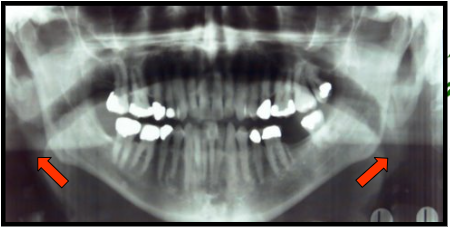
Air Spaces​

The air spaces on a panoramic radiograph appear as wide radiolucent bands which form an arch shape spanning across or above the roots of the maxillary teeth then downward toward the angle of the mandible.

New cards
36

palatoglossal airspace

  • is the space between the palate and the tongue.​

  • appears as a horizontal well-defined radiolucent band located superior to the apices of the maxillary teeth.
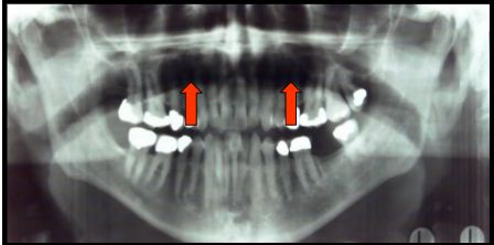
<ul><li><p>is the space between the palate and the tongue.​</p></li><li><p>appears as a horizontal well-defined radiolucent band located superior to the apices of the maxillary teeth.</p></li></ul><p></p>
New cards
37

glossopharyngeal air space

  • is an outline of the pharnx.​

  • appears as a vertical radiolucent band superimposed over the ramus of the mandible.
<ul><li><p><span>is an outline of the pharnx.​</span></p></li><li><p><span>appears as a vertical radiolucent band superimposed over the ramus of the mandible.</span></p></li></ul><p></p>
New cards
38
New cards
robot