Topic 6: Cell function
1/76
There's no tags or description
Looks like no tags are added yet.
Name | Mastery | Learn | Test | Matching | Spaced |
|---|
No study sessions yet.
77 Terms
Properties of plasma membrane
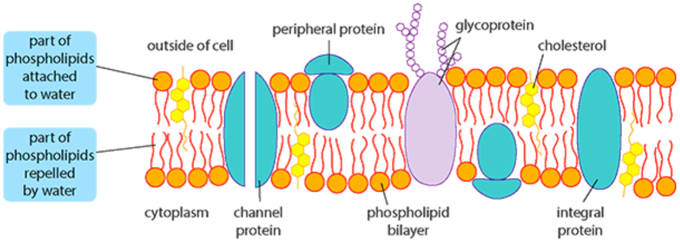
7-10 nm thick, made of phospholipids, selectively permeable, fluid (molecules can move everywhere, even switch layers)
Phospholipids
amphipathic molecules (polar hydrophilic heads, nonpolar hydrophobic tails)
What is the role of cholesterol in the membrane
in animal cells (plant cells have sterol), one end associates with the polar heads whilst the other with the nonpolar fatty acid chains, makes the membrane less fluid/permeable and more rigid
What is the role of integral proteins in the membrane
either embedded in the membrane (fully hydrophobic) or spanning the whole length (amphipathic), can be immobilised enzymes, carry out sequences of metabolic reactions
What is the role of peripheral proteins in the membrane
hydrophilic, surface of membrane (often attached to integral proteins), often glycoproteins (proteins w/ carbohydrates), can be hormone binding sites (recognise hormones/antigens) or used in cell communication/cell adhesion
What is the role of channel proteins in the membrane
integral proteins spanning the bilayer, let ions and molecules pass by passive transport, form pumps for active transport
What is the role of aquaporins in the membrane
channel proteins allowing transfer of water and small solutes
What is the role of glycolipids in the membrane
lipids with carbohydrates attached (glycosidic bonds), maintain cell membrane stability, used for cell recognition (self-antigens)
Membrane fluidity
affected by temperature (fluidity increases with temp), cholesterol (stabilises and stiffens, prevents phospholipids aggregating), also saturated and unsaturated fatty acids (unsaturated have kinks, ↑fluid) - ratio of saturated to unsaturated fatty acids is changed by cells based on temperature
Importance of membrane fluidity
binding of peripheral proteins, movement of enzymes within the membrane, cell signalling, exocytosis and endocytosis
CAMs
cell adhesion molecules are surface glycoproteins
Junctions made by CAMs
• cells to cells
• cells to extracellular matrix (chains of sugar and proteins surrounding cells to make tissues more stable - eg. collagen)
• extracellular matrix to cell cytoskeleton
Role of CAMs
organised tissue structure (adhesion of cells to one another), transmission of cues and signals from outside the cell (through membrane), movement of certain cells (contact inhibition - regulating what cell they adhere to), apoptosis
Types of CAMs
• integrins (connect cells to extracellular matrix for integration) • immunoglobulins (adhesion molecules in the nervous system) • cadherins (cell to cell adhesion) • selectins (involved in inflammation, bind to cell surface carbohydrates)
Homophobic vs heterophilic binding
binding to same or different CAMs
How does increased protein production affect cell composition
it increases rough endoplasmic reticulum content in cells
Difference between free ribosomes and membrane-bound ribosomes
free ribosomes make proteins for cell use, membrane-bound ribosomes produce proteins for Golgi apparatus
What are polyribosomes
ribosomes attached to an mRNA strand as they are translating it, and they can bind to the RER
Role of the Golgi apparatus
receives proteins for modification, packaging, and release in lysosomes (within vesicles = exocytosis, in vesicle membranes = part of plasma membrane)
Lysosomes
small spherical organelles containing hydrolytic enzymes (proteases, amylases, nucleases, lipases) with acidic pH, engulf and break down macromolecules, destroy invading particles, burst causing apoptosis (“suicide packets”)
Describe nuclear structure
two membranes (inner and outer phospholipid bilayers) which connect to the RER lumen (cisternae), inner membrane has protein lining binding to chromatin and other nuclear contents (structural framework), nuclear envelope acts as a barrier with cytoplasm containing nuclear pores (entry of DNA/RNA nucleotides, ATP and histones/other large proteins, regulating mRNA and ribosome subunit export from nucleus to cytoplasm)
Contents of the phospholipid bilayer
cholesterol, integral proteins, peripheral proteins, channel proteins, aquaporins, glycolipids
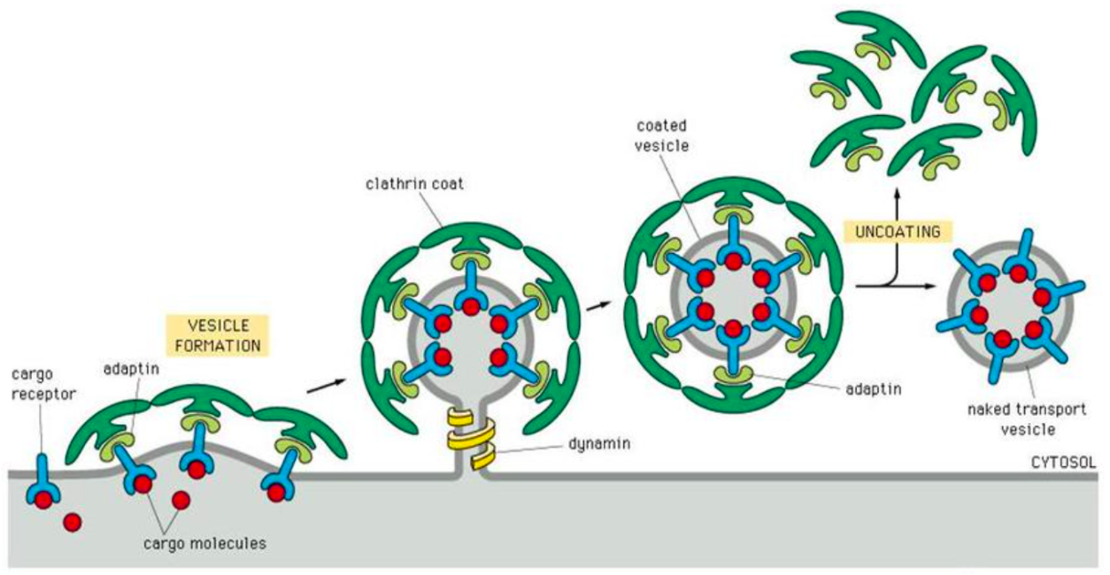
Coated vesicles
vesicles coated with clathrin protein complex can bud from the Golgi apparatus or the membrane, and proteins in the coat determine the destination in the cell
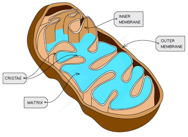
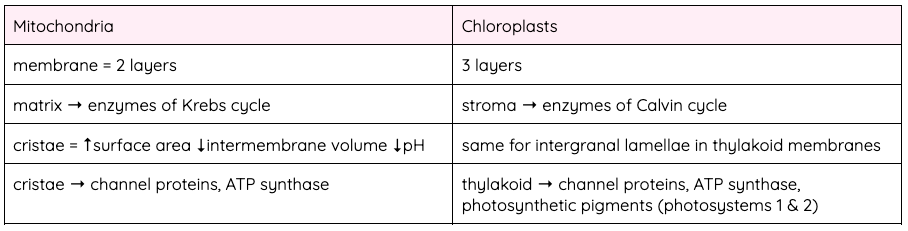
Describe mitochondrial structure
inner membrane highly folded into cristae to increase surface area (with ATP synthase - H+ ions pass through), also the cristae create compartments in which the reactions for aerobic respiration take place, inner volume called the matrix separates the enzymes in the reactions of the Krebs cycle, the intermembrane space (between inner and outer membranes) has a small volume and high concentration of H+ ions (created by Krebs cycle - lower pH)
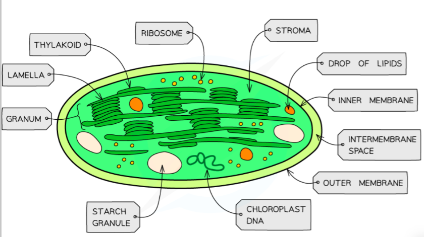
Describe chloroplast structure
inner volume is the stroma (liquid with enzymes for the Calvin cycle - light-independent reactions), thylakoids fold the membrane to produce grana and lamellae (the light-dependent reactions) and have a large surface area and small intermembrane space with the membrane proteins pumping H+ ions into the inner thylakoid space (lowering pH), passed through ATP synthase into the stroma
Compare mitochondria and chloroplasts
Concentration gradient
difference in concentration of a substance between two regions
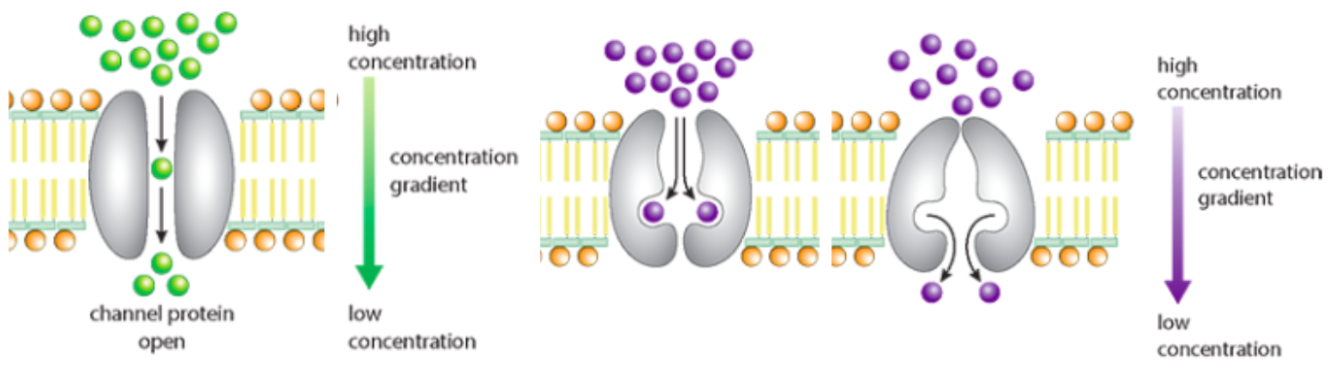
Simple diffusion
net movement of molecules from an area of higher concentration to an area of lower concentration (only occurs if the membrane is fully permeable to that substance)
Passive movement
net movement of molecules down a concentration gradient from higher to lower concentration without the need for energy (eg. simple or facilitated diffusion, osmosis)
Osmosis
net movement of water across a semi-permeable plasma membrane from an area of lower water potential to an area of higher water potential
What cannot move by active transport or simple/facilitated diffusion?
large chemical molecules, thus moved in vesicles
General endocytosis
uptake of molecules into a cell via vesicles, requiring ATP and fluidity as part of the plasma membrane is pulled inwards surrounding the molecules and pinching off making a vesicle
Two types of endocytosis
phagocytosis (substances taken in are particles - eg. bacteria) and pinocytosis (substances are in solution - eg. end products of digestion)
Exocytosis
export of molecules from a cell via vesicles, requiring ATP and fluidity as materials for export (like digestive enzymes) made in RER are transported to the Golgi apparatus where they’re enclosed in vesicles and moved to the plasma membrane along microtubules, fusing with it and releasing contents outside
Facilitated diffusion
diffusion across a membrane allowing large and/or charged molecules to pass via channel proteins forming pores (hydrophilic interior, specific only allowing one substance through, some permanently open while others gated) or carrier proteins (combine with diffusing molecules and release them on the other side of the membrane)
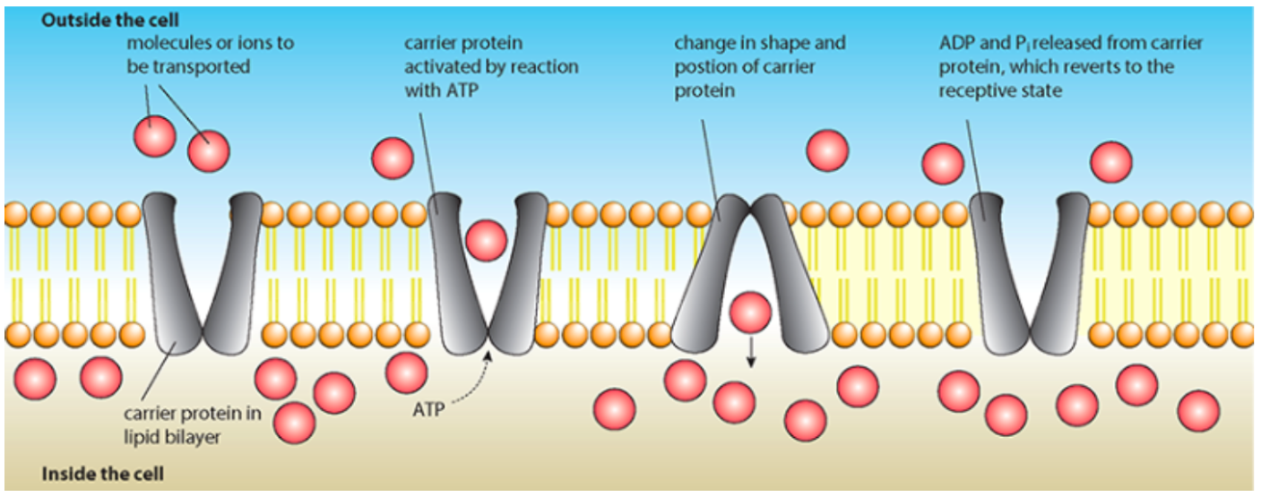
Active transport
movement of molecules from an area of lower to higher concentration against a concentration gradient, using metabolic energy (from breakdown of ATP) and pump proteins, it is specific
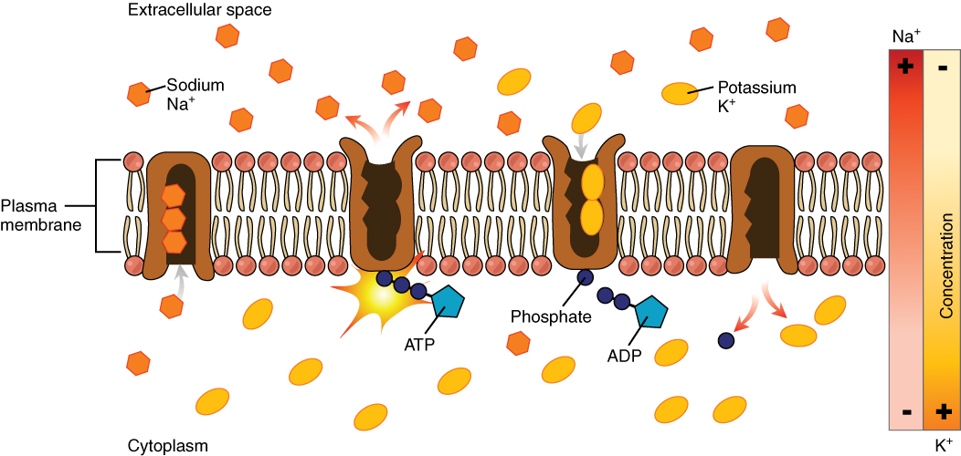
Sodium-potassium pump
protein pump doing active transport binding 3 Na+ and ATP, hydrolysing ATP to change shape and releasing the Na+ ions in the extracellular space binding 2 K+, release of the phosphate allows the channel to revert to the original position and release the K+ ions intracellularly
Water potential
potential energy of water per unit volume (unit = kPa)
Values of water potential
impossible to measure absolute quantity so values are relative to pure water (0 kPa) at atmospheric pressure and 20°C
Factors affecting water potential
concentration of solute molecules and pressure of H2O molecules in the solution
Water potential formula
water potential (ψw) = solute potential (ψs) + pressure potential (ψp)
Describe the solute potential
goes from 0 downwards and the more solute is present the lower the solute potential, since water molecules make hydrogen bonds and surround solute molecules leaving fewer molecules free to move
Describe the pressure potential and how it is affected by extracellular water potential
generally positive inside cells, if placed in a solution with higher/lower water potential then water will move out/in respectively affecting the pressure potential until equilibrium is reached
Example of negative pressure potential
transpiration stream: in the xylem which pulls up the water via capillary action
Hypotonic solution
higher water potential in the solution compared to the cell (water moves into the cell, animal cells will often lyse bcs no cell wall, plant cells will fill up and exert turgor pressure on the cell wall)
Isotonic solution
same osmotic concentration so same water potential (no net movement of water, used in hospitals for dehydrated people and storage of organs for transplantation)
Hypertonic solution
lower water potential in the solution (water moves out of the cell, animal cells will shrink via crenation, plant cells will become flaccid or even plasmolysed as the plasma membrane pulls away from the cell wall)
Limitation to cell size
surface area to volume ratio (affects distance and so diffusion rate)
Specialised structures to increase surface area
folds, microvilli, flattened or elongated cells - all increase SA to V ratio
What happens to cells that are too large (SA:V)?
cells divide
Chemicals regulating cell growth
growth factors (proteins which regulate cell growth and survival)
Chemicals regulating mitosis
mitogens (peptides or small proteins that cause cells to enter mitosis by binding to receptors on plasma membranes)
Exceptions to normal cell cycle
grow without dividing (neurons, fat, muscle fibres) vs divide without growing (fertilised egg cell)
Cell cycle duration
about 24 hours
Cell cycle generates new cells
for growth, repair of damaged tissues (or to replace old cells) and asexual reproduction
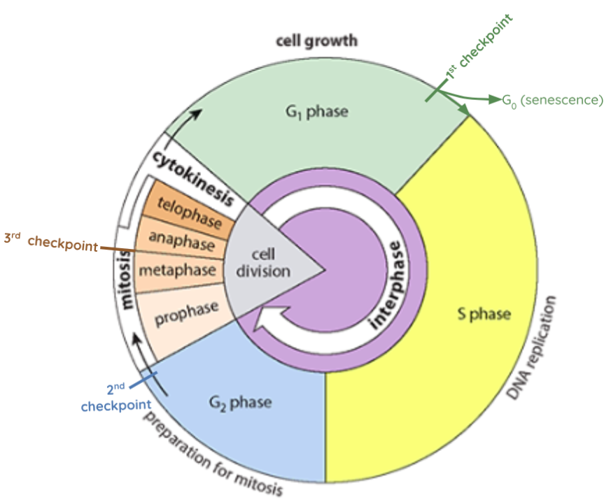
Interphase
number of mitochondria increases so the respiratory rate is sufficient for cell division energy 1. G1 gap phase 1 → cell growth, DNA transcription, protein synthesis S synthesis phase → DNA replication G2 gap phase 2 → cell growth, some organelles are duplicated, cell prepares for division
Cytokinesis in animal cells
two sides of the plasma membrane meet and split the cell into two, ring of contractile actin and myosin pinches the cell membrane together
Cytokinesis in plant cells
two sides of the plasma membrane meet and split the cell into two, cell plate forms along the centre of the cell membrane separating it into two regions, then vesicles filled with cellulose and pectin accumulate at the edges of the cell plate, before bursting and releasing their contents which are then used to build the cell wall for each new daughter cell
Examples of asymmetrical cytokinesis
oogenesis (one cell all cytoplasm + polar bodies), yeast (new cells budding off original parent cell, smaller and longer cell cycle)
Proto-oncogenes
functioning genes which help regulate normal cell growth, signalling to initiate cell division or apoptosis. Most are important in embryo development. However, these genes can become permanently overactive or reactivate at a later stage they become oncogenes and can cause cancer (uncontrolled and abnormal mitosis). These can be caused by gene amplification/duplication (accidental repetitions of the gene), mutations (often by environmental factors - such as UV light, viral infections) or chromosomal rearrangement (moved to a different area of the chromosome)
Tumour suppressor genes
functioning genes which slow down cell division, repair DNA or control apoptosis. Without them, cells can grow excessively and out of control
Cancer differences from tumour
primary tumour undergoes metastasis and migrates to other tissues, becoming a secondary tumour or cancer
Apoptosis
controlled cell death (very different to necrosis - death by damage/injury), for example caused by damaged DNA which can’t be repaired by DNA polymerases
Role of apoptosis
removal of cells during development, cancerous cells or infected cells (by viruses)
Mechanism of apoptosis
cell will shrink and develop ‘blebs’ (bubble-like extensions) on its surface. The DNA will break down and organelles/plasma membrane will be fragmented and packaged into membranes (apoptotic bodies) which have signals on their own membranes to get engulfed by phagocytic macrophages
Meiosis definition
reduction division which produces gametes (haploid nucleus)
Sources of genetic variation in sexual reproduction
random mating, crossing over, independent assortment and random fertilisation
Non-disjunction of homologous chromosomes
Sometimes in anaphase II the centromere doesn’t split and the pair doesn’t separate properly (more common in women and esp. when old), so some eggs have too few or too many chromosomes (trisomy on chromosome 21 causes Down syndrome)
Karyotyping
cells from unborn child collected through chorionic villus sampling (CVS) or amniocentesis, then grown in lab and karyotype performed to check chromosomes
Mendel’s 1st law (Law of segregation)
individuals possess two alleles and parents pass only one allele to offspring
Mendel's 2nd law (Law of independent assortment)
during gamete formation the segregation of the alleles of one gene is independent from the segregation of the alleles of other pairs (metaphase I, bivalents line up randomly) - aka one gene does not influence any other gene as alleles are sorted into gametes, making all allele combinations equally likely and increasing variation
Main stages of the cell cycle
interphase, mitosis (division of nucleus) and cytokinesis (division of cytoplasm)
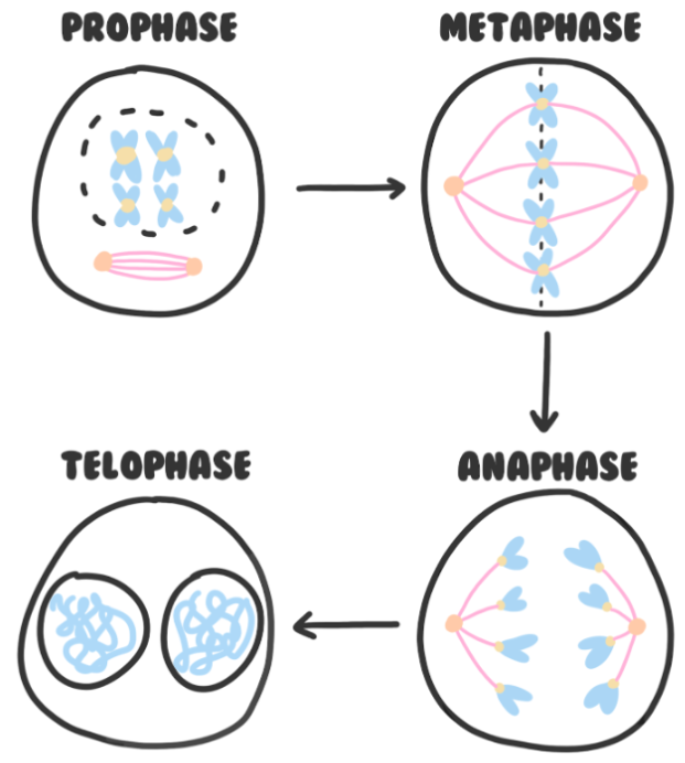
Mitosis
PMAT: Prophase (chromosomes supercoil condense preventing transcription, centrioles move to opposite sides of cell and spindle fibres start forming, nuclear envelope breaks down)
Metaphase (sister chromatids align on the equator, microtubules attach to centromeres)
Anaphase (centromeres split and sister chromatids are pulled to opposite poles of the cell as spindle fibres shorten)
Telophase (spindle fibres break down, nuclear envelopes and nucleoli reform, chromosomes uncoil)
Control of the cell cycle
1st checkpoint (G1) → received signals from other cells (growth factors), large enough, sufficient nutrients - if not goes into senescence
2nd checkpoint (G2) → large enough, chromosomes have been duplicated
3rd checkpoint (Metaphase) → chromosomes are attached to the spindles

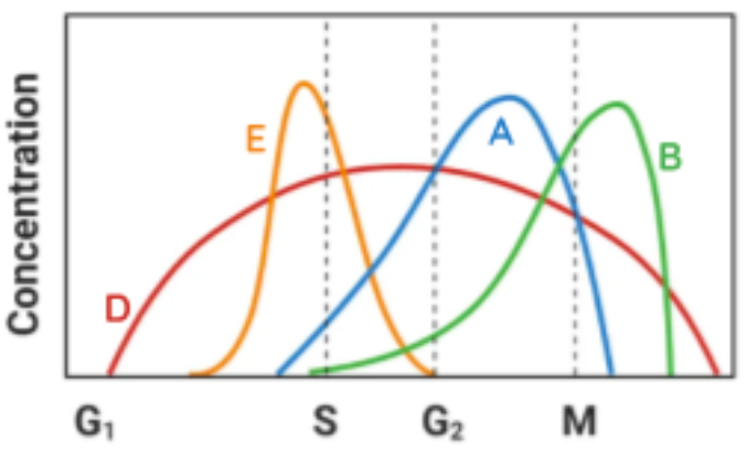
Chemicals regulating cell cycle
cyclin-dependent kinases (CDKs - kinase proteins which phosphorylate necessary proteins when cyclins bind to them) control microtubule formation and chromatid alignment:
D → influenced by growth-regulating signals, coordinates cell growth + start new cycle
E → starts the initial process of DNA replication, promotes centrosome duplication
A → induces DNA replication, promotes early events in mitosis
B → influences the formation of mitotic spindles and alignment of sister chromatids
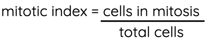
Mitotic index
ratio of cells undergoing mitosis to cells not undergoing mitosis in a population of cells. If it is higher than normal, it could be an indicator of cancer
Meiosis steps
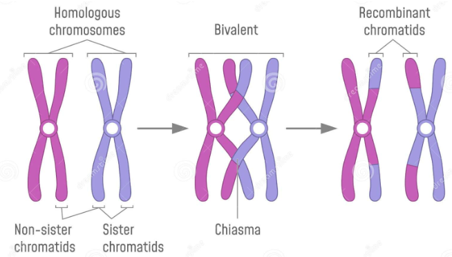
Meiosis I: Prophase I → chromosomes are supercoiled, homologous chromosomes pair up into a bivalent (2 chromosomes, 4 chromatids), crossing over at chiasma forming new combinations of alleles
Crossing over occurs at the same locus (p: loci) when two chromosomes touch and break. They then rejoin on the other chromosome, forming a chiasma (p: chiasmata) to make new combinations. This doesn’t occur on the X and Y chromosomes, centrioles move to the poles of the cell and spindles begin to form, nuclear envelope is broken down
Metaphase I → bivalents line up on the equator (alignment is random – aka independent assortment or random orientation) and spindle fibres attach to the centromeres (1 spindle per centromere, 2 in mitosis)
Anaphase I → microtubules contract to opposite poles and the homologous chromosomes are separated (stay pairs of sister chromatids as the centromeres don’t divide) – this is the reduction division (cell becomes haploid)
Telophase I → spindle fibres break down nuclear envelopes reform around each set of chromosomes and nucleoli reform chromosomes reach opposite sides of the cell, then cytokinesis
Meiosis II: all same as mitosis