Mycology Lecture - Dr. Abramovitch
 Call Kai
 Learn
Learn Practice Test
 Spaced Repetition
 Match
Match Flashcards
 Knowt Play
1/53

There's no tags or description
Looks like no tags are added yet.
Name | Mastery | Learn | Test | Matching | Spaced | Call with Kai | Chat |
|---|
No analytics yet
Send a link to your students to track their progress
54 Terms
⭐ 1. Q: What are the defining characteristics of fungi?
Fungi are non-photosynthetic eukaryotes that often have a cell wall composed of chitin and other polysaccharides.
🔎 What this means:
Eukaryotic means fungal cells belong to the group of cells with complex internal organization, unlike prokaryotic bacteria.
The lecture emphasizes two big clues:
Fungus = eukaryote + non-photosynthetic + often has a chitin-containing cell wall.
⭐ 2. Q: How does the lecture distinguish fungi from eubacteria, plants, and animals?
Fungi are eukaryotes whereas eubacteria are prokaryotes; fungi are non-photosynthetic and form a kingdom separate from plants; fungi often have chitin-containing cell walls, and their plasma membranes contain ergosterol, which is absent from animal cells.
🔎 What this means:
Ergosterol is a type of lipid found in Fungi cell membranes. It acts similar to how cholesterol does in animals
Q: In the lecture's simplified tree of life, which eukaryotic kingdom branches most closely with fungi?
Animals branch most closely with fungi in the simplified tree shown in the lecture.
🔎 What this means:
The figure is another reminder that fungi are not plants even though fungi can have cell walls.

🟡 LOWER PRIORITY 3. Q: Are most fungal species pathogenic to animals?
No. The lecture states that most described fungal species are non-pathogenic, with approximately 400 described as pathogenic to animals.
🔎 What this means:
More than 80,000 fungal species were listed in the slide, but only a relatively small fraction were described as animal pathogens.
⭐ 4. Q: How do yeasts and molds differ in fungal morphology?
Yeasts are unicellular fungi that divide by budding, whereas molds are multicellular fungi that grow from the tips of branching filaments.
🔎 What this means:
Yeast → individual cells
Mold → branching filamentous growth

⭐ 5. Q: What are a hypha and a mycelium?
A hypha is a branching thread-like fungal filament, whereas a mycelium is a mass of intertwined hyphae.
🔎 What this means:
One filament = hypha
Many hyphae growing together = mycelium
💡 Think: individual thread versus the whole tangled network.

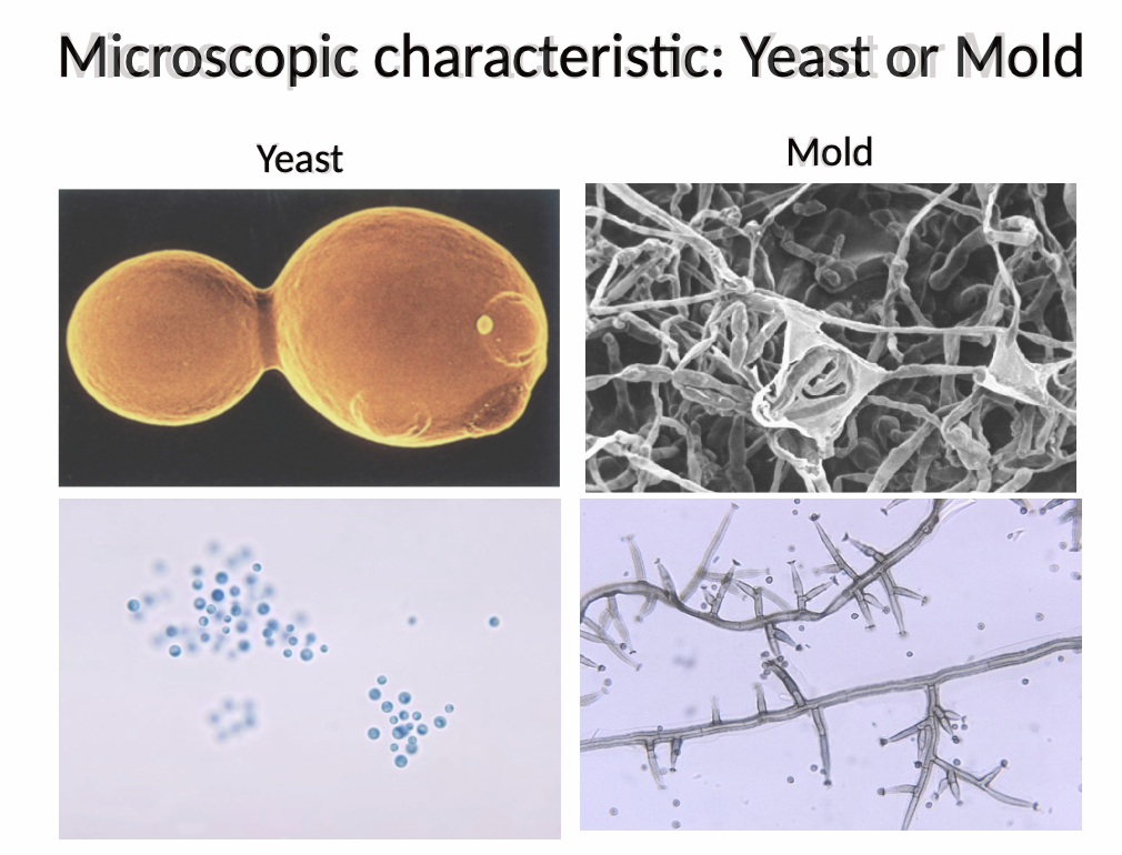
⭐ 6. Q: Which microscopic characteristics can be used to characterize different fungi?
Fungi can be characterized microscopically by whether they are yeast or mold, whether their hyphae are septate or aseptate, the characteristics of their spores and spore heads, and their size.
🔎 What this means:
Septate hyphae have internal walls called septa that divide the long fungal filament into compartments.
Aseptate hyphae lack those septa, so the filament looks more like one continuous tube. The lecture’s diagram shows exactly that visual difference.
Septate = has little dividers inside the hypha
Aseptate = no internal dividers

⭐ 7. Q: How do septate and aseptate hyphae differ?
Septate hyphae contain cross-walls called septa, whereas aseptate hyphae lack these cross-walls.
🔎 What this means:
A septum divides a hypha internally.
Septate → visible internal divisions
Aseptate → continuous filament
This distinction becomes especially important when identifying fungal phyla.

⭐ 8. Q: How do fungal spores contribute to fungal spread?
Fungi can spread by producing and disseminating spores, including spores called conidia and sporangiospores in some fungal species.
🔎 What this means:
Spores are an important dispersal form.
Their appearance and method of production can also help characterize the fungus.

⭐ 9. Q: What is the difference between a conidiophore and a sporangiophore?
A conidiophore is a specialized hyphal stalk associated with conidia, whereas a sporangiophore is a specialized hyphal stalk associated with sporangiospores.
🔎 What this means:
These are spore-bearing structures.
Their shape and the spores associated with them can help identify fungi microscopically.

⭐ 10. Q: How does fungal size generally compare with bacterial size?
Fungi are generally larger than bacteria.
🔎 What this means:
The lecture compares:
Saccharomyces cerevisiae yeast: about 5–10 μm
Staphylococcus aureus: about 0.5–1.5 μm
Because fungi are larger, they are often easier to resolve at lower microscope magnification.

⭐ 11. Q: Why does filamentous growth alone not prove that a microorganism is a fungus?
Filamentous growth alone does not prove an organism is fungal because some bacteria, including Nocardia and Actinomyces, can also appear filamentous.
🔎 What this means:
“Looks like threads” is not enough.
You need additional microscopic characteristics.

⭐ 12. Q: Why can potassium hydroxide be used when examining a specimen for fungi?
Fungal cell walls resist degradation by potassium hydroxide, allowing fungal hyphae or yeast to remain visible during microscopic examination.
🔎 What this means:
Potassium hydroxide is abbreviated KOH.
The tough fungal wall remains while the specimen is prepared for examination.

Q: How is a KOH preparation performed for microscopic fungal examination?
A specimen is placed on a slide, one to two drops of 10% KOH are added with a coverslip, the preparation sits for 15–30 minutes, and it is then examined for yeast or hyphae.
🔎 What this means:
The lecture lists possible samples such as:
skin scraping, nail, hair, swab, or body fluid.

⭐ 13. Q: Which stains are used in the lecture to improve microscopic visualization of fungi?
Lactophenol cotton blue and calcofluor white are used to improve fungal visualization.
🔎 What this means:
These are additional methods for making fungal structures easier to see.

Q: What is distinctive about calcofluor white staining?
Calcofluor white specifically stains fungi and fluoresces under ultraviolet light.
🔎 What this means:
UV = ultraviolet light.
The fluorescence makes fungal structures easier to recognize microscopically

⭐ 14. Q: Which macroscopic characteristics can be used to characterize fungal colonies?
Fungal colonies can be characterized macroscopically by their texture, topography, and color.
🔎 What this means:
These are characteristics you can observe at the colony level, rather than by examining individual fungal structures under a microscope.
💡 Macroscopic = texture + topography + color.
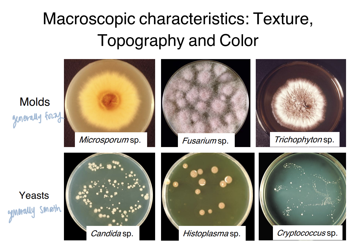
⭐ 15. Q: How do microscopic and macroscopic fungal characterization differ?
Microscopic characterization examines fungal form, hyphal septation, spores, spore heads, and size, whereas macroscopic characterization examines colony texture, topography, and color.
🔎 What this means:
Microscope → cells and fungal structures
Colony → visible growth characteristics
⭐ 16. Q: What is a dimorphic fungus?
A dimorphic fungus is a fungus that can exist in either a mold form or a yeast form.
🔎 What this means:
Dimorphic = two forms.
Which form is present depends on the conditions described in the lecture.
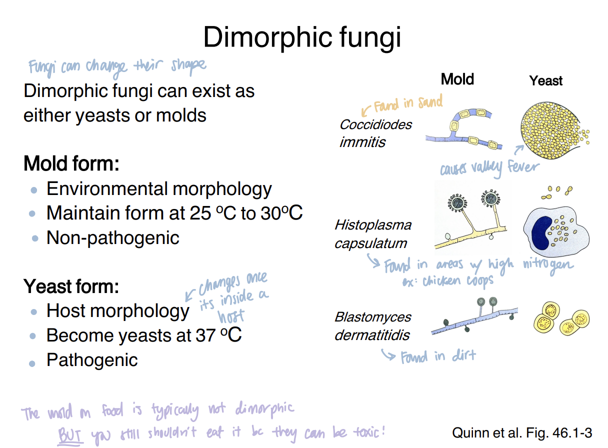
⭐ 17. Q: How do the environmental and host forms of dimorphic fungi differ?
The lecture describes the environmental form as a non-pathogenic mold maintained at approximately 25–30°C and the host form as a pathogenic yeast that develops at approximately 37°C.
🔎 What this means:
Course framework:
Environment, 25–30°C → mold
Host, 37°C → yeast

⭐ 18. Q: What are the three broad mechanisms by which fungi can cause disease in animals?
Fungi can cause disease through mycotoxins that cause mycotoxicosis, spores that cause Type I respiratory allergies, and virulence factors that cause mycoses.
🔎 What this means:
These are three different disease mechanisms:
Toxin-mediated disease
Allergic disease
Fungal infection
⭐ 19. Q: How are mycoses categorized in the lecture?
Mycoses are categorized according to the location of infection as cutaneous, subcutaneous, or systemic mycoses.
🔎 What this means:
Mycosis = fungal infection.
⭐ 20. Q: How do pathogenic fungi and opportunistic fungal pathogens differ?
Pathogenic fungi can cause disease in immunocompetent individuals and commonly show thermal dimorphism, whereas opportunistic fungi primarily cause disease in immunocompromised individuals and less commonly show dimorphism.
🔎 What this means:
The important distinction is the host immune status needed for disease to develop.
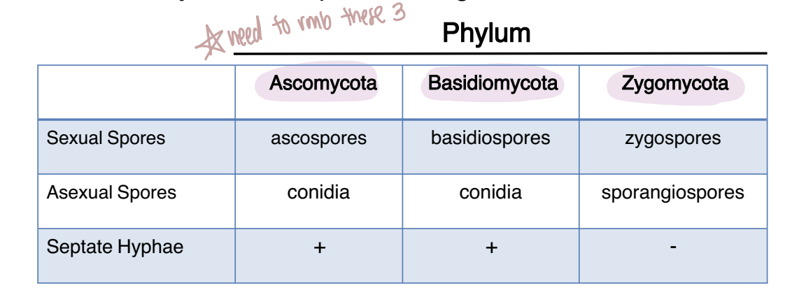
⭐ 21. Q: Which three fungal phyla are used in the lecture's taxonomy?
The three phyla are Ascomycota, Basidiomycota, and Zygomycota.
🔎 What this means:
The course distinguishes these groups mainly through:
sexual spores
asexual spores
whether the hyphae are septate
⭐ 22. Q: What are Deuteromycota, or “imperfect fungi”?
Deuteromycota is an informal grouping for fungi that do not have a known sexual stage.
🔎 What this means:
The lecture does not present Deuteromycota as one of the three main sexually reproducing phyla.
⭐ 23. Q: How do Ascomycota, Basidiomycota, and Zygomycota differ in spores and hyphal septation?
Ascomycota have septate hyphae, produce ascospores sexually, and conidia asexually;
Basidiomycota have septate hyphae, produce basidiospores sexually, and conidia asexually;
Zygomycota have aseptate hyphae, produce zygospores sexually, and sporangiospores asexually.

⭐ 24. Q: How are sexual spores organized in Ascomycota?
Sexual reproduction in Ascomycota produces an ascocarp containing spore sacs called asci, and each ascus contains ascospores.
🔎 What this means:
Think from outside to inside:
Ascocarp → asci → ascospores
A cleistothecium is given as an example of an ascocarp.

⭐ 25. Q: How does sexual reproduction occur in Zygomycota?
Sexual reproduction occurs when two hyphae come together to form a zygosporangium containing zygospores.
🔎 What this means:
Two hyphae meet → zygosporangium → zygospores
This phylum also has aseptate hyphae in the lecture's classification.

⭐ 26. Q: How are sexual spores produced in Basidiomycota?
Sexual reproduction in Basidiomycota produces a club-like structure called a basidium that bears basidiospores.
🔎 What this means:
Basidium = structure
Basidiospores = sexual spores
Mushrooms are also identified as members of Basidiomycota.


⭐ 27. Q: What is this phylum?
The fungus belongs to Zygomycota.
🔎 What this means:
The key visual clue is the zygosporangium formed between hyphae.


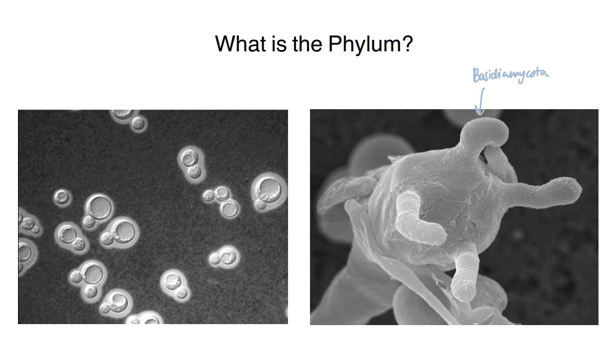
⭐ 28. Q: What is this phylum?
The fungus belongs to Basidiomycota.
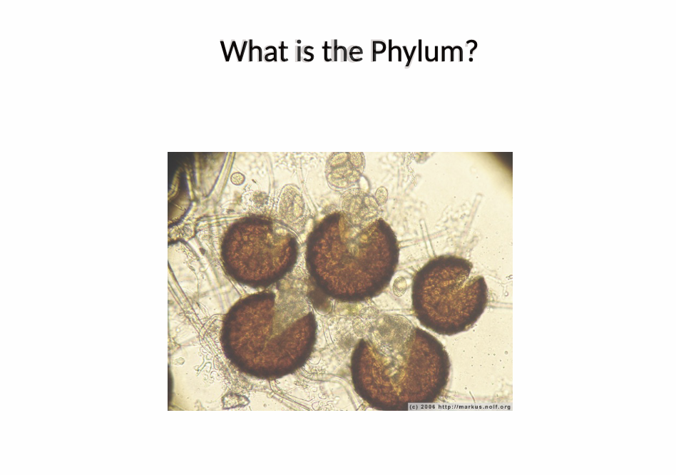
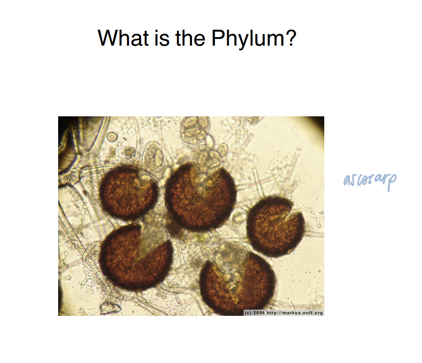
⭐ 29. Q: What is this phylum?
The fungus belongs to Ascomycota.
⭐ 30. Q: What structures make up the fungal cell envelope?
The fungal cell envelope consists of the plasma membrane, the surrounding cell wall, and sometimes an external capsule.
🔎 What this means:
From inside toward outside:
Plasma membrane → cell wall → optional capsule
The lecture specifically identifies these as the major envelope structures.

⭐ 31. Q: Where is a fungal capsule located, and what is its major function?
A fungal capsule is a polysaccharide layer outside the cell wall that protects the fungus from environmental stresses and the host immune system.
🔎 What this means:
Not every fungus has a capsule.
When present, it provides an additional protective outer layer.
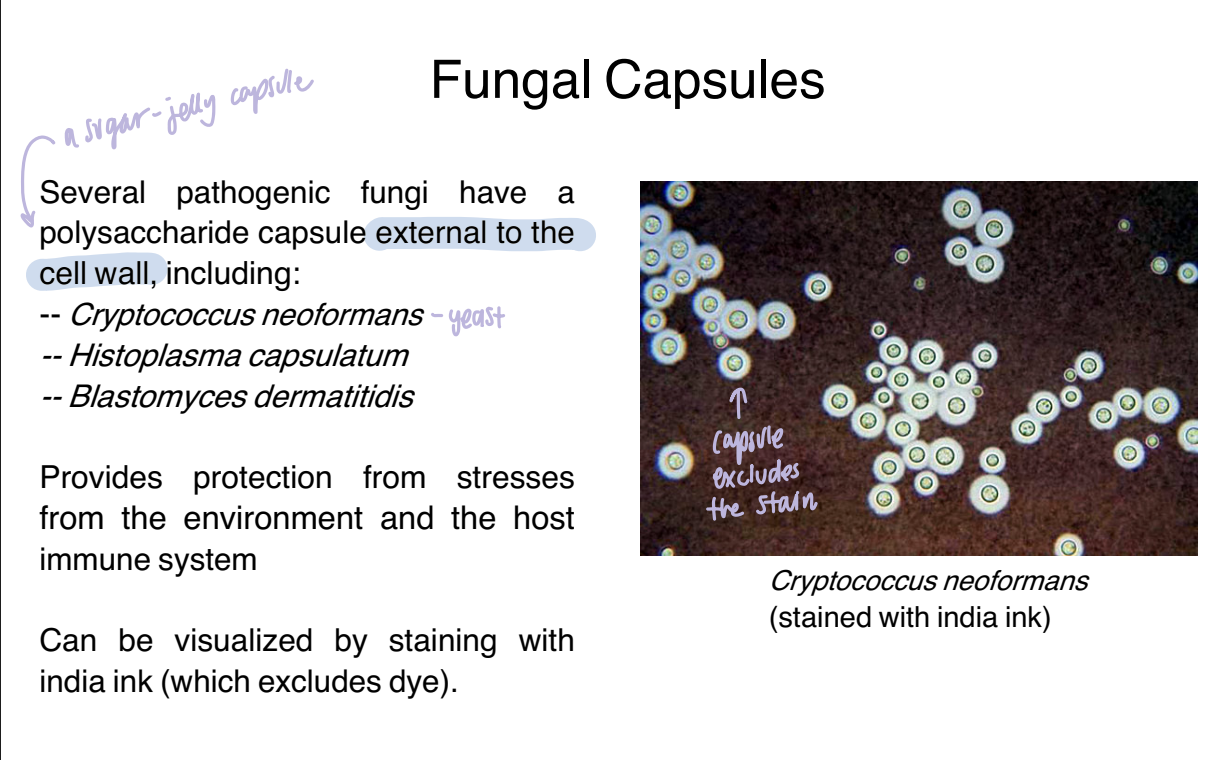
⭐ 32. Q: How can a fungal capsule be visualized with India ink?
India ink excludes dye from the capsule, allowing the unstained capsule to be visualized around the fungal cell.

⭐ 33. Q: What is ergosterol, and where is it found?
Ergosterol is a sterol found in the fungal plasma membrane that is absent from animal cells.
🔎 What this means:
This makes ergosterol a useful fungal-specific target.
💡 Ergosterol → membrane.
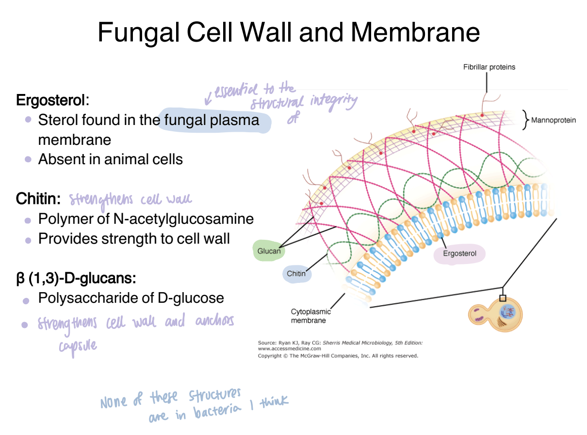
⭐ 34. Q: What is chitin, and what is its role in the fungal cell wall?
Chitin is a polymer of N-acetylglucosamine that provides strength to the fungal cell wall.
🔎 What this means:
Polymer means a large molecule composed of repeating smaller units.
💡 Chitin → structural strength.

⭐ 35. Q: What are β(1,3)-D-glucans?
β(1,3)-D-glucans are fungal cell-wall polysaccharides composed of D-glucose.
🔎 What this means:
Like chitin, these are carbohydrate components of the fungal wall.

⭐ 36. Q: How do ergosterol, chitin, and β(1,3)-D-glucans differ in the fungal envelope?
Ergosterol is a sterol in the plasma membrane, chitin is an N-acetylglucosamine polymer that strengthens the cell wall, and β(1,3)-D-glucans are D-glucose polysaccharides in the cell wall.
🔎 What this means:
This is the major envelope comparison:
Ergosterol → membrane
Chitin → wall
β-glucan → wall
The three molecules and their characteristics are shown together on the fungal-envelope slide.

⭐ 37. Q: Why are fungal cell-wall and plasma-membrane components useful drug targets?
Fungal-specific features of the cell wall and plasma membrane provide targets that antifungal drugs can act on.
🔎 What this means:
The lecture specifically points to molecules such as ergosterol, chitin, and glucans as important fungal-envelope targets.
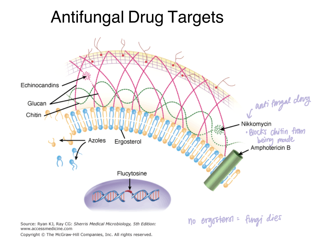
Q: According to the antifungal-target diagram, which major fungal targets are associated with different antifungal drugs?
The diagram associates echinocandins with glucan, nikkomycin with chitin, azoles and amphotericin B with the ergosterol-containing membrane, and flucytosine with fungal genetic material.

⭐ 38. Q: How can the differences between fungi and bacteria affect microscopic diagnosis?
Fungi are generally larger than bacteria, can retain recognizable structures during KOH preparation, and can be visualized with fungal stains such as lactophenol cotton blue and calcofluor white.
🔎 What this means:
The lecture gives you multiple clues rather than one perfect distinguishing feature.
Remember that filamentous growth alone is not enough, because some bacteria are also filamentous.
⭐ 39. Q: How can fungal cell-envelope differences affect treatment?
Fungal-specific cell-wall and plasma-membrane molecules provide targets for antifungal drugs.
🔎 What this means:
Examples from the slides include:
ergosterol, chitin, and β(1,3)-D-glucans.
⭐ 40. Q: A fungus is multicellular, grows from filament tips, and forms a network of intertwined hyphae. Is it a yeast or mold?
It is a mold.
🔎 What this means:
Multicellular filamentous growth is the mold form.
The network of hyphae is the mycelium.
⭐ 41. Q: A fungus has septate hyphae and produces ascospores during sexual reproduction. Which phylum is it?
It belongs to Ascomycota.
🔎 What this means:
Ascospores + septate hyphae → Ascomycota.
⭐ 42. Q: A fungus has aseptate hyphae and produces sporangiospores asexually. Which phylum is it?
It belongs to Zygomycota.
🔎 What this means:
Zygomycota are the odd ones out in the lecture table:
aseptate + sporangiospores.
⭐ 43. Q: A fungus has septate hyphae and produces conidia. Can those features alone distinguish Ascomycota from Basidiomycota?
No. Both Ascomycota and Basidiomycota have septate hyphae and produce conidia, so their sexual reproductive structures are needed to distinguish them using the lecture's taxonomy.
🔎 What this means:
You would then look for:
Ascospores/ascus → Ascomycota
Basidiospores/basidium → Basidiomycota
⭐ 44. Q: A fungus changes from an environmental mold at 25–30°C to a pathogenic host-associated form at 37°C. What fungal characteristic is being described?
The characteristic is thermal dimorphism.
🔎 What this means:
Temperature is associated with switching between the two fungal forms in the course framework.
⭐ 45. Q: A fungal structure is made of N-acetylglucosamine and gives the cell wall strength. What is it?
The structure is chitin.
🔎 What this means:
Don't confuse the three major envelope molecules:
Chitin → N-acetylglucosamine
β-glucan → D-glucose
Ergosterol → membrane sterol