Head and Neck Anatomy Test 1 Ch 3 pt 1 overview
 Call Kai
 Learn
Learn Practice Test
 Spaced Repetition
 Match
Match Flashcards
 Knowt Play
1/118

There's no tags or description
Looks like no tags are added yet.
Name | Mastery | Learn | Test | Matching | Spaced | Call with Kai | Chat |
|---|
No analytics yet
Send a link to your students to track their progress
119 Terms
Bony Prominences
A general term for any prominence on a bony surface is a process.
Condyle
an oval prominence usually involved in joints. Large convex rounded at the end of a bone.
Tuberosity
A large, often rough prominence on a bony surface that serves as an attachment for muscles or tendons.
Arch
prominence shaped on a bony surface like a bridge with a bowlike outline.
Cornu
is a small hornlike prominence on a bony surface.
Tubercle/Eminence
is a small, rounded elevation on a bony surface.
Crest
a prominent, often roughened border or ridge on a bony surface. (Thin and wedge shaped)
Spine
An abrupt small prominence on a bony surface that may be a blunt or sharply pointed projection
Groove
A furrow on a bony surface.
Sinus
A cavity within a bone.
Incisura/Notch
An indentation at the edge of a bone.
Sulcus/Sulci
A shallow depression or groove on a bony surface that usually marks the course of blood vessels or nerves.
Fossa/Fossae
A generally deeper depression or concavity on a bony surface. Can be parts of joints, be attachment sites for muscles, or have other functions.
Plate
A flat bony structure of a bone. not a prominence or depression
Foramen/Foramina
A short and small windowlike opening(s) in a bone.
Canal
A longer, narrow tubelike opening in a bone.
Meatus
A type of canal in a bone.
Fissure
A narrow cleflike uneven opening in a bone.
Articulation
An area of the skeleton where the bones are joined to each other. (junction)
Joint
A site of a junction or union between two or more bones.
Suture
The union of bones joined by fibrous tissue that appears on the dry skull as a jagged line. (immoveable)
Cone Beam Computed Tomography (CBCT)
A type of imaging technique that provides detailed 3D images of the head and neck bones.
Skull Bones
The bones of the braincase, divided into the cranium (contains the brain) and the face (inner support by facial bones), totaling 22 bones excluding auditory ossicles.
Coronal Suture
The suture extending across the skull between the frontal bone and each parietal bone.

Sagittal Suture
A single suture extending from the anterior to posterior of the skull at the midline between the parietal bones.

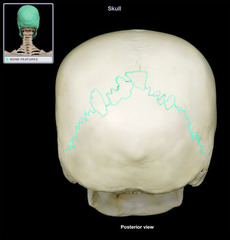
Lambdoidal Suture
A single suture located between the occipital bone and the parietal bone, representing an immovable articulation.
Orbit
A feature of the anterior skull containing and protecting the eyeballs, formed by certain skull bones like frontal, ethmoid, lacrimal, maxilla, and zygomatic bones.
Optic Canal
The round opening in the orbital apex lying between the two roots of the lesser wing of the sphenoid bone.
Superior Orbital Fissure
A curved slit-like opening located between the greater wing and lesser wing of the sphenoid bone, allowing passage for several cranial nerves and veins.
Inferior Orbital Fissure
An opening between the greater wing of the sphenoid bone and the maxilla, allowing passage for the inferior ophthalmic vein to join the pterygoid plexus of veins.
Orbital Rim
The sharp edge of the orbital opening, forming the peripheral border of the pyramid-shaped orbit.
Zygomaticomaxillary Suture
A depression in the midpoint of the infraorbital rim formed by the zygomatic bone with its maxillary process and the maxilla with its zygomatic process.
Nasal Cavity
The cavity on the skull's anterior aspect with lateral walls and a floor, mainly composed of bone and cartilage.
Nasion
A cephalometric midpoint landmark located at the junction of the frontal and nasal bones.

Piriform Aperture
The large triangular anterior opening of the nasal cavity. (large and triangular)

Nasal Conchae
Three projecting structures on each lateral wall of the nasal cavity extending inward, forming the superior, middle, and inferior ____________________

Turbinates
The lateral boundaries of the nasal cavity formed by the maxillae.
Nasal Meatus
A channel protected by each nasal concha, allowing communication between paranasal sinuses or nasolacrimal duct with the nasal cavity.

Nasal Septum
The vertical partition dividing the nasal cavity into two parts, formed by bone and cartilage.

Deviated Septum
A condition where the thin wall forming the nasal septum is displaced to one side, separating the right and left nasal cavities.
Temporal Fossa
Space on the lateral skull containing the temporalis muscle
Infratemporal Fossa
Area below the temporal fossa on the lateral skull
Pterygopalatine Fossa
Deeper area under the infratemporal fossa on the lateral skull
Zygomatic Arch
Cheekbone formed by the union of temporal and zygomatic bones
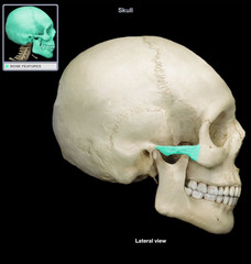
Temporozygomatic Suture
Suture between temporal and zygomatic bones in the skull
Masseter Muscle
Muscle originating from the zygomatic arch
TMJ
Movable joint between temporal bone and mandible
Hard Palate
Anterior part of the skull's inferior aspect, formed by maxillary bones
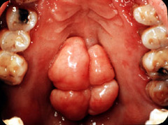
Palatal Torus
Midline variation on the hard palate
Pterygoid Process
Part of the sphenoid bone forming lateral borders of nasal apertures

Pterygoid Fossa
Depression between medial and lateral plates of pterygoid process

Hamulus
Thin curved process at the inferior part of the medial plate of pterygoid process

Foramen Ovale
Oval opening on sphenoid bone for mandibular nerve
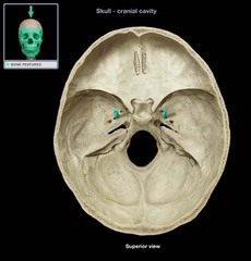
Foramen Spinosum
Opening for middle meningeal artery on sphenoid bone

Foramen Lacerum
Irregularly shaped opening on the external skull surface
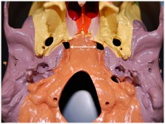
Carotid Canal
Round opening in the petrous part of the temporal bone
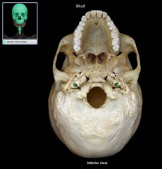
Styloid Process
Pointed bony projection posterior to the carotid canal

Stylomastoid Foramen
Opening for facial nerve exit from the skull

Jugular Foramen
Opening for internal jugular vein and cranial nerves passage

Foramen Magnum
Largest opening on occipital bone for spinal cord passage

Cribriform Plate
Perforated plate with foramina for olfactory nerve
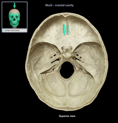
Paranasal Sinuses
Air-filled cavities in bones including frontal, sphenoidal, ethmoidal, and maxillary sinuses
epicondyl
small prominence that is located superior to or upon a condyle.
occipital condyle
projections of occipital bone that articulate with lateral masses of first cervical vertebra.

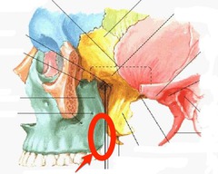
maxillary tuberosity
Connects our medial pterygoid muscle.
head of bone
rounded surfaces projected by a neck on bone
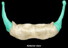
greater cornu of hyoid bone
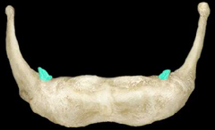
lesser Cornu
ridge
raised margin- internal oblique ridge
ethmoid, frontal, occipital, parietal, sphenoid, and temporal bones
What bones makeup the cranium?
inferior nasal conchae, lacrimal bones, mandible, maxilla, vomer, zygomatic bones
what bones make up the facial bones?
22
how many bones does the skull have?
parietal and temporal
What cranial bones are paired?
inferior nasal conchae, lacrimal bones, maxillae, zygomatic
what facial bones are paired?
the mandible with the TMJ
all skull bones are immovable except?
cervical region
where is the moveable articulation with the vertebral column?
frontal and parietal bones
what bones does the coronal suture attach?
frontonasal suture
Suture between the nasal and frontal bones

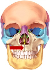
intermaxillary suture
the suture between the two maxillae of the upper jawbone
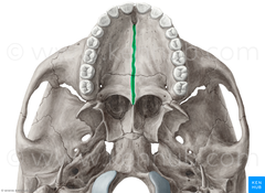
median palatine suture
the union between the palatine bones
squamosal sutures
Between parietal and temporal bones

Temporozygomatic sutures (paired)
temporal bones and zygomatic bones

transverse palatine suture
maxillae and palatine bones
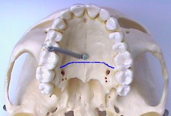
Zygomaticomaxillary suture
Connects the zygomatic to the maxilla

frontal bone, parietal bone, occipital bone
what cranial bones can you see from the superior view?
coronal suture, sagittal suture, and the lambdoidal suture
what sutures can you see from the superior view?
1. Nasal bone
2. Lacrimal bone
3. Zygomatic bone
4. Inferior nasal concha
5. Vomer
6. Maxilla
7. Mandible
what facial bones can you see from the anterior view?
frontal, ethmoid, lacrimal, maxilla, and zygomatic bones.
what bones form the orbit?
frontal bone
what bone forms the roof or superior wall of the orbit?
ethmoid and lacrimal bones
what bone forms the medial wall of the orbit?
zygomatic and sphenoid bones
what bone forms the lateral wall of the orbit?
sphenoid bone, maxilla, and palatine bones
what bone forms the apex or base of the orbit?
orbital apex
deepest part of orbit

orbital canal
round opening in the orbital apex

superior orbital fissure
Lateral to the optic canal is the curved and slit like _________

inferior orbital fissure
fissure in the orbit floor between maxilla and greater wing of sphenoid
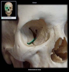
orbital rim

supraorbital rim
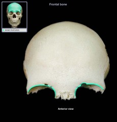
infraorbital rim

- the occipital, frontal, parietal, temporal bones, as well as the sphenoid bone and ethmoid bone.
what cranial bones can you see from the lateral view?