The Nervous System - Chp 33
 Call Kai
 Learn
Learn Practice Test
 Spaced Repetition
 Match
Match Flashcards
 Knowt Play
1/44

There's no tags or description
Looks like no tags are added yet.
Name | Mastery | Learn | Test | Matching | Spaced | Call with Kai | Chat |
|---|
No analytics yet
Send a link to your students to track their progress
45 Terms
What is the nervous system divided into?
CNS - Central Nervous System
brain + spinal cord
PNS - Peripheral Nervous System
network of nerves that bring signals between the CNS and the rest of the body
Sensory Neuron Diagram
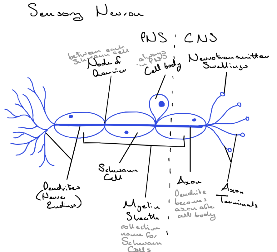
Where is the cell body of a sensory cell?
outside the CNS/ in the PNS
Motor Neuron Diagram
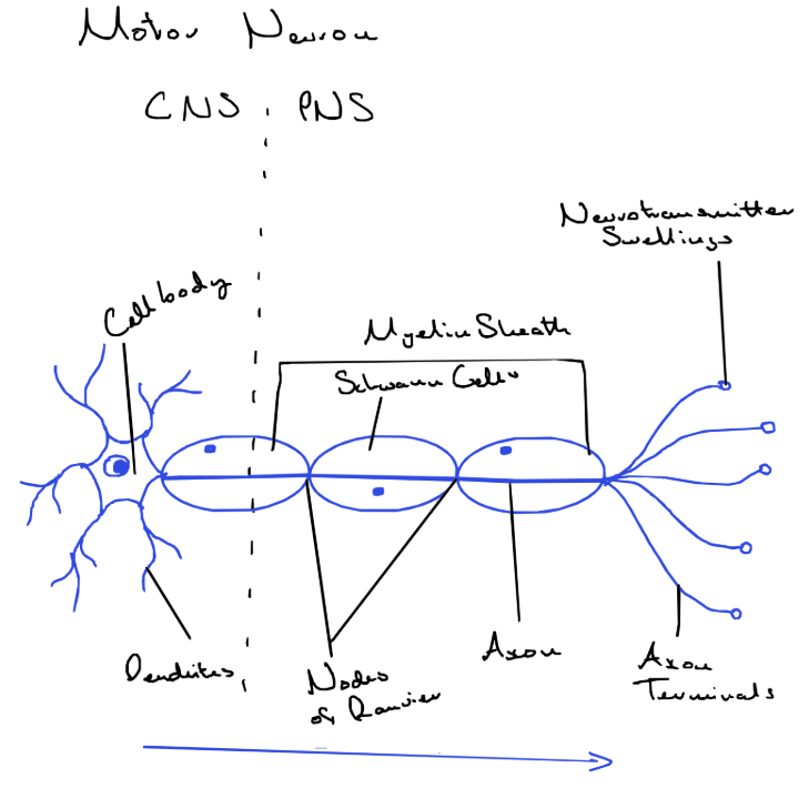
Where is the cell body of a motor cell?
in the CNS
Neuron
functional cells of the nervous system, that carry electrical signals to and from the CNS to bring about a response
Receptor
a cell or group of cells that detects a stimulus
Nerve endings
connect sensory neurons to receptor cells or sense organs
Dendrites
fibres that carry impulses towards the cell body of the neuron
Schwann cells
Resting Neuron
are neurons where no impulse is being carried
Threshold
an electrical impulse will only be carried along a neuron when it meets its threshold
All or Nothing Law
is the threshold is not reached there will be no impulse carried, and if the threshold is met the impulse will be carried
Describe the movement of an impulse through a neuron
a resting neuron has a higher negative ion concentration inside, and a higher positive ion concentration outside
when the threshold is met, the neuron changes its permeability to ions
this allows positive ions to move into the neuron, and the concentration changes to positive on the inside and negative on the outside
this happens section by section as the impulse travels down the neuron
What affects the speed of an impulse?
the myelin sheath
if present - the impulse jumps from each node of ranvier to the next, making the impulse travel faster
if not present - the impulse will be a lot slower
the larger the diameter of the neuron, the faster the impulse will be passed along
Synapse
the region where 2 neurons come into close contact with each other
Synaptic Cleft
the tiny gap between the 2 neurons
Neurotransmitter
convert electrical signals to chemical signals so they can cross over the cleft to the next neuron, where they are converted back into electrical signals
Name 2-3 neurotransmitters
Acetylcholine
Noradrenaline
Dopamine
Synapse Diagram
How do neurotransmitters move from one neuron to another?
diffusion
What happen to the neurotransmitters after they cross the synaptic cleft?
enzymes in the post-synaptic neuron digest the neurotransmitters
they are moved back to the swellings in the pre-synaptic neuron and can be used again
What are the functions of the synapse?
transports impulses from one neuron to the next
controls the direction of the impulse
prevents overstimulation - with constant stimulation over a long period of time neurotransmitter stop being produced
How do painkillers work?
they stop the impulse by blocking the neurotransmitters crossing a synapse
What is the function of the brain?
sorts all incoming impulses from sensory neurons + decides a response
What bones protect the brain + spinal fluid?
skull
spine
What does the brain consist of?
Grey matter - made up of cell bodies and synapses
White matter - made up of nerve fibres/axonz
What protects the brain inside the skull + spinal cord?
3 membranes called the meninges
cerebrospinal fluid between the membranes + around the brain
What is the function of the cerebrospinal fluid?
acts as a shock absorber
is an exchange medium between the brain and the blood
Cerebrum - function
controls voluntary muscles
receives impulses from sense organs
controls intelligence, memory + personality
Cerebellum - function
Controls muscular coordination and balance
Medulla - function
controls involuntary actions
Thalamus- function
sends messages to different parts of the brain
Hypothalamus - function
controls internal environment of the body
Disorder of the Nervous System - cause, symptoms + treatment
Parkinsons disease
caused by a lack of dopamine in the brain
trembling limbs, rigid body, inability to walk
cannot be prevented, treatments include drugs + physiotherapy
Where is the spinal cord located?
in the neural canal
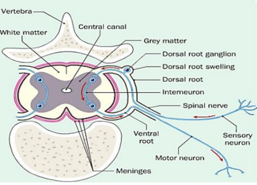
Draw + describe the structure of the spinal cord
outer white ring made of white matter
inner H shaped tissue made up of grey matter
central canal contains cerebrospinal fluid
the dorsal root carries sensory neurons to the spinal cord
the ventral root carries motor neurons out of the spinal cord
Reflex action
an automatic response to a stimulus, they are fast and designed to protect the body from harm
What is the reflex arc?
if a message is sent to the spine and it is very strong, the response happens before the message has been received and processed in the brain
Examples of reflex actions
grasp reflex in young children
blinking the eyes
breathing
knee jerk
Diagram of a withdrawal reflex action
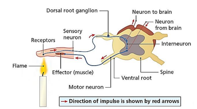
Describe the process of a withdrawal reflex
receptors in the finger are stimulated by the hot flame
sensory neurons carry impulse through the dorsal root to spinal cord
in the spinal cord numerous synapses are made with other neurons
and interneuron carries the impulse to a motor neuron
another neuron takes the impulse to the brain
motor neurons take the impulse through the ventral root to the effector
at the same time that the hand withdraws, the impulse reaches the brain, this makes us aware of what happened and some pain, however, the actions was not caused by the brain