Week 4: Pediatric Orthopedic/MSK disorders
 Call Kai
 Learn
Learn Practice Test
 Spaced Repetition
 Match
Match Flashcards
 Knowt Play
1/73

There's no tags or description
Looks like no tags are added yet.
Name | Mastery | Learn | Test | Matching | Spaced | Call with Kai | Chat |
|---|
No analytics yet
Send a link to your students to track their progress
74 Terms
what information needs to be assessed when taking the clinical history during the pediatric orthopedic exam?
full vs premature
vaginal delivery vs C section
oligohydramnios
“packaging defitics”
multiples
family history
sleeping/seating positions
what is oligohydramnios?
condition characterized by low amniotic fluid levels surrounding the fetus during pregnancy, which can lead to complications such as growth issues or cord compression.
what fetal presentation is typically recommended for c-section?
The breech presentation, where the baby is positioned feet or buttocks first, is typically recommended for cesarean section.
what is the vertex presentation?
refers to the typical position of the fetus during delivery, where the head is down and presents first in the birth canal, facilitating a vaginal delivery.
what is the weakest link in the bone ligament complex in children that results in differences in mechanism of injury and healing pattern?
the “physis” or growth plate
what LE positioning is normal at infancy?
normal to see genu varus (physiological bowing)
when might genu valgum/knock knees develop in children and be considered normal?
around 3.5
when should leg position be normalized?
by age 7
what is SCFE?
slipped capital femoral epiphysis, is the most common hip disorder in adolescents in which there is a high incidence of avascular necrosis of the femoral head
what age would you commonly see onset of SCFE?
for females 11-13 (avg 12), for males 13-15 (avg 13.5)
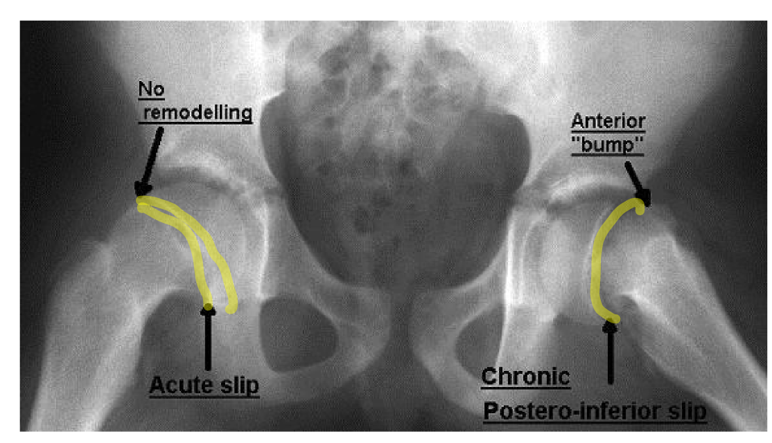
what does the image show?
SCFE X-ray
describe the acute SCFE clinical presentation
immediate, severe pain and restricted hip abduction and IR due to posterior and inferior displacement
describe the chronic SCFE clinical presentation
most frequently seen, history of pain and limp for weeks or months, limited hip abduction and IR
describe the acute-on-chronic SCFE clinical presentation
chronic aching in the hip, thigh, or knee due to a chronic slip, with trauma the epiphysis slips acutely, causing a marked increase in symptoms
when might there be a stable slipped capital femoral epiphysis?
with chronic clinical presentation- there is minimal change in alignment of the femoral head
what is the PT emphasis on SCFE after surgical correction?>
not many precautions, emphasis on maintaining the hip, knee, and ankle ROM and strength, restoration of muscular balance in LE, keep displacement to a minimum, joint protection strategies to avoid degenerative arthritis, proprioceptive and balance training
what is legg-calve-perthes disease?
A childhood hip disorder (osteochondrosis) that occurs when blood supply to the femoral head is temporarily reduced, leading to bone death and subsequent regrowth. will have a limp with pain referred to the groin, thigh, or knee. See characteristics decrease of hip IR and abduction and a hip flexion contracture
what is the demographic of LCP disease?
occurs between ages of 3-11, with average onset between 5-7. more common in males (4:1 ratio to females)
how long does LCP disease take to heal generally?
1-3 years

what does the image show?
LCP disease (notice the flattened femoral head)
what is the treatment for LCP when there is avascular necrosis?
non weight bearing and joint protection, surgical osteotomy
what is the treatment for LCP during revascularization?
spica cast is post surgical, or bracing with scottish rite brace achieves containment by abduction while allowing free knee motion
what is the treatment for LCP during bone healing?
guide bone remodeling for spherical femoral head in acetabulum, restore ROM and muscle symmetry
what is the treatment for LCP during residual deformity?
restore functional play skills, movement/recruitment patterns, and achievement of motor milestones
what is the presentation of internal tibial torsion?
most common cause of intoeing or “pigeon toed”, usually self correcting due to the nature of lateral drift with bone growth, see compensatory foot eversion and hip ER
what is the presentation of external tibia torsion?
one cause of outtoeing, usually worsens with time, the foot with invert and the hip IR to compensate
what is genu varum?
common torsional deformity that occurs secondary to normal in utero positioning, tight posterior hip capsule causes an ER of the thigh at the hip combined with internal tibial torsion, normally resolves spontaneously by the time the child is aged 2
what are the characteristics of genu valgum?
out-toeing foot progression angle, usually due to child being overweight, leads to awkward gait and flat feet
what is blout’s disease/tibia vara?
a progressive growth disorder characterized by disordered ossification of the medial aspect of the proximal tibia, is most common in children of african american jamaican descent- looks like extreme bowleggedness
what is the typical presentation of blout’s disease?
bilateral or unilateral (see unilateral with later onset)
what is the demographic of blout’s disease?
Most commonly affects African American children, particularly those of Jamaican descent. can be early onset (infantaile in children less than 3), late onset (juvenile form 4-10 and adolescent form 11 and over)
what is the treatment for blout’s disease?
under 2 years, progressive bracing (HKAFO) to be worn 23 hours per day, infantile to juvenile (3-6): if deformity continues to worsen, proximal tibial osteotomy with external fixation is recommended
what are some differentiating characteristics between blout’s disease and genu varum?
genu varum usually due to obesity and increase compressive force at the medial tibia, presents as angular deformity from the femur and the tibia, will typically correct itself whereas blout’s see prominent metaphyseal break, internal tibial torsion, and leg length discrepancy, progresses to severe deformity without medical intervention
what is discoid lateral meniscus?
A type of congenital knee abnormality characterized by a thicker, disc-shaped lateral meniscus instead of the usual crescent shape. It can lead to joint pain, clicking, or locking, and may require surgical intervention if symptomatic (10-20% bilateral findings). common in children 5-10, with complaints of “snapping knee”
what is talar dome syndrome?
An injury to the cartilage on the talar dome, often caused by ankle trauma or severe ankle sprains, leading to pain and swelling. It can result in osteochondral lesions and may require surgical treatment if conservative measures fail.
what do anterolateral lesions in talar dome syndrome result from?
from IV and DF forces, causing the talar dome to impact the fibular, these lesions are usually shallow and wafer shaped
what are medial lesion in talar dome syndrome cause by?
results from a combination of IV, PF, and ER forces causing the talar dome to impact the tibia, these lesions are usually deeper and cup shaped
what is the clinical presentation of talar dome injuries?
chronic ankle pain along with intermittent swelling and weakness, stiffness, instability. Severe pain with palpation behind the medial malleolus when ankle in DF with posterior medial lesions, and tenderness at the anterolateral ankle joint when the joint is in max PF with anterolateral lesions
what is the treatment for talar dome injuries?
usually conservative and non operateive- immobilization, graded restoration of AROM, strength, and proprioception, if no improvement in 6 weeks, MRI may be necessary to rule out intra-articular derangement.
what are symptoms of talar dome injuries?
effusion, catching or locking of the ankle, instability preceded by pain, and ankle pain relieved with diagnostic lidocaine
what are the pediatric ortho limping conditions from birth to 5 years?
osteomyelitis
septic arthritis
transient synovitis
occult fractures
kholer syndrome
what are the pediatric ortho limping conditions from 4-10 years?
transient synovitis
leg-calve-perthes disease
discoid lateral meniscus
sever’s disease
growing pains
what are the pediatric limping conditions from 11-15 years?
SCFE
Osgood Schlatter syndrome
osteochondritis dissecans
tarsal coalition
frediberg disease
accessory navicular
what are the two periods of rapid spinal growth?
birth-5 years, and then the adolescent growth spurt (females at 8-14 with avg 12, males at 11-16 with avg at 14)
what constitutes mild scoliosis?
10-20 degrees of deviation
what constitutes moderate scoliosis?
20-40 degrees of deviation
what constitutes severe scoliosis?
greater than 50 degrees of deviation
what are the three different types of scoliosis?
ideopathic, neuromuscular, and congenital
how is scoliosis named?
based on the convexity of the curvature (Right or Left) and location (cervical/thoracic/lumbar)
describe ideopathic scoliosis
lateral curvature with unknown cause
most common cause in children
early onset from birth to 10 years
can also have late on set
describe congenital scoliosis
caused by an anomalous vertebral development in utero, failure of the vertebral segmentation, failure of vertebrae formation, mixed defect
describe neuromuscular scoliosis
systemic or chronic with a rapid progression, direct cause is not understood
may be due to asymmetrical paraplegia, mechanical forces, intraspinal/congenital abnormalities, sensory feedback, control of spinal balance, central pathways
what is scheuermann disease?
A condition characterized by abnormal curvature of the spine due to wedged thoracic vertebrae (due to the posterior vertebral column growing much faster than the anterior), typically seen in adolescents. It can lead to back pain and postural issues. presents as severe thoracic kyphosis. becomes apparent during growth periods of pre-puberty and most common in lower thoracic segments
what does early intervention of spinal disorders look like?
not aggressive, increase the kinesthetic awareness of “good” alignment, restore neutral muscle balance in the spine, correct faulty postural habits
what are the exercise goals of spinal disorder treatment?
stabilization fo the curves
mobilization of stiff body areas
improve postural awareness and alignment
teach ADLs and ergonomics
enhance NM control
increase muscle strength and endurance
pain reduction
improve cardio-pulmonary function
restore normal movement patterns and spinal coupling mechanics
what is juvenile idiopathic arthritis?
the most common rheumatological disease in children and one of the most common chronic diseases of childhood, more common in females (3.5-4/5:1), symptoms may start in infancy but not dx until 6 weeks of symptoms in 4 or more joints, is classified by the number of joints affected and type
what is the clinical presentation of JIA?
morning limp or stiffness in the affected joints, generalized joint and muscle pain
what should be assessed in the PT eval of a pt with JIA?
comprehensive full body assessment
ROM and MMT of all extremities
movement and recruitement patterns
scapular and shoulder and core stability
joint integrity and stability with functional movements
standardized testing for objective outcomes
current physical activities
pt and parent goals for therapy
for JIA, what should PT intervention look like?
more activity generally see better prognosis, allow older children to self-limit their activities based on pain level. include GENTLE stretching, pain modalities, joint protection, energy efficiency strategies, and HEP

what are the images consistent with?
juvenile idiopathic arthritis
what is osteogenesis imperfecta?
an inherited disorder of connective tissue that results in bone fragility, muscle weakness, and hypermobility leading to frequent fractures, aka Brittle bone disease, there are four types categorized by severity and genetic inheritance patterns.
what is the pathophysiology of osteogenesis imperfecta?
defects of collagen synthesis leads to brittle bones, collagen fibers fail to mature beyond the reticular fiber stage, formation of endochondral and intramembranous bone affected, osteoblasts have normal or increased activity but do not produce collagen
what is the presentation of mild osteogenesis imperfecta?
ability to ambulate without AD for community distances
what is the presentation of moderate osteogenesis imperfecta?
can ambulate household distances, may use alternative mobility and AD for longer distances
what is the presentation of severe osteogenesis imperfecta?
W/C as the primary mobility, potential ability to weightbear during transfers
is there a cure for OI?
no, treatment focuses on management of symptoms (with medications such as biophosphonates, pamidronate infusions, zoledronic acid, and vitamin D)
what is fracture healing like for those with OI?
generally heal within normal timeframes but once fractured is much more susceptible to future fractures, immobilization during healing can cause disuse osteoporosis and greater risk of fracture, pseudoarthrosis may occur when not immobilized so is managed with internal fixation with IM rods
what is arthrogryposis multiplex congenita?
A congenital condition characterized by multiple joint contractions, muscle weakness, and limb deformities that limits mobility. can be due to intrinsic or extrinsic factors like genetics. exposure to viral infections, fetal crowding, myasthenia gravis, toxoplasmosis, alcohol, phenytoin, cocaine
what are the three groups of arthrogryposis multiplex congenita?
amyoplasia
CNS
heterogenous
what is the clinical manifestation of arthrogyroposis multiplex congenita?
there is variability but constant is severe contractures and lack of muscle development (amyoplasia), can affect any joint but typically the foot, hip, wrist, knee, elbow, and shoulder, can present “jackknifed” or “froglike”
what are other associated characteristics of arthrogyroposis multiplex congenita?
scoliosis
dimpling of skin over joints
hemangiomas
absent or decreased finger crease
congenital heart disease
facial abnormalities
respiratory problems
abdominal hernias
what does medical management of arthrogyroposis multiplex congenita look like?
surgical management, prevent of correct hip dysplagia, orthotics and or splinting to prevent contractures, osteotomies and or guided growth may be needed, also conservative management of scoliosis, potential need for growing rods and PSF later in life
are there any specific developmental tests for AMC?
no, some gross motor skills may never be attained but should note movement patterns
when is stretching and splinting most critical in kids with AMC?
during the first 2 years of life and growing years