Forearm&Hand + Cutaneous Innervation

0.0(0)
Studied by 0 people
call kaiCall Kai
Locked
learnLearn
examPractice Test
spaced repetitionSpaced Repetition
heart puzzleMatch
flashcardsFlashcards
GameKnowt Play
Card Sorting

1/20

encourage image

There's no tags or description

Looks like no tags are added yet.

Last updated 5:11 PM on 9/22/26
Name
Mastery
Learn
Test
Matching
Spaced
Call with Kai
Chat

No analytics yet

Send a link to your students to track their progress

21 Terms

1
New cards

upper limb dermatomes

-regions of skin that correspond to 1 spinal nerve; single spinal nerve providing sensory

-adjacent dermatomes overlap

-more conceptual than physical

<p>-regions of skin that correspond to 1 spinal nerve; <mark data-color="blue" style="background-color: blue; color: inherit;">single spinal nerve providing sensory</mark></p><p>-adjacent dermatomes overlap</p><p>-more conceptual than physical</p>
2
New cards

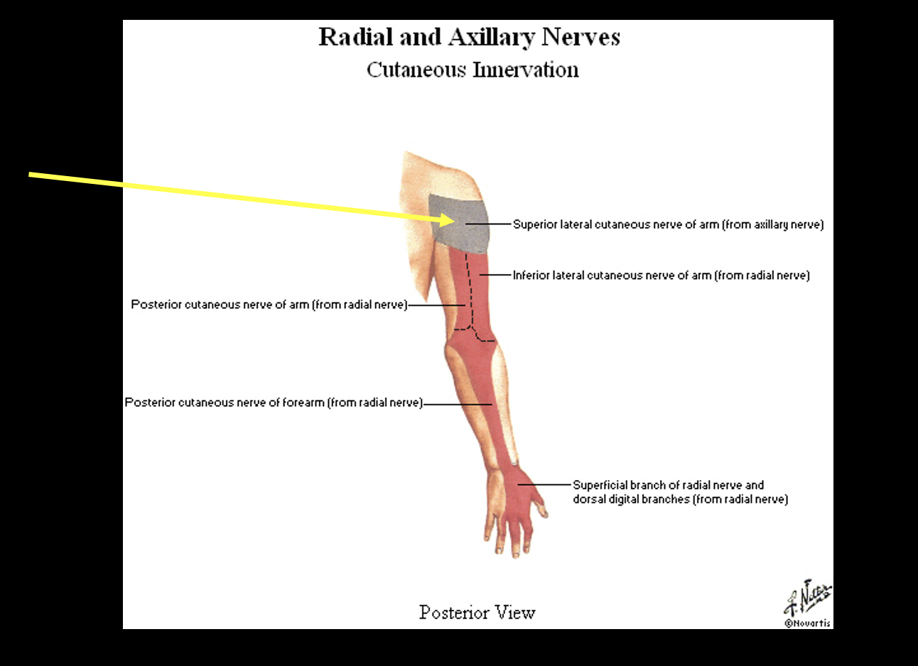
Cutaneous innervation: axillary N.

-C5, C6

-post and lateral shoulder
<p>-C5, C6</p><p>-post and lateral shoulder</p>
3
New cards

Cutaneous innervation: musculucutaneous nerve

→ lateral antebrachial cutaneous nerve

<p>→ lateral antebrachial cutaneous nerve</p>
4
New cards

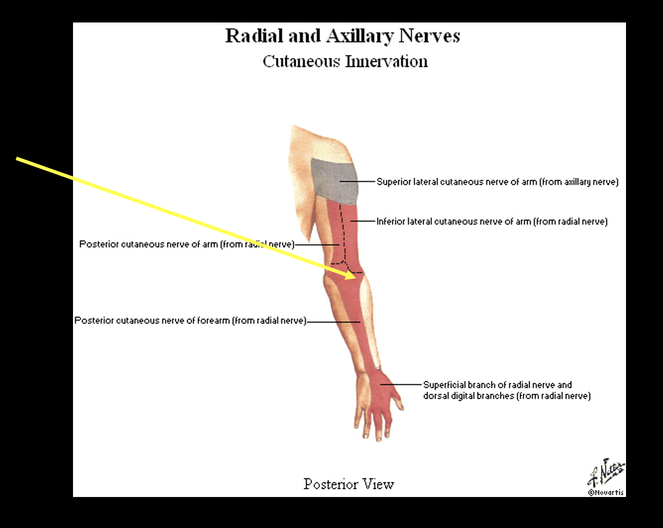
Cutaneous innervation: radial nerve

posterior arm, forearm, and hand
<p>posterior arm, forearm, and hand</p>
5
New cards

Cutaneous innervation: median nerve

lateral hand palmar side, nail beds 1-3, ~4

<p>lateral hand palmar side, nail beds 1-3, ~4</p>
6
New cards

Cutaneous innervation: ulnar nerve

-medial hand pamlar and dorsal

<p>-medial hand pamlar and dorsal</p>
7
New cards

cutaneous innervation: limb overview

knowt flashcard image
8
New cards

cutaneous innervation of hand

-parashesia were no overlap

<p>-parashesia were no overlap</p>
9
New cards

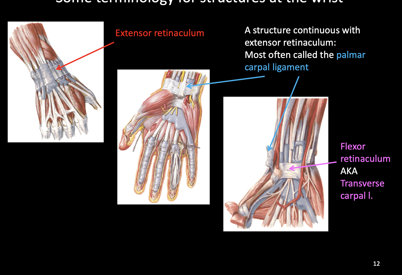
structures at the wrist

-Extensor retinaculum: keeps tendosn in place

-Palmar carpal ligament: continuous with extensor retinaculum

-Flexor retinaculum/transverse carpal l: forms part of carpal tunnel
<p><strong>-Extensor retinaculum</strong>: keeps tendosn in place</p><p><strong><mark data-color="red" style="background-color: red; color: inherit;">-Palmar carpal ligament:</mark></strong><mark data-color="red" style="background-color: red; color: inherit;"> continuous with extensor retinaculum</mark></p><p><strong><mark data-color="yellow" style="background-color: yellow; color: inherit;">-Flexor retinaculum/transverse carpal l</mark></strong>: forms part of carpal tunnel</p>
10
New cards

 retinacula

•The retinacula are like a bracelet

•Specialized parts of deep fascia

•Creates tunnels for tendons / other structures

•Holds tendons in place to create lever system for wrist

11
New cards

Extensor retinaculum: attachment + tunnel for…

Attaches at:

–pisiform, hook of hamate, AND the distal end of the radius

Tunnel for, lateral to medial, the tendons of:

1.abductor pollicis longus and extensor pollicis brevis muscles

2.extensor carpi radialis longus and brevis muscles

3.extensor pollicis longus muscle

4.extensor digitorum and extensor indicis muscles

5.extensor digiti minimi muscle

6.extensor carpi ulnaris muscle

*”Adults Believe, Children Citerently, Love, Do Infects, Make Us”

<p><u>Attaches at:</u></p><p>–pisiform, hook of hamate, <mark data-color="red" style="background-color: red; color: inherit;">AND the distal end of the radius</mark></p><p><u>Tunnel for, lateral to medial, the tendons of:</u></p><p>1.abductor pollicis longus and extensor pollicis brevis muscles</p><p>2.extensor carpi radialis longus and brevis muscles</p><p>3.extensor pollicis longus muscle</p><p>4.extensor digitorum and extensor indicis muscles</p><p>5.extensor digiti minimi muscle</p><p>6.extensor carpi ulnaris muscle</p><p><mark data-color="purple" style="background-color: purple; color: inherit;">*”Adults Believe, Children Citerently, Love, Do Infects, Make Us”</mark></p>
12
New cards

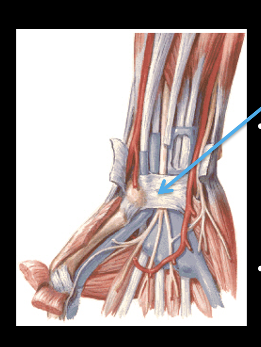
Flexor retinaculum: attaches to + tunnel for…

Attaches to:

•pisiform bone and hook of hamate bone (medially)

•tubercles of scaphoid and trapezium bones (laterally)

Forms a tunnel for 10 things: (carpal tunnel)

•4 tendons of the flexor digitorum superficialis m.

•4 tendons of the flexor digitorum profundus m.

•tendon of the flexor pollicis longus m.

•median nerve
<p><u>Attaches to:</u></p><p>•pisiform bone and hook of hamate bone <mark data-color="purple" style="background-color: purple; color: inherit;">(medially)</mark></p><p><mark data-color="red" style="background-color: red; color: inherit;">•tubercles of scaphoid and trapezium bones</mark> <mark data-color="purple" style="background-color: purple; color: inherit;">(laterally)</mark></p><p><u>Forms a tunnel for 10 things: </u>(carpal tunnel)</p><p>•4 tendons of the flexor digitorum superficialis m.</p><p>•4 tendons of the flexor digitorum profundus m.</p><p>•tendon of the flexor pollicis longus m.</p><p>•median nerve</p>
13
New cards

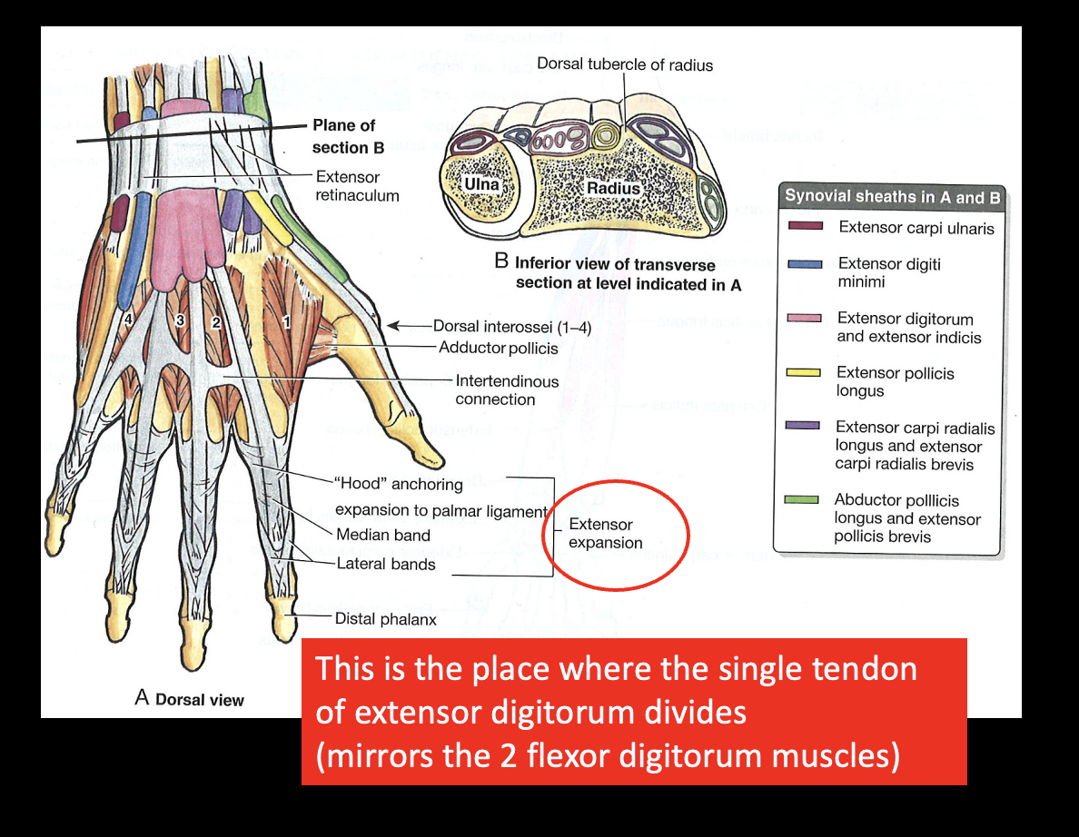
Dorsal Hand: Extensor Digitorum muscle

-acts on all IP; to distal phalanx

-Extensor Expansion: dorsal; place where the single tendon of extensor digitorum divides
<p>-acts on all IP; to <mark data-color="red" style="background-color: red; color: inherit;">distal </mark>phalanx</p><p><strong>-Extensor Expansion:</strong> dorsal; place where the single tendon of extensor digitorum divides</p>
14
New cards

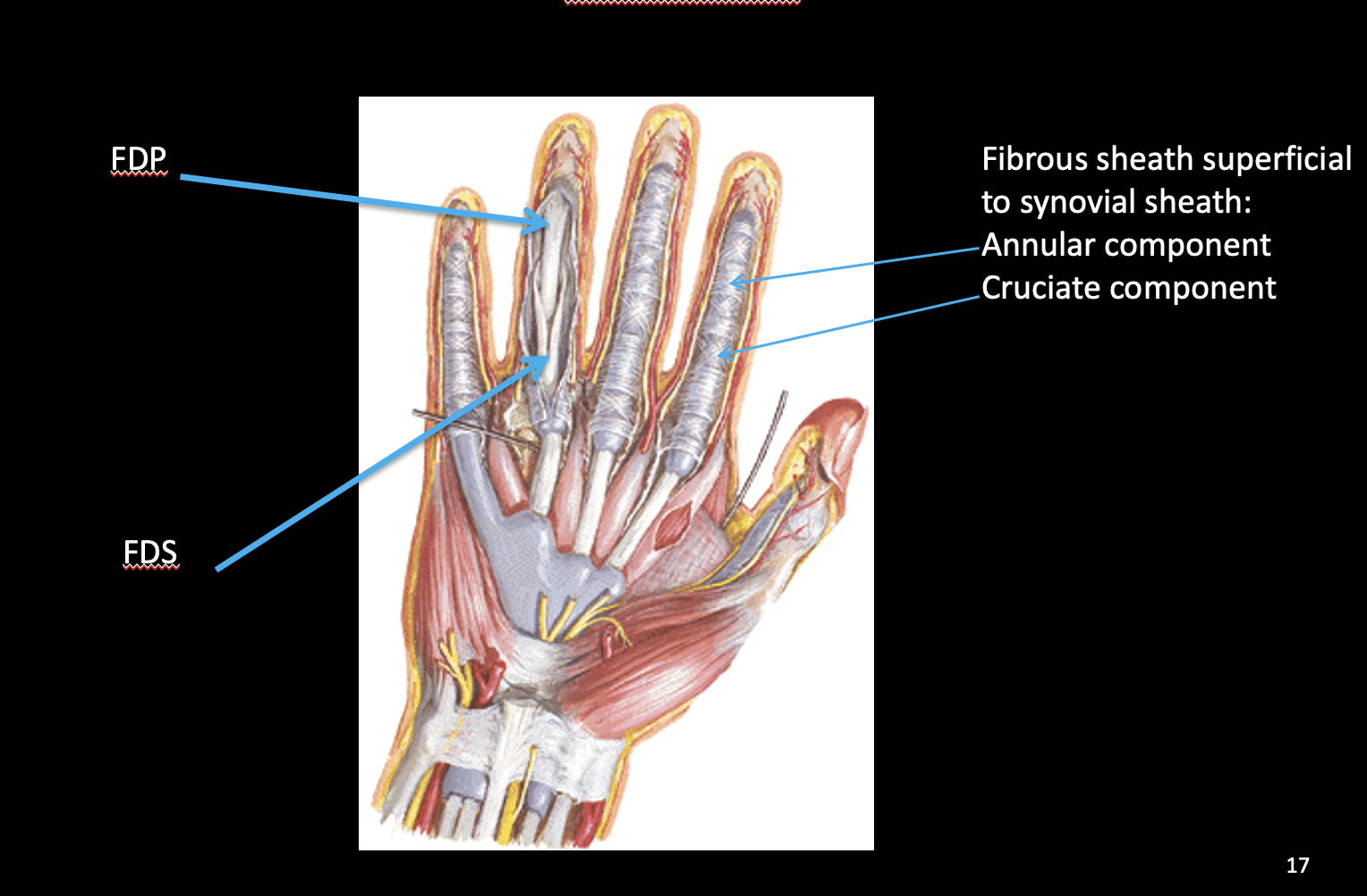
Palmar Hand: Flexor Digitorum Superficialis and Profundus

different insertions so different actions…

-FDS: tendon splits, FDP tendon pass through it; insert middle phalanx and flex PIP

-FDP: inserts on distal phalanx so flexes PIP and DIP

-+Annular ligament circular, cruciate component x
<p>different insertions so different actions…</p><p><u>-FDS:</u> tendon splits, FDP tendon pass through it; insert <em>middle </em>phalanx and flex PIP</p><p><u>-FDP:</u> inserts on <em>distal</em> phalanx so flexes PIP and DIP</p><p>-+Annular ligament circular, cruciate component x</p>
15
New cards

tendon for extensor digitorum + lumbricals

knowt flashcard image
16
New cards

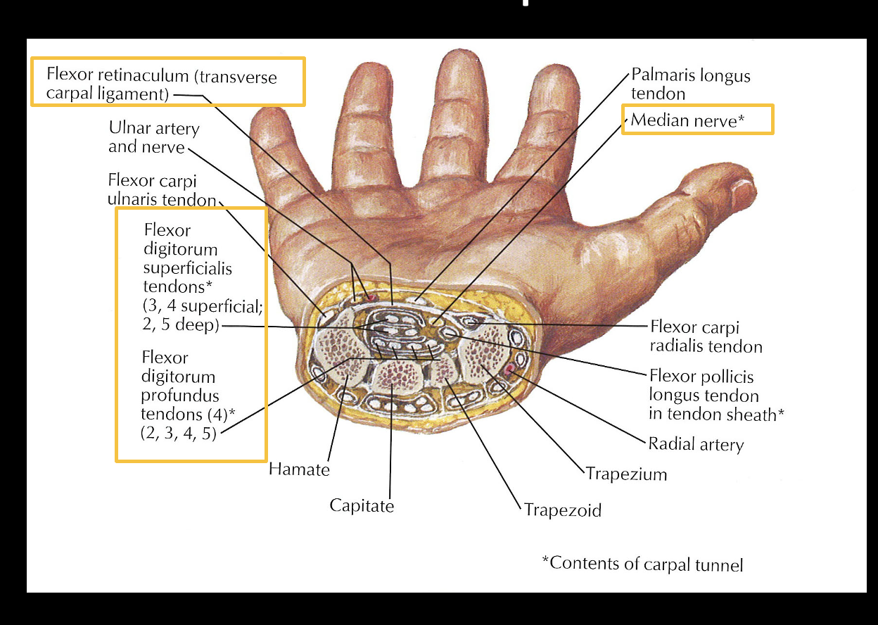
carpal tunnel

-Cylindrical space tendons and median nerve pass through

Boundaries

-Floor: Hamate, capitate, trapezoid

-Medial wall: Hamate

-Lateral wall: Trapezium

-Roof: Hook of hamate, flexor retinaculum attached to trapezium (*retinaculum rigid)

Content

-10 contents=4 tendons FDS, 4 tendons FDP, tendon Flexor Pollicis Longus, median n

Injury

-inflammation or overuse impose on median n

-Phalen’s test or Tinel’s test reproduce symptoms
<p>-Cylindrical space tendons and median nerve pass through</p><p><u>Boundaries</u></p><p>-Floor: Hamate, capitate, trapezoid</p><p>-Medial wall: Hamate</p><p>-Lateral wall: Trapezium</p><p>-Roof: Hook of hamate, flexor retinaculum attached to trapezium (*retinaculum rigid)</p><p><u>Content</u></p><p><mark data-color="green" style="background-color: green; color: inherit;">-10 contents</mark>=4 tendons FDS, 4 tendons FDP, tendon Flexor Pollicis Longus, median n</p><p><u>Injury</u></p><p>-inflammation or overuse impose on median n</p><p>-Phalen’s test or Tinel’s test reproduce symptoms</p>
17
New cards

Radial foveola

-A depression found on lateral surface of wrist

Boundaries:

-Medial: by tendon of extensor pollicis longus muscle

-Lateral: by tendons of abductor pollicis longus and extensor pollicis brevis muscles

Structures palpated:

  • Radial artery

  • radial styloid process

  • 1st metacarpal bone

Why care?

-radial pulse

-Test for inflammation of tendons due to repetitive stress / infection (de Quervain tenosynovitis)= “Finkelstein Test”

-Palpate the scaphoid for fracture: most commonly fractured, not always on x-rays; interrupt blood supply to bone if fracture → sepsis

<p>-A depression found on lateral surface of wrist</p><p><u>Boundaries:</u></p><p><mark data-color="red" style="background-color: red; color: inherit;">-Medial: by tendon of extensor pollicis longus muscle</mark></p><p><mark data-color="red" style="background-color: red; color: inherit;">-Lateral: by tendons of abductor pollicis longus and extensor pollicis brevis muscles</mark></p><p><u>Structures palpated:</u></p><ul><li><p>Radial artery</p></li><li><p><mark data-color="red" style="background-color: red; color: inherit;">radial styloid process</mark></p></li><li><p>1st metacarpal bone</p></li></ul><p><u>Why care?</u></p><p>-radial pulse</p><p>-Test for inflammation of tendons due to repetitive stress / infection (de Quervain tenosynovitis)= “Finkelstein Test”</p><p><mark data-color="purple" style="background-color: purple; color: inherit;">-Palpate the scaphoid for fracture:</mark> most commonly fractured, not always on x-rays; interrupt blood supply to bone if fracture → sepsis</p>
18
New cards

what type of joint is the radiocarpal joint? what bones make up the joint?

condyloid joint

-radius + schapoid, lunate, and triquetrum

<p>condyloid joint</p><p><mark data-color="blue" style="background-color: blue; color: inherit;">-radius + schapoid, lunate, and triquetrum</mark></p>
19
New cards

motions of the wrist joint

•Flexion

•Extension

•Adduction

•Abduction

•Circumduction

–This is a combination of the others

20
New cards

muscle table: wrist flexion and extension

knowt flashcard image
21
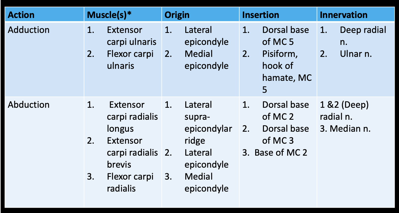
New cards

muscle table: wrist adduction and abduction
knowt flashcard image



