General surgery
 Call Kai
 Learn
Learn Practice Test
 Spaced Repetition
 Match
Match Flashcards
 Knowt Play
1/73

There's no tags or description
Looks like no tags are added yet.
Name | Mastery | Learn | Test | Matching | Spaced | Call with Kai | Chat |
|---|
No analytics yet
Send a link to your students to track their progress
74 Terms
What are the THREE essential components of any hernia, and what makes up each part?
- Sac: peritoneal diverticulum (may be complete/incomplete; has a neck at the defect). 2. Contents: omentum (omentocle), small/large intestine (enterocele), bladder, ovaries, ascites; every organ except pancreas can herniate. 3. Coverings: derived from the abdominal wall layers the hernia passes through.
What are the FIVE main causes/risk factors for abdominal wall hernias?
- Increased intra-abdominal pressure (chronic cough, straining, heavy lifting, ascites, pregnancy). 2. Congenital weakness (collagen disorders, patent processus vaginalis). 3. Age-related degeneration (elderly muscle/fibrous tissue weakness). 4. Trauma (sharp/blunt, previous surgery → incisional). 5. Genetic/acquired collagen abnormalities (smoking impairs collagen maturation).
Classify hernias by complexity from simple to complicated. Which stage is a painful irreducible lump with overlying erythema?
Occult → Reducible → Irreducible → Incarcerated → Obstructed → Strangulated → Infarcted. The case described (painful, irreducible, erythema) is STRANGULATED – requires emergency surgery. Overlying cellulitis = content is strangulating.
Compare INDIRECT (lateral) vs DIRECT (medial) inguinal hernias across key features.
Indirect: congenital, lateral to inferior epigastric vessels, narrow neck, often descends to scrotum, controlled by internal ring pressure, younger patients. Direct: acquired, medial (Hesselbach's triangle), wide neck, rarely scrotal, NOT controlled by internal ring pressure, elderly patients.
A 72yo woman with SBO has a tender lump below and lateral to the pubic tubercle. What is it and why is it dangerous?
Femoral hernia. Dangerous because: 50% present as emergencies; narrow neck → high strangulation risk; easily missed (no cough impulse, mistaken for lymph node); delay in diagnosis. Location is below & lateral to pubic tubercle (inguinal is above & medial).
What 4 steps differentiate inguinal hernia, femoral hernia, and lymph node using landmarks and tests?
- Pubic tubercle: inguinal = above/medial; femoral = below/lateral. 2. Zieman's test (3 fingers): deep ring, superficial ring, saphenous opening – feel cough impulse location. 3. Ring occlusion: pressure on deep ring controls indirect, not direct. 4. Reducibility/cough impulse: inguinal usually reducible with impulse; femoral often irreducible with NO impulse.
What is the pathophysiology of hernia formation and why is it called a 'collagen disease'?
Straining usually REVEALS rather than causes hernia. Collagen disease theory: inherited imbalance in collagen types. Evidence: histological changes, association with aortic aneurysm, Ehlers-Danlos makes repair difficult, smoking impairs collagen maturation, age-related degeneration, and surgical closure technique for incisional hernias.
A 45yo overweight male has a painful, non-reducible, doughy bulge near the umbilicus. Diagnosis and pathophysiology?
Epigastric hernia. Arises through linea alba (transverse split <1cm). Contains only extraperitoneal fat (no peritoneal sac). Narrow neck causes fat ischemia/partial strangulation → sharp pain. Irreducible, no cough impulse; may mimic a lipoma. Small ones may infarct and disappear.
How do you differentiate a hernia containing intestine from one containing omentum on examination?
Intestine: soft elastic, last part reduces with gurgling, resonant (tympany) on percussion, peristaltic sounds heard. Omentum: doughy granular, first part reduces easily but last part adheres to fundus (difficult), dull on percussion, no bowel sounds.
When is hernia repair indicated and when is 'watchful waiting' appropriate? What is the conversion rate?
Repair: all femoral, symptomatic/irreducible, contains bowel, acute pain, enlarging, younger adults. Watchful waiting: asymptomatic inguinal in elderly, small umbilical/epigastric with fat/omentum only. Conversion rate ~10% per year to surgery. Trusses not recommended.
List complications of inguinal hernia surgery by timing. Which complication can mimic recurrence after laparoscopy?
Early: pain, bleeding/haematoma, urinary retention. Medium (week 1): seroma, wound infection. Late: chronic pain, testicular atrophy (injury to spermatic artery). Laparoscopic seroma may be misdiagnosed as early recurrence (but resolves spontaneously).
A 6-month-old infant has an umbilical bulge that appears when crying. Management and prognosis?
Umbilical hernia in children. Conservative with parental reassurance – 95% resolve spontaneously by age 2. Surgery only if persists beyond 2 years. Obstruction/strangulation is extremely rare under age 3. Higher incidence in premature and black infants.
What is a sliding hernia and which organs can be involved?
Visceral wall forms part of the hernia sac wall (not completely enclosed). Left side: colon. Right side: caecum. Either side: bladder. Belongs to irreducible hernia category. Requires careful surgical dissection to avoid organ injury.
What are the principles of modern hernia repair and when is mesh contraindicated?
Principles: reduce contents, excise non-viable tissue/repair bowel, excise/close sac, close defect, reinforce with mesh (bridge, plug, or augment). Mesh types: synthetic/biological, large/small pore, non-adhesive if intraperitoneal. LIMITATION: infection is primary contraindication; avoid in gross contamination.
What are the risk factors for incisional hernia and how is it managed?
Risk factors: patient (obesity, collagen disorders, steroids, malnutrition, cough, cancer), wound (tension, infection), surgical (poor closure technique – major factor). Management: asymptomatic may not need treatment; abdominal binder for relief; repair (open or laparoscopic) covering whole incision, minimal tension, use mesh to reduce recurrence.
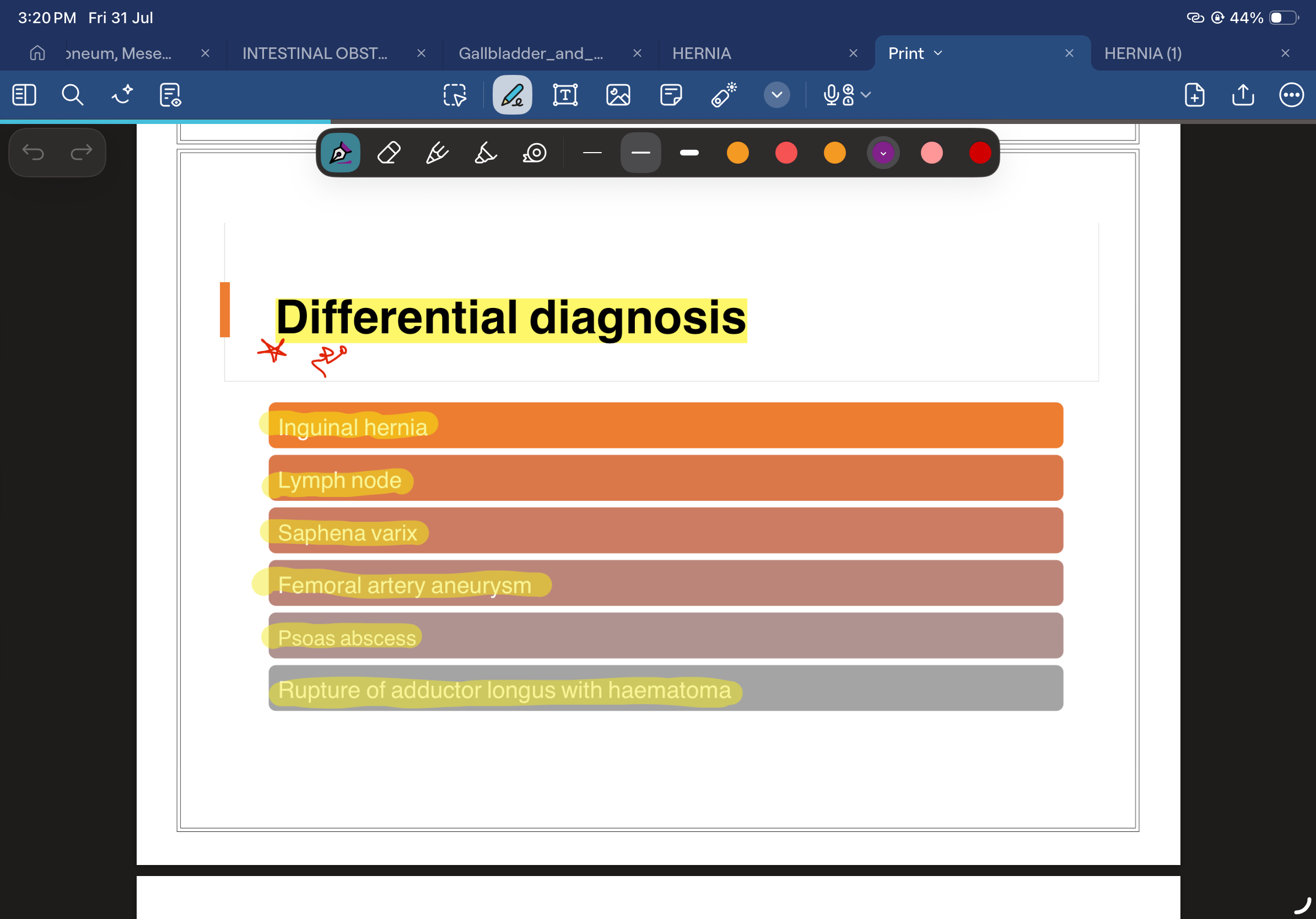
List the differential diagnoses of a groin lump.
Inguinal/femoral hernia, lymphadenopathy, saphena varix, hydrocele (cannot get above it), lipoma (spermatic cord/subcut), femoral artery aneurysm, undescended testis, psoas abscess, neuroma, muscle tumours. Scrotal: hydrocele, hernia, varicocele, testicular tumour.
Which imaging modality is best for each hernia scenario?
US: irreducible hernia, mass vs fluid, lymph node vs hernia, saphena varix (operator dependent). CT: complex ventral/incisional hernias (defects, contents, adhesions, excludes other pathology, plans reconstruction). MRI: sportsman's (Gilmore's) groin. Laparoscopy: occult defects but NOT intraparietal (e.g. spermatic cord lipoma). Plain X-ray: little value except SBO in emergency.
Describe the boundaries and contents of the inguinal canal.
Boundaries: Roof = conjoint tendon; Posterior = transversalis fascia; Anterior = external oblique aponeurosis; Floor = inguinal (Poupart's) ligament. Contents (male): testicular artery, veins, vas deferens, lymphatics, covered by cremasteric muscle. (Female): round ligament. Nerves: ilioinguinal, iliohypogastric, genital branch of genitofemoral.
What is the difference between herniotomy and herniorrhaphy, and in whom is each used?
Herniotomy: excision/closure of sac ONLY – used in children (persistent processus vaginalis is the primary defect). Herniorrhaphy: muscle-strengthening repair (with or without mesh) – used in adults because herniotomy alone has high recurrence. Types: Bassini, Shouldice (suture), Lichtenstein (mesh), TEP/TAPP (laparoscopic).
What are the clinical features of strangulation and the emergency management?
Features: severe constant pain, irreducible, tense, overlying cellulitis/erythema, no cough impulse, signs of obstruction, systemic upset. Management: EMERGENCY surgery – do NOT delay with taxis. Resect non-viable bowel (~20% of emergency cases), may need laparotomy. Synthetic mesh acceptable if minimal contamination with antibiotics.
Early management of acute pancretitis

Ranson score of pancreatitis sevirity

Complication of pancreatitis
Manigment of chronic pancreatitis

etiology of chronic pancreatitis

Investigation of chronic pancreatitis

A patient has a hernia that was previously reducible but has become painful, tender, irreducible, and has lost its cough impulse. What complication should you assume, and why is it an emergency?
Strangulated hernia. Strangulation means the blood supply to the herniated contents is compromised. Typical findings are a painful, tender, irreducible swelling, absent cough impulse, and possibly vomiting or intestinal obstruction. It is a surgical emergency because continued vascular compromise can lead to bowel ischemia, necrosis, and perforation.
Ddx of femoral hernia
During surgery, where would you expect the critical point of vascular compromise in a strangulated hernia, and why?
At the neck of the hernial sac. The neck is the narrowest part of the hernia and is therefore the common site where vessels supplying the herniated contents become compressed. Venous obstruction occurs first, followed by arterial compromise, ischemia, and potentially bowel necrosis.
A young man has a groin swelling that enters through the deep inguinal ring and extends into the scrotum. Is it direct or indirect, and what anatomical relationship confirms your answer?
Indirect inguinal hernia. It enters through the deep inguinal ring, travels through the inguinal canal, and may descend into the scrotum. It lies lateral to the inferior epigastric vessels and is classically associated with a patent processus vaginalis.
An older man develops an acquired groin hernia through a weakened posterior inguinal wall. Where is it located relative to the inferior epigastric vessels, and through what region does it protrude?
Direct inguinal hernia. It protrudes through the posterior wall of the inguinal canal in Hesselbach's triangle and lies medial to the inferior epigastric vessels. It is usually caused by acquired weakness of the abdominal wall.
You compress the deep inguinal ring and ask the patient to cough. The swelling still appears. What type of inguinal hernia is most likely, and why?
Direct inguinal hernia. Compression of the deep inguinal ring prevents an indirect hernia from entering through the ring. A direct hernia passes through the posterior wall of the inguinal canal, so it can still appear despite deep-ring occlusion.
A patient has a scrotal swelling. What examination findings favor a hydrocele rather than an indirect inguinoscrotal hernia?
Hydrocele is favored by absence of a cough impulse, inability to reduce the swelling, positive transillumination, a smooth cystic tense consistency, and the ability to get above the swelling. An inguinoscrotal hernia usually has an expansile cough impulse, may be reducible, does not transilluminate, and you usually cannot get above it.
An elderly woman has a small groin lump below and lateral to the pubic tubercle. What is the likely diagnosis, and why should it usually be repaired even if asymptomatic?
Femoral hernia. Femoral hernias occur below the inguinal ligament and below and lateral to the pubic tubercle. Their neck is narrow and rigid, giving them a high risk of incarceration and strangulation. Therefore, surgical repair is generally recommended even when symptoms are minimal.
How can the position of a groin swelling relative to the pubic tubercle help distinguish an inguinal hernia from a femoral hernia?
An inguinal hernia is typically above and medial to the pubic tubercle, whereas a femoral hernia is below and lateral to the pubic tubercle. This relationship is especially useful when assessing a small groin swelling in an elderly woman.
A 3-year-old child has a 1.5 cm painless, completely reducible umbilical hernia that becomes prominent when crying. What is the initial management and why?
Watchful waiting. Most congenital umbilical hernias in children close spontaneously, usually by approximately 2–5 years of age. Surgery is considered if the hernia persists beyond about 5 years, is large, or develops complications. Routine mesh repair is not indicated in a young child.
An adult develops a bulge through an old abdominal surgical incision. What is the diagnosis, what is the most important risk factor, and what is the standard treatment?
Incisional hernia. It is herniation through a previous surgical scar. Wound infection is the most important risk factor. Other factors include obesity, poor surgical technique, malnutrition, and increased intra-abdominal pressure. Mesh repair is generally the gold-standard definitive treatment.
A patient has a small painful midline swelling between the xiphoid process and the umbilicus. What type of hernia is most likely, and what does it usually contain?
Epigastric hernia. It occurs through the linea alba between the xiphoid and umbilicus. It usually contains preperitoneal fat rather than bowel. Incarceration of this fat may produce significant localized pain despite a relatively small swelling.
A patient has a tender strangulated hernia but continues to pass stool and does not have complete intestinal obstruction. What special type of hernia should you suspect, and why can strangulation occur without obstruction?
Richter's hernia. Only part of the bowel wall, usually the antimesenteric border, is trapped in the hernial defect. Because the entire circumference and lumen of the bowel are not incarcerated, the bowel wall can become ischemic and strangulated while intestinal contents continue to pass.
During hernia surgery, part of the cecum is found to form part of the wall of the hernial sac. What type of hernia is this, and what is the major operative danger?
Sliding hernia. In a sliding hernia, part of the hernial sac wall is formed by a retroperitoneal organ such as the cecum, sigmoid colon, or urinary bladder. The major surgical danger is accidental injury to the organ while dissecting what appears to be the hernial sac.
An elderly, thin, multiparous woman presents with intestinal obstruction and medial thigh pain but no obvious groin swelling. What diagnosis should you suspect, and what causes the thigh pain?
Obturator hernia. It passes through the obturator canal and may cause bowel obstruction without a visible external swelling. Medial thigh pain is the Howship-Romberg sign and results from compression of the obturator nerve. CT is useful because the hernia is often clinically occult.
A patient has persistent lateral abdominal-wall pain and a small or clinically hidden swelling along the semilunar line. What hernia should you suspect, and what investigation can help confirm it?
Spigelian hernia. It occurs through the Spigelian fascia near the lateral border of the rectus abdominis along the semilunar line. It is often interparietal and therefore difficult to detect clinically. CT can demonstrate the hidden abdominal-wall defect.
An alcoholic patient with known cirrhosis and ascites presents with fever and diffuse abdominal pain. Ascitic fluid analysis shows elevated neutrophils. What is the diagnosis and primary modality of treatment?
Diagnosis is Primary (Spontaneous) Peritonitis. Because there is no perforation, treatment primarily involves targeted antibiotics rather than surgical intervention.
A patient presents with a rigid abdomen, absent bowel sounds, and rebound tenderness. Upright chest X-ray reveals free air under the diaphragm. What is the underlying pathophysiology and immediate next step in management?
The pathophysiology is Secondary Peritonitis due to a perforated viscus (most commonly a perforated duodenal ulcer). Immediate management requires ABC resuscitation, broad-spectrum antibiotics, and urgent surgery for source control.
A patient who underwent emergency laparotomy for a ruptured appendix 48 hours ago continues to exhibit signs of systemic infection and peritoneal inflammation despite appropriate initial source control. What is this condition called?
Tertiary Peritonitis. It is characterized by persistent or recurrent infection after 48 hours of apparent resolution of primary or secondary peritonitis.
An immigrant from a developing nation presents with ascites, significant weight loss, and omental thickening. Ascitic fluid analysis reveals high protein and a lymphocytic predominance. What is the most likely diagnosis and what can it mimic?
Tuberculous Peritonitis. Clinically and on imaging, it frequently mimics peritoneal malignancy (carcinomatosis). It requires anti-TB therapy.
A patient undergoes laparotomy for a suspected abdominal tumor, but the surgeon discovers "jelly-like" mucinous ascites throughout the peritoneal cavity. Where is the most likely primary source of this pathology and what is the treatment?
This is Pseudomyxoma Peritonei, and the most likely primary source is an appendiceal tumor. Management typically involves cytoreductive surgery and HIPEC (Hyperthermic Intraperitoneal Chemotherapy).
A female patient with a history of ovarian cancer presents with new-onset diffuse abdominal nodules on CT. What is the mechanism of spread and what other primary cancers commonly cause this?
The mechanism is direct spread via peritoneal implantation (Peritoneal Carcinomatosis). Besides ovarian cancer, other common origins include gastric and colorectal cancers.
An elderly patient with a history of atrial fibrillation presents with sudden, agonizing abdominal pain. On early physical examination, the abdomen is soft and non-tender. What is the most likely diagnosis based on this discrepancy?
Acute Mesenteric Ischemia (AMI). The classic clinical hallmark is sudden, severe abdominal pain that is entirely out of proportion to the physical exam findings.
In a patient with sudden-onset acute mesenteric ischemia, what is the most common etiology and where did it likely originate?
The most common etiology is an arterial embolism. Because of the patient's atrial fibrillation, the embolism most likely originated from the heart and lodged in the mesenteric vessels.
A physician suspects acute mesenteric ischemia and notes elevated serum lactate and metabolic acidosis on the patient's ABG. Are these findings reliable for early diagnosis, and what is the gold-standard diagnostic step?
No, elevated lactate and metabolic acidosis are late, unreliable signs that often indicate bowel necrosis has already occurred. The first-line diagnostic investigation of choice is a CT angiography.
A 10-year-old boy presents with right lower quadrant pain and a mild fever. His mother notes he had a severe cold and sore throat last week. He appears non-toxic. What is the likely diagnosis and the typical causative organism?
Mesenteric Lymphadenitis. It frequently follows a viral upper respiratory tract infection and is classically associated with the bacteria Yersinia enterocolitica.
How can ultrasound or CT imaging help differentiate between acute appendicitis and mesenteric lymphadenitis in a child with right lower quadrant pain?
Imaging for mesenteric lymphadenitis will show enlarged mesenteric lymph nodes but, crucially, a normal-appearing appendix. This confirms that treatment should be conservative observation rather than surgery.
A patient is diagnosed with a mesenteric tumor on imaging. Regardless of whether it is a benign lipoma or a malignant liposarcoma, what is the most common clinical complication and presentation?
The most common complication and clinical presentation of a mesenteric tumor is a bowel obstruction, requiring surgical resection.
A patient on warfarin therapy presents with severe flank pain and signs of hypovolemic shock. Examination reveals a soft abdomen with no signs of peritonitis (no rigidity or rebound). What is the likely diagnosis?
Retroperitoneal Hemorrhage. The lack of early peritonitis is a hallmark of hidden retroperitoneal bleeding, as the blood is not irritating the peritoneal cavity. It is confirmed via CT scan.
A patient presents with vague back pain and newly diagnosed acute renal failure. Ultrasound reveals bilateral hydronephrosis. What retroperitoneal pathology could explain these findings, and what is the initial medical therapy?
Retroperitoneal Fibrosis (which can be idiopathic, drug-induced, or malignancy-related). It causes ureteric obstruction leading to renal failure. Initial medical therapy involves steroids.
An asymptomatic patient is found to have a massive abdominal mass on routine exam. CT imaging reveals the mass is displacing the kidneys and duodenum rather than invading them. What is the most likely diagnosis and most common malignant type?
A Retroperitoneal Tumor. Their anatomical location allows them to grow very large and displace fixed organs early on. The most common malignant type is a Liposarcoma.