CBNS 106 Final Lecture 1 (Systems Neuroscience + Retina)
1/12
There's no tags or description
Looks like no tags are added yet.
Name | Mastery | Learn | Test | Matching | Spaced | Call with Kai |
|---|
No analytics yet
Send a link to your students to track their progress
13 Terms
Systems Neuroscience
Systems Neuroscience
a) Study of networks/circuits of neurons that carry out specific functions
b) Focuses on how groups of neurons work together
c) Neural circuits = interconnected neurons that process information
Neural Circuits
a) Individual neurons form functional circuits
b) Different neuron types contribute differently:
PYR = pyramidal neurons
PV+ interneurons = parvalbumin interneurons
SST+ interneurons = somatostatin interneurons
VIP+ interneurons = vasoactive intestinal peptide interneurons
Why Study Neural Circuits?
a) Brain processing relies on population coding
b) Single action potentials usually have small effects
c) Many neurons firing together create meaningful signals
Neural Coding
AP Frequency (Rate Coding)
a) Strength of sensory stimulus coded by firing rate
b) More action potentials = stronger stimulus
Example: Cold-sensing neurons increase firing frequency as the temperature drops more
Coordinated activity (Synchrony Coding)
a) Information can also be coded by timing precision between neurons
b) Coordinated firing = synchrony
c) Higher synchrony often linked to perception and cognition
Example: EEG studies show stronger synchrony during face recognition task
Graded Potentials
a) Some neurons encode stimulus strength using membrane potential amplitude
b) Larger depolarization → more neurotransmitter release
c) Important in photoreceptor cell
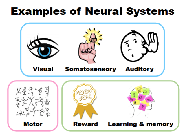
Neural Systems (YK this)
Examples of Neural Systems
Visual system
Somatosensory system
Motor system
Auditory system
Reward system
Brain Functional Specialization
Specific brain areas perform specific functions
Example:
Visual cortex processes vision
Somatosensory cortex processes touch/pain
Subsystems
Each sensory system contains specialized subsystems
Example in vision:
Shape processing
Color processing
Motion processing
Properties of Sensory Systems
Central Pathway
Sensory information travels:
Peripheral receptors
Subcortical structures
Cerebral cortex
Sensory Receptors (part of Peripheral receptors)
Characteristics:
Modality specific
Convert energy into electrical activity (transduction)
Specialized for detecting particular stimulus features
3. Receptive Field
Area of sensory space that changes a neuron’s activity
Can refer to:
Visual space
Skin surface
Auditory frequency range
Topographic Map
Nearby neurons represent nearby sensory locations
Maintains orderly spatial organization in the brain
Example:
Mouse whisker system has organized spatial mapping
Visual system overview
Main Visual Pathway
Light → Eye → Retina → LGN → Primary Visual Cortex
Light
Electromagnetic radiation visible to humans
Converted into neural activity through phototransduction
(add pic)
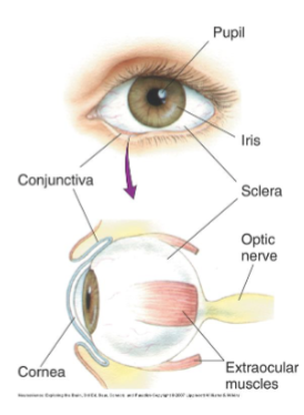
Structure of the Eye
Pupil
Opening that lets light enter eye
Iris
Colored part of eye
Controls pupil size
Cornea
Transparent covering
Refracts light
Sclera
“White of the eye”
Tough outer wall
Extraocular Muscles
Move the eyeball
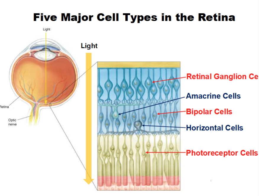
Retina
Overall
Neural tissue lining back of eye
Site of phototransduction
Five Major Retinal Cell Types
Photoreceptor cells (rods and cones)- where phototransduction occurs
Bipolar cells
Retinal ganglion cells
Horizontal cells
Amacrine cells
Retinal Information Linear and Lateral Pathway combined:
Light enters eye
Photoreceptors detect the light, begin phototransduction
Horizontal cells connect neighboring photoreceptors (compare, enhance contrast, and sharpen edges)
Bipolar Cells relay information and sends forward
Amacrine cells modify timing and regulate motion processing
RGCs axons from optic nerve and carry visual information to the brain
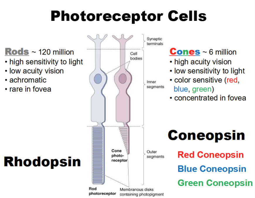
Photoreceptors
Rods
a) Rod Characteristics
~120 million rods
Highly sensitive to light
Function in dim light
Low visual acuity
Achromatic (no color)
Rare in fovea
Cones
a) Cone Characteristics
~6 million cones
Less sensitive to light
High visual acuity
Detect color
Concentrated in fovea
b) Cone Types
Red cones
Green cones
Blue cones
Fovea
What is it
Center of visual field
Light directly reaches photoreceptors
Produces:
Sharp vision
High acuity
Best color vision
Blind Spot
Region lacking photoreceptors
Optic nerve exits eye here
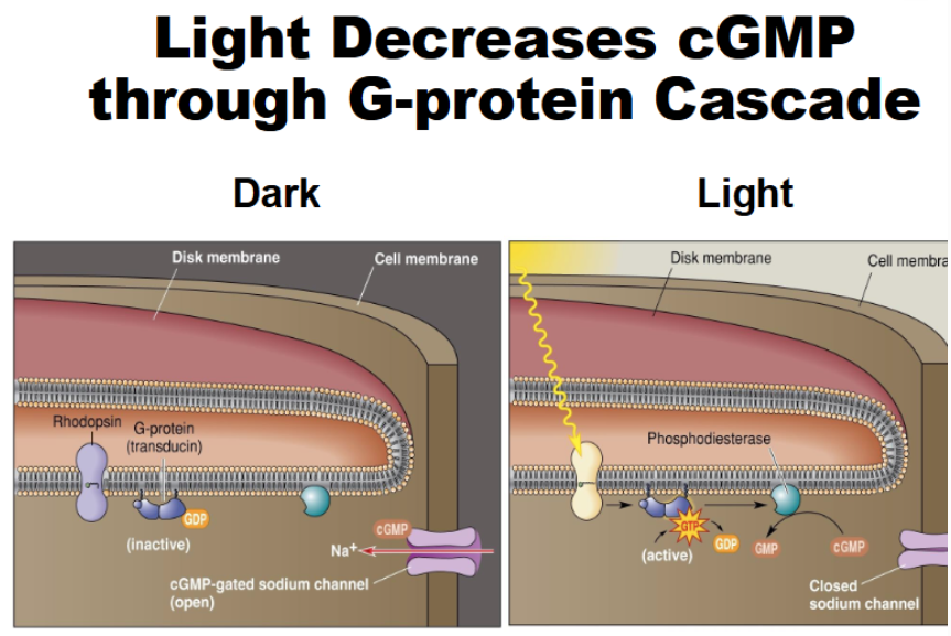
Phototransduction
Dark Current
In darkness, photoreceptors are actually active
Rhodopsin is inactive
Transducin (G protein is inactive)
cGMP levels are high and keep sodium channels open
cGMP-gated Na channels stay open
Na continues to flow into the cell (dark current)
Cell stays depolarized (-30mV)
Glutamate is continuously released
Light Response
When light hits the photoreceptors, the opposite occurs
Light activates rhodopsin
Activated rhodopsin activates transducin (G protein)
Transducin activates phosphodiesterase (PDE)
PDE converts cGMP to GMP
cGMP decreases
Na channels close and don’t flow into cell (influx stops)
Cell is hyperpolarized (-65mV)
Glutamate release stops
***Photoreceptors signal light by REDUCING neurotransmitter release
(add pics)
Opsins
Opsin
G protein-coupled receptor (GPCR)
Combines with retinal to detect light
Rhodopsin
Found in rods
Cone Opsins
Different opsins detect different wavelengths/colors
Color Vision
Red Cones
Hyperpolarize most strongly to red light
Blue Cones
Respond best to blue light
Green Cones
Respond best to green light
Color Detection
Based on comparing activity across cone types
Light intensity coding
Graded Response to Light
Increasing light intensity causes:
Greater hyperpolarization
Less glutamate release
Relationship
Bright light:
Lowest glutamate release
Greatest hyperpolarization
Darkness:
Highest glutamate release
Most depolarized state