PERIO817: How to Measure FGM to CEJ with W.H.O Probe & Periodontal/Gingival Health
 Call Kai
 Learn
Learn Practice Test
 Spaced Repetition
 Match
Match Flashcards
 Knowt Play
1/18

There's no tags or description
Looks like no tags are added yet.
Name | Mastery | Learn | Test | Matching | Spaced | Call with Kai | Chat |
|---|
No analytics yet
Send a link to your students to track their progress
19 Terms
Probing Pocket Depth (PPD)
distance from the free gingival margin to the base of the pokcet
Clinical Attachment Level/Loss (CAL)
defined as the distance from the cementoenamel junction of the tooth to the base of the pocket.
You must ask the question "Can I see the CEJ?" Is there any bone loss?. If not the FGM - CEJ measurement is at least ___ mm
minus (-) 1 mm
Gingivitis Dental Biofilm Induced by
- Associated with __ __ alone
- Mediated by systemic or local ___ __
- __ gingival enlargement
dental biofilm
risk factors
drug-induced
When is FGM - CEJ = Negative # ?
FGM is coronal to the CEJ
(occurs in health, no bone loss)
When is FGM - CEJ = 0?
FGM is at the CEJ
(may occur with/without bone loss)
When is FGM-CEJ = Positive # ?
FGM is apical to the CEJ
(occurs when bone loss is present and recession )
The ___ mm ball of the WHO "engages" the CEJ
0.5 mm
Mucogingival Pathology
straight buccal and lingual surfaces, not interproximal
Mucogingival Defect
probing to or beyond the mucogingival junction (no attached gingiva)
What is the Attached Gingiva Attached to?
Junctional Epithelium
Connective tissues fibers inserted into the cementum
periosteum/bone
When is a Mucogingival defect necessary for treatment?
progressive recession
interferes with oral hygiene
absence of keratinized tissue
Furcation Involvement
bone loss apical to the area where the roots divide, thus making it accessible to bacteria
What do you use to detect furcation involvement?
Nabers probe
Glickman Class 1
slight bone loss
feel convexity of roots
feel concavity of furcation entrance
no horizontal component
no radiographic changes
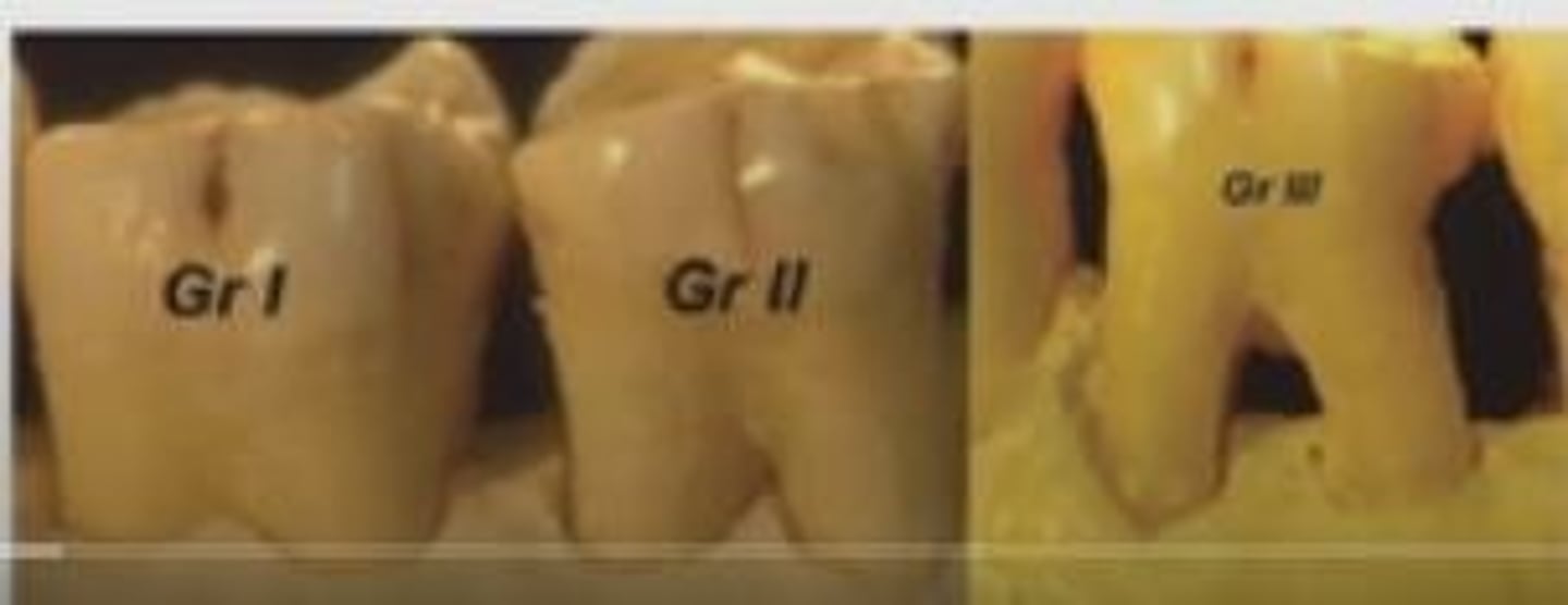
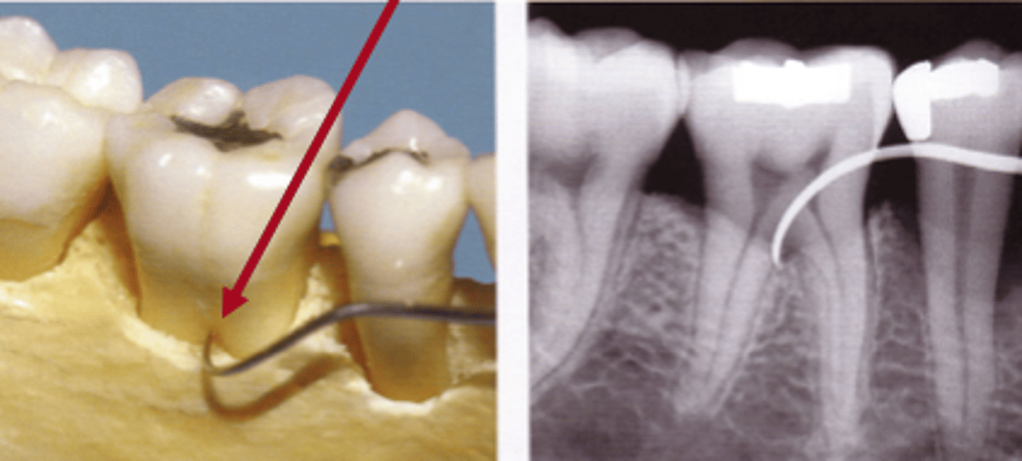
Glickman Class II
definite horizonal component
not through and through
radiographic involvement possible
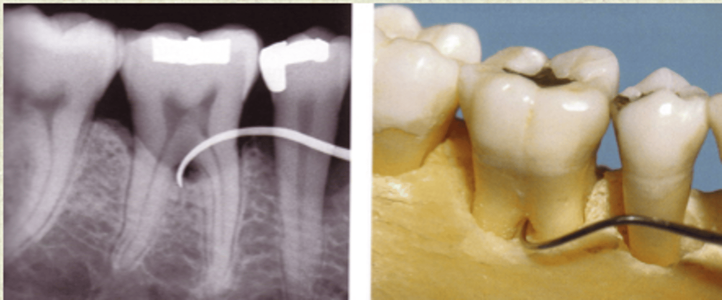
Glickman Class III
bone loss through and through
radiographic evidence
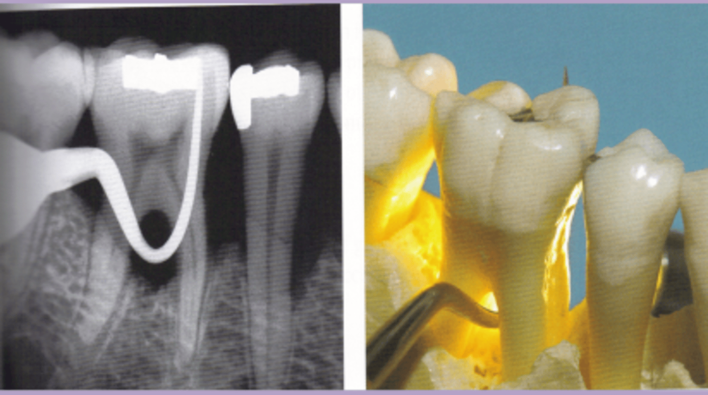
Glickman Class IV
bone loss through and through
clinically can see
radiographic evidence
What stage of Periodontitis must an SRP be done?
Stage III