Functional Anatomy 1 - Lecture 3: Muscles of the Back and Neck
 Call Kai
 Learn
Learn Practice Test
 Spaced Repetition
 Match
Match Flashcards
 Knowt Play
1/47

There's no tags or description
Looks like no tags are added yet.
Name | Mastery | Learn | Test | Matching | Spaced | Call with Kai | Chat |
|---|
No analytics yet
Send a link to your students to track their progress
48 Terms
superficial back muscles
extrinsic
connect upper limb to trunk
attach to the clavicle/humerus/scapula
innervated by names peripheral nerves/ventral rami
intermediate back muscles
extrinsic
serratus posterior superior
serratus posterior inferior
innervated by intercostal nerves
deep back muscles
intrinsic
function
move vertebral column
posture
shock absorbers
all innervated by the posterior/dorsal rami of the segments they span
4 groups (superficial to deep)
splenius muscles (capitis, cervicis) (S)
erector spinae (I)
transversospinales (D)
segmental (D)
splenius capitis
O: ligamentum nuchae and spinous process of C7-T4
I: lateral 1/3 of superior nuchal line and mastoid process
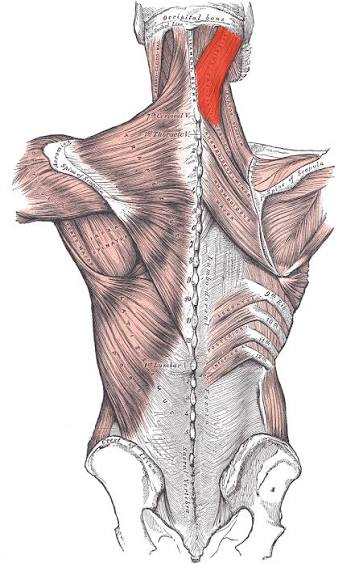
splenius cervicis
O: spinous processes of T3-T6
I: transverse process of C1-C3
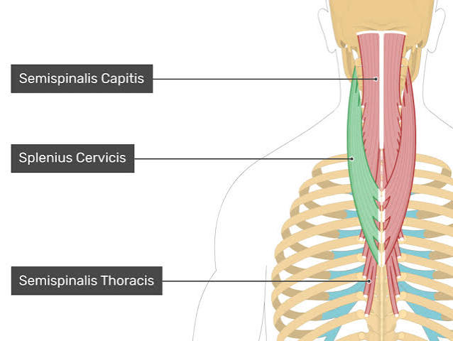
splenius muscles actions
unilaterally: ipsilateral lateral flexion and rotation of head (capitis) and neck (cervicis)
bilaterally: extend head (capitis) and neck (cervicis)
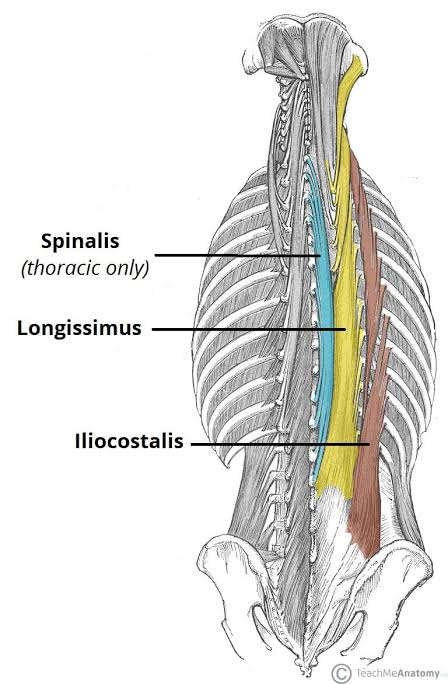
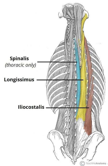
erector spinae
lateral to medial
iliocostalis
longissimus
spinalis
intermediate layer!
iliocostalis
O: iliac crest/lumbar fascia, posterior sacrum
I: angles of ribs (lumbar and thoracic), transverse processes C4-6 (cervical)
longissimus
O: iliac crest, lumbar fascia, lumbar TPs (thoracis), TPs upper thoracic (cervicis/capitis)
I: thoracic TPs/ribs (thoracis), cervical TPs (cervicis), mastoid (capitis)
spinalis
O: spinous process upper lumbar and thoracic
I: spinous processes upper thoracic and cervical
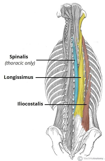
erector spinae group action
bilaterally: extend head and vertebral column
unilaterally: ipsilateral lateral flexion of vertebral column
more lateral attachment, more oblique fiber direction, more contribution to SB
transversospinales
deep layer, note the superior and medial fiber direction for all three
semispinalis
most superficial of deep layer
prominent in cervical region
fibers span 4-6 levels
O: TP C4-T10
I: SPs upper thoracic/cervical and superior nuchal line
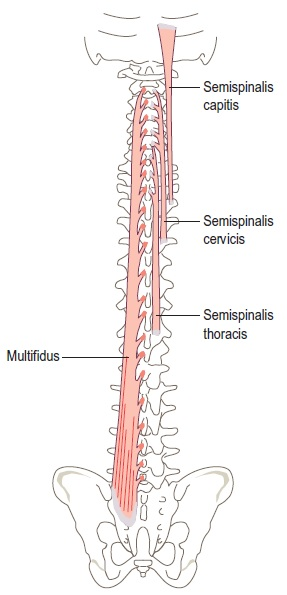
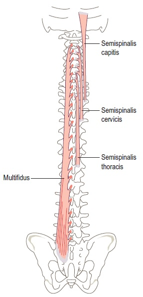
multifidus
prominent in lumbar region
O: posterior sacrum/pelvis, lumbar superior articular processes, TP thoracic
I: SP 2-4 levels above
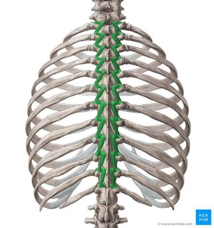
rotatores
deepest of deep layer
prominent in thoracic region
O: transverse processes
I: lamina and SP 1-2 above
transversopinales group action
bilaterally: extend vertebral column
unilaterally: contralateral rotation
pull the spinous process of adjacent/superior vertebrae towards the transverse process of their origin
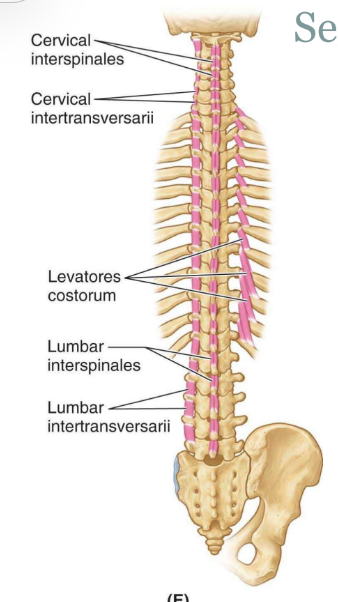
segmental (deepest) muscles
interspinous and intertransversus
run between spinous processes or transverse processes
provide stability
minimal movement
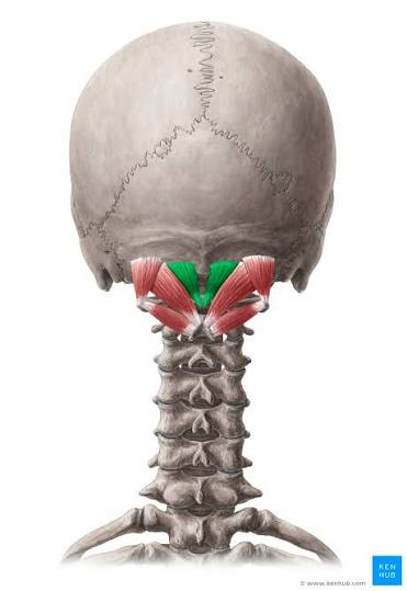
rectus capitis posterior major
O: SP of C2
I: lateral inferior nuchal line
A: extend head; ipsilateral rotation of head
N: posterior ramus of C1 (suboccipital nerve)
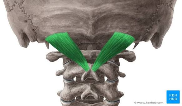
rectus capitis posterior minor
O: posterior tubercle of C1
I: medial aspect of inferior nuchal line
A: extend head
not part of sub-occipital triangle
N: posterior ramus of C1 (suboccipital nerve)
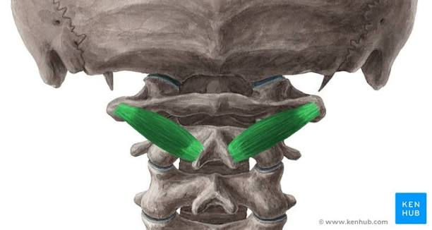
obliquus capitis inferior
O: SP of C2
I: TP of C1
no attachment to skull
A: ipsilateral rotation of C1 on C2
N: posterior ramus of C1
obliquus capitis superior
O: TP of C1
I: occiput between superior and inferior nuchal lines
A: extend head, ipsilateral lateral flexion
N: posterior ramus of C1 (suboccipital nerve)
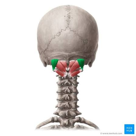
suboccipital nerve (C1)
emerges in middle of triangle
motor innervation to all 3 muscles of the suboccipital triangle
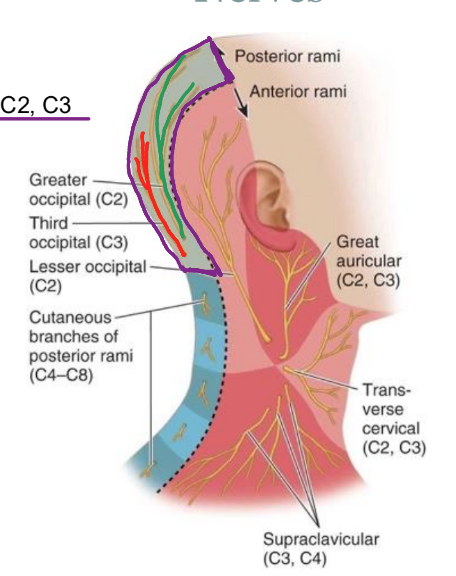
greater occipital nerve (C2)
emerges inferior to triangle
passes through superficial muscles
innervates skin on posterior scalp
cervicogenic headaches
cutaneous innervation of C1-C3
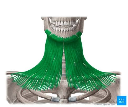
platysma
very superficial
thin and small muscle fibers
O: fascia in the anterior neck and mandible
I: fascia covering pectoralis major
A: muscle of facial expression
N: facial nerve 7
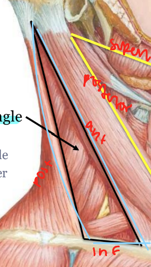
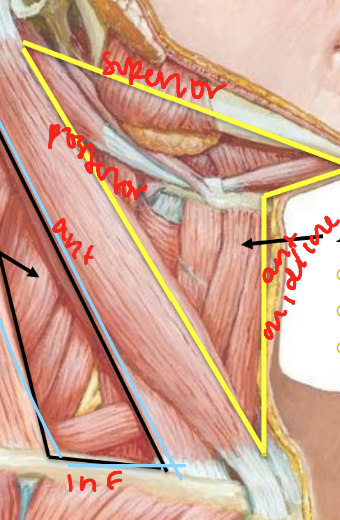
posterior triangle
anterior: SCM
inferior: clavicle
posterior: upper trapezius
anterior triangle
anterior: midline
superior: mandible
posterior: SCM
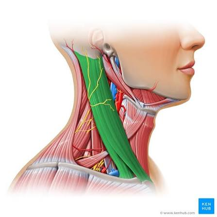
sternocleidomastoid
O: mastoid process/lateral superior nuchal line
I: manubrium of sternum and medial clavicle
A: ipsilateral lateral flexion, contralateral rotation
N: spinal accessory nerve
posterior triangle
floor
splenius capitis
levator scapulae
posterior scalene
anterior scalene
middle scalene
structures within
external jugular vein
subclavian artery and vein
roots of brachial plexus
suprascapular nerve
omohyoid muscle
cervical plexus
anterior rami C1-C4
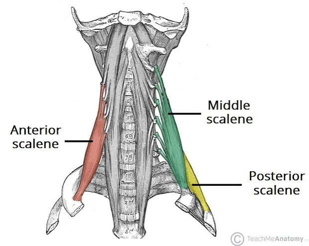
anterior scalene
O: transverse process of C3-C6
I: 1st rib
A:
bilaterally: cervical flexion
unilaterally: laterally flex the cervical spine
alternate: accessory respiratory muscle by raising 1st rib
N: C4-C6 ventral rami
anterior to phrenic nerve, posterior to subclavian artery
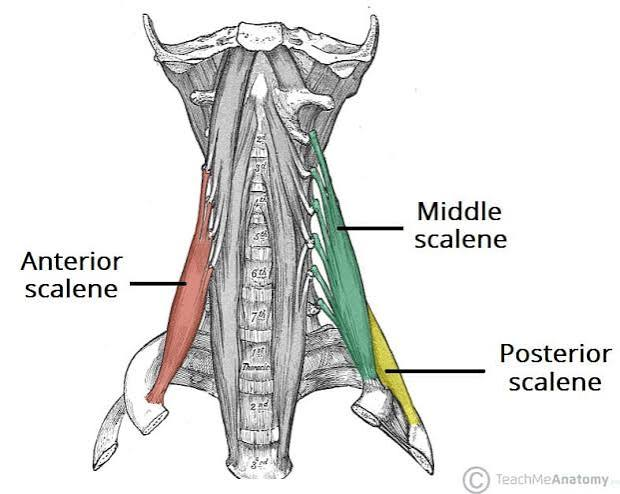
middle scalene
O: TP of C4-C7
I: superior surface of 1st rib
A: ipsilateral lateral flexion cervical spine, elevate ribs
N: mid-lower cervical ventral rami
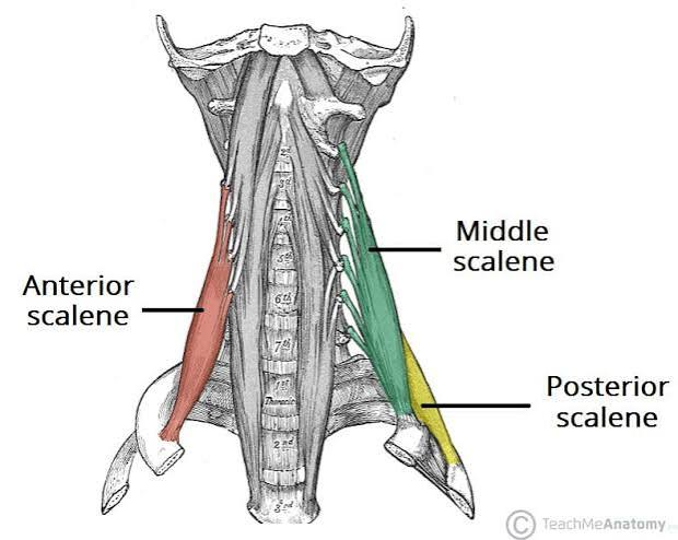
posterior scalene
O: TP C4-C6
I: lateral superior surface of 2nd rib
A: ipsilateral lateral flexion cervical spine, elevate ribs
N: mid-lower cervical ventral rami
suprahyoid muscles
floor of mouth, stable base for tongue
digastric muscle
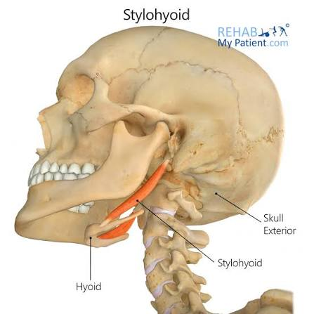
stylohyoid
mylohyoid
geniohyoid
infrahyoid muscles
more hyoid during speech and swallowing actions
omohyoid
thyrohyoid
sternothyroid
hyoid muscles
supra vs infra
defined by their position relative to the hyoid bone
as a whole, aid in movements of swallowing and fine movements of eating
suprahyoids innervated by cranial nerves
infrahyoids innervated by branches off the ansa cervicalis
suprahyoids
floor of the mouth
provide stable base for tongue movements
elevate hyoid to assist in speech and swallow
include
digastric muscle, stylohyoid, mylohyoid, geniohyoid
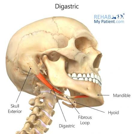
digastric muscle
2 muscle bellies
O: mastoid process
intermediate tendon attached to hyoid
I: anterior mandible
A:
hyoid fixed: depress and retract mandible
mandible is fixed: elevate hyoid
N:
posterior belly: facial nerve
anterior belly: trigeminal nerve, mandibular division
stylohyoid
O: styloid process of skull
I: hyoid
A: elevates hyoid
N: facial nerve
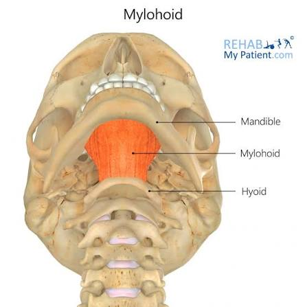
mylohyoid
O: mandible
I: hyoid
forms the floor of the mouth
A:
hyoid fixed: depress mandible
mandible fixed: elevate hyoid
N: trigeminal nerve, mandibular division
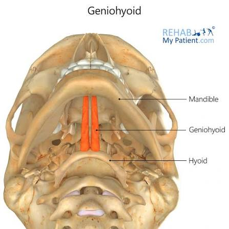
geniohyoid
deep to mylohyoid
O: mandible
I: hyoid
A:
hyoid fixed: depress mandible
mandible fixed: elevate hyoid
N: C1 via hypoglossal nerve (CN 12)
infrahyoids
“strap muscles” because of appearance
omohyoid, thyrohyoid, sternothyroid
A: stabilize the hyoid with the suprahyoid muscles, depress hyoid to assist with speech and swallow
N: ansa cervicalis
except thyrohyoid innervated by C1 via hypoglossal (CN12)
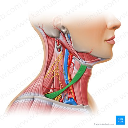
omohyoid
O: hyoid
I: superior border of scapula (near notch)
superior and inferior bellies
superficial to internal jugular vein
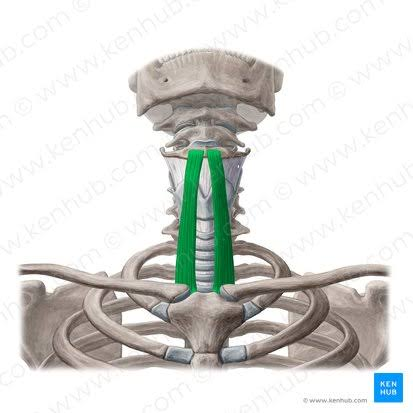
thryohyoid
O: hyoid
I: thyroid cartilage
A: stabilizes/depresses hyoid
if hyoid is fixed it will elevate the larynx
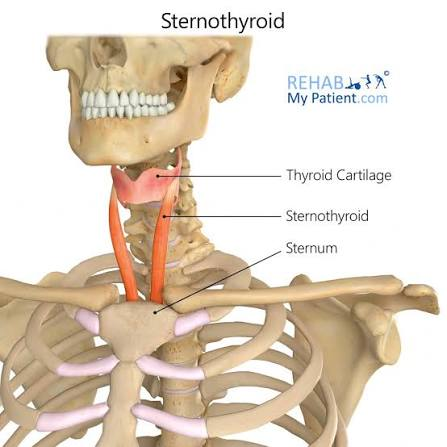
sternothyroid
O: thyroid cartilage
I: manubrium of sternum
not attached to hyoid bone
A: depress larynx
deep anterior muscles
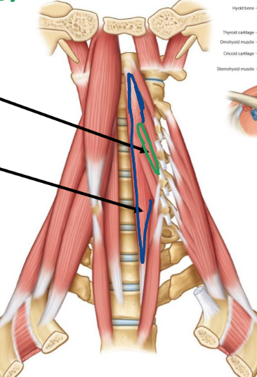
longus colli
oblique portion
O: bodies/transverse process of C3-5
I: C5, C6 vertebrae
linear portion
O: bodies of C5-T3
I: bodies of C2-C4 (linear)
A:
bilaterally: flexes cervical spine
unilaterally: ipsilateral lateral flexion cervical spine
N: C2-C6 ventral rami
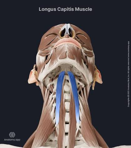
longus capitis
O: TP C3-C6
I: basilar occipital bone
A: flexion and ipsilateral lateral flexion of head
N: C1-C3 ventral rami
veins of the neck
retromandibular
anterior jugular
posterior auricular
external jugular
transverse cervical
internal jugular
arteries of the neck
superior thyroid
superior laryngeal
common carotid
inferior laryngeal
inferior thyroid
left recurrent laryngeal
left subclavian